Medical Pharmacology Chapter 43: Adult Cardiac Procedures
|
|
|
|
|
|
|
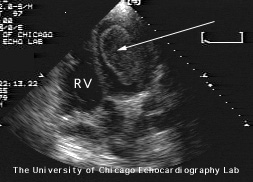
Valvular Disease and Ventricular Function Analysis
-
Echocardiography is useful in detecting
-
Valvular dysfunction
-
Ventricular dilatation/hypertrophy
-
Ventricular dysfunction -- wall motion anomalies; abnormally low ejection fraction
-
-
Large tricuspid valve vegetation (left); Left Atrial Myxoma (right) 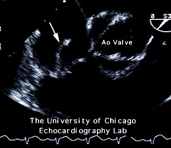
-
Large Left Ventricular Thrombus 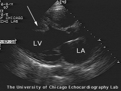

-
"Very large LV thrombus. Initially visible in the parasternal axes.
-
In the apical four chamber it occupies a substantial portion of the LV apex and when the transducer is angulated more inferior the enormous size of the thrombus is evident"
-
Courtesy of University of Chicago Echocardiography Laboratory
-
-
Radionuclide Angiocardiography
-
Pulmonary vascular anomalies, such as pulmonary emboli
-
Rationale for imaging to detect pulmonary emboli
-
Pulmonary emboli common (25-50,000 deaths per year; 25 percent autopsy incidence)
-
Most reliable signs: sudden dyspnea/hypoxemia [less reliable signs: sub-sternal pressure; hemoptysis; pleuritic pain]
-
Pulmonary emboli: maybe lethal, but treatable
-
Imaging techniques confirm clinical sign and screen high-risk patients
-
Diagnostic methodology:
-
Nuclear medicine (isotope tagging) Ventilation/Perfusion Scan (V/Q)
-
Based on IV injection of isotope-tagged microspheres, which "embolize" an insignificant number of capillaries-- as a result "MAP PERFUSION"
-
Ventilation images are obtained by patient inhalation of radioactive Xe 133
-
Ventilation images and perfusion images are then compared
-
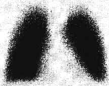
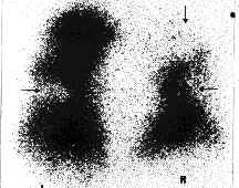
Normal pulmonary ventilation Corresponding abnormal perfusion image -
The presence of several large focal perfusion defects not managed by ventilation defects indicates a high probability of pulmonary embolism.
-
Images courtesy of Creighton University School of Medicine, Special Pulmonary Imaging
-
-
-
-
-
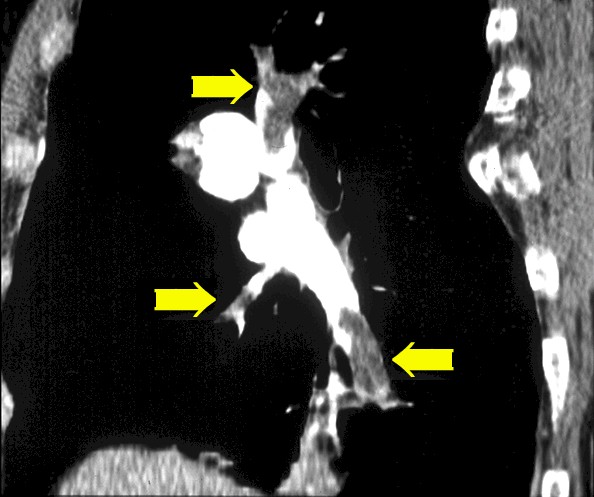
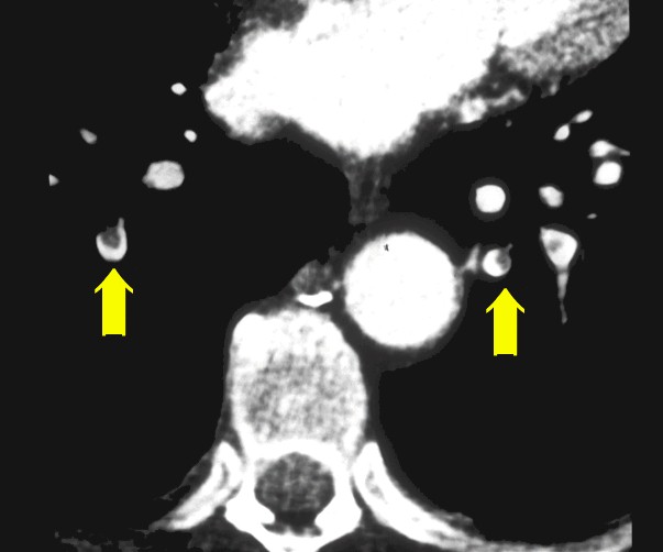
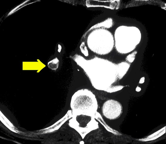
Electron Beam Tomography
-
-
Electron beam capture tomography and visualization of pulmonary emboli (yellow arrows); images courtesy of Dr. Patrick Sheedy, Mayo Clinic, MN
-
-
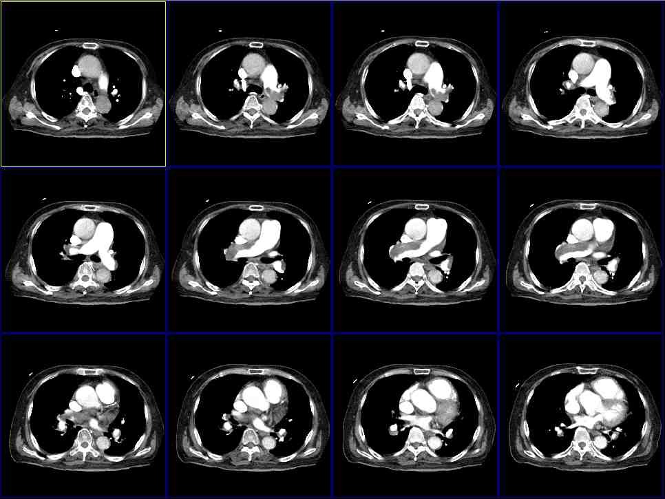
Electron Beam Tomography: Pulmonary Emboli (images courtesy of Imatron )
-
-
-
Primary Reference: Ross, AF, Gomez, MN. and Tinker, JH Anesthesia for Adult Cardiac Procedures in Principles and Practice of Anesthesiology (Longnecker, D.E., Tinker, J.H. Morgan, Jr., G. E., eds) Mosby, St. Louis, Mo., pp. 1659-1698, 1998.
-
Primary Reference: Shanewise, JS and Hug, Jr., CC, Anesthesia for Adult Cardiac Surgery, in Anesthesia, 5th edition, vol 2, (Miller, R.D, editor; consulting editors, Cucchiara, RF, Miller, Jr.,ED, Reves, JG, Roizen, MF and Savarese, JJ) Churchill Livingston, a Division of Harcourt Brace & Company, Philadelphia, pp. 1753-1799, 2000.
-
Primary Reference: Wray Roth, DL, Rothstein, P and Thomas, SJ Anesthesia for Cardiac Surgery, in Clinical Anesthesia, third edition (Barash, PG, Cullen, BF, Stoelting, R.K, eds), Lippincott-Raven Publishers, Philadelphia, pp. 835-865, 1997.
|
|
|
|
|
|
|
|
This Web-based pharmacology and disease-based integrated teaching site is based on reference materials, that are believed reliable and consistent with standards accepted at the time of development. Possibility of human error and on-going research and development in medical sciences do not allow assurance that the information contained herein is in every respect accurate or complete. Users should confirm the information contained herein with other sources. This site should only be considered as a teaching aid for undergraduate and graduate biomedical education and is intended only as a teaching site. Information contained here should not be used for patient management and should not be used as a substitute for consultation with practicing medical professionals. Users of this website should check the product information sheet included in the package of any drug they plan to administer to be certain that the information contained in this site is accurate and that changes have not been made in the recommended dose or in the contraindications for administration. Advertisements that appear on this site are not reviewed for content accuracy and it is the responsibility of users of this website to make individual assessments concerning this information. Medical or other information thus obtained should not be used as a substitute for consultation with practicing medical or scientific or other professionals. |