CHAPTER 8 · ANTIARRHYTHMIC DRUGS

Section 1

Use-Dependent Sodium Channel Blockade

The shared mechanism of Class I agents and the kinetic property that defines their three subclasses

All Class I antiarrhythmic agents block voltage-gated fast sodium channels in cardiac muscle, slowing the rate of phase 0 depolarization and reducing conduction velocity. What distinguishes the three subclasses from each other — and determines their clinical safety profiles — is not whether they block sodium channels but how quickly they release them.

Use-Dependence: What It Means and Why It Matters

Sodium channels cycle through three states during each action potential: resting (closed and available), open (during phase 0 depolarization), and inactivated (immediately after opening, before recovery). Class I drugs bind preferentially to channels in the open or inactivated state. This means that at higher heart rates — when more channels are cycling through these states per unit time — more drug binding occurs per unit time and channel blockade intensifies. This property is called use-dependence or frequency-dependence.

The practical consequence is that Class I agents become more potent during tachycardias, which can be therapeutically useful, but also more dangerous in diseased myocardium where channels may already be partially inactivated due to membrane depolarization from ischemia or scarring. In that setting, use-dependent blockade can convert heterogeneous conduction slowing into a stable, fatal re-entrant circuit.

Three Subclasses Defined by Recovery Kinetics

The clinically important distinction among Class I agents is the speed with which channels recover from drug-bound blockade during the diastolic interval between beats. This recovery kinetics is the basis of the Ia, Ib, and Ic subclassification.

Class Ia agents — quinidine, procainamide, and disopyramide — have intermediate recovery kinetics. They slow conduction moderately, prolong action potential duration and the QT interval (because they also block repolarizing potassium channels), and produce both QRS widening and QT prolongation on the electrocardiogram.

Class Ib agents — lidocaine and mexiletine — recover rapidly, faster than the diastolic interval at normal heart rates. This means their blockade is largely relieved between beats in normal tissue. Their net effect is conduction slowing that is most pronounced in ischemic or rapidly firing tissue where channels spend more time in inactivated states. They shorten action potential duration and cause minimal electrocardiogram changes at rest.

Class Ic agents — flecainide and propafenone — recover very slowly, slower than the diastolic interval even at slow heart rates. Channel blockade is not relieved between beats, producing tonic conduction depression at all heart rates that intensifies further during tachycardia. They produce marked QRS widening with minimal change in action potential duration. This combination of properties makes them highly effective in structurally normal hearts but profoundly dangerous when myocardial scarring is present.

The Key Clinical Distinction Within Class I

Class Ib agents are the safest Class I drugs because rapid recovery kinetics limit their activity to ischemic or tachycardic tissue. Class Ic agents are the most dangerous in diseased hearts because slow recovery kinetics produce persistent, rate-independent conduction slowing that can stabilize re-entrant circuits in scar tissue. Class Ia agents occupy the middle ground and carry their own proarrhythmic risk through QT prolongation.


Section 2

Class Ia Agents

Quinidine, procainamide, and disopyramide — intermediate kinetics with QT prolongation

Class Ia agents block sodium channels with intermediate recovery kinetics and additionally block repolarizing potassium channels, producing both QRS widening and QT interval prolongation. All three carry torsades de pointes risk. They differ from each other primarily in their additional receptor effects, which determine their unique indications and toxicity profiles.
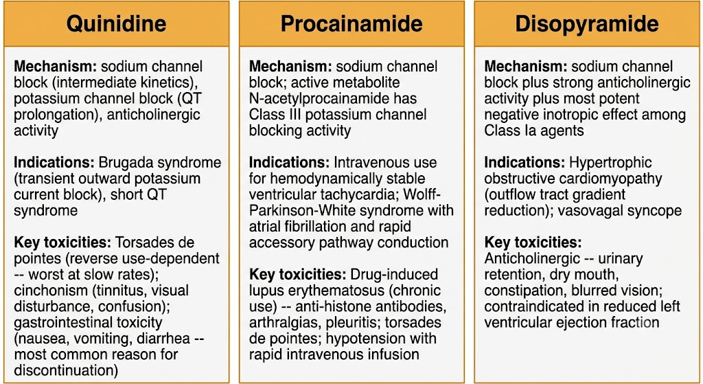
Three-panel comparison diagram showing the three Class Ia antiarrhythmic agents — quinidine, procainamide, and disopyramide — with mechanism, primary indications, and key toxicities for each drug.
Source: AI-generated figure (Gemini). Educational use.

Quinidine

Quinidine is the prototype Class Ia agent and the oldest antiarrhythmic drug in clinical use. It blocks sodium channels with intermediate kinetics, blocks multiple potassium channels producing QT prolongation, and has significant anticholinergic activity that can paradoxically accelerate atrioventricular nodal conduction and partially offset direct atrioventricular nodal slowing. The net electrocardiogram effect is modest QRS widening combined with QT prolongation.

Contemporary indications for quinidine are narrow. It retains a role in Brugada syndrome, where its blockade of the transient outward potassium current that exaggerates the phase 1 notch can normalize the electrocardiogram pattern and reduce ventricular fibrillation burden. It is also used in short QT syndrome to prolong an abnormally short QT interval. Its prior use for maintaining sinus rhythm in atrial fibrillation has been largely replaced by safer agents.

Quinidine Toxicity — Three Patterns to Know

Quinidine syncope (torsades de pointes): Paradoxically more common at low drug concentrations and slow heart rates — a manifestation of reverse use-dependence. Requires immediate discontinuation and intravenous magnesium. Occurs in roughly 1 to 3 percent of patients.

Cinchonism: A dose-related syndrome of tinnitus, hearing loss, visual disturbance, headache, and confusion caused by quinidine's central nervous system effects. Resembles salicylate toxicity.

Gastrointestinal toxicity: Nausea, vomiting, and diarrhea in up to 30 to 40 percent of patients — the most common reason for discontinuation.

Procainamide

Procainamide shares quinidine's sodium channel blocking properties but has weaker anticholinergic activity and weaker potassium channel blockade. Its most important contemporary use is intravenous administration for hemodynamically stable wide-complex tachycardia when ventricular tachycardia is confirmed or strongly suspected, and for Wolff-Parkinson-White syndrome with atrial fibrillation and rapid accessory pathway conduction — a situation where atrioventricular nodal blocking agents are contraindicated. Intravenous procainamide must be infused slowly; rapid infusion causes hypotension from vasodilation.

Procainamide is converted by hepatic acetylation to an active metabolite called N-acetylprocainamide, which is a pure potassium channel blocker with Class III activity. This metabolite accumulates in patients with renal impairment and contributes to QT prolongation risk independent of the parent drug.

Long-term oral procainamide carries a serious risk of drug-induced lupus erythematosus, occurring in 15 to 40 percent of patients on chronic therapy. The syndrome presents with arthralgias, pleuritis, and pericarditis, and is characterized by antinuclear antibodies with an anti-histone pattern. It resolves on drug discontinuation. Because of this toxicity, oral procainamide is rarely used today.

Disopyramide

Disopyramide is the most potently anticholinergic of the Class Ia agents and has the most pronounced negative inotropic effect among them. These properties that limit its use in most settings give it a unique therapeutic niche in hypertrophic obstructive cardiomyopathy, where the negative inotropic effect reduces the left ventricular outflow tract gradient. It is also used in vasovagal syncope, where its anticholinergic and negative inotropic effects can reduce reflex bradycardia and peripheral vasodilation.

Anticholinergic toxicity is the dominant adverse effect: urinary retention (particularly in men with prostatic enlargement), dry mouth, constipation, blurred vision, and precipitation of narrow-angle glaucoma. Like all Class Ia agents, disopyramide causes QT prolongation and carries torsades de pointes risk. It is contraindicated in patients with reduced left ventricular ejection fraction outside the hypertrophic obstructive cardiomyopathy indication, because its negative inotropic effect can precipitate heart failure.

Class Ia

Quinidine

  • Sodium channel + potassium channel + anticholinergic
  • Brugada syndrome, short QT syndrome
  • Torsades de pointes, cinchonism, gastrointestinal toxicity

Class Ia

Procainamide

  • Sodium channel block; active metabolite has Class III activity
  • Intravenous use: stable ventricular tachycardia, Wolff-Parkinson-White with atrial fibrillation
  • Drug-induced lupus (oral, long-term); torsades de pointes

Class Ia

Disopyramide

  • Sodium channel block + strong anticholinergic + negative inotrope
  • Hypertrophic obstructive cardiomyopathy, vasovagal syncope
  • Urinary retention, dry mouth; contraindicated in reduced ejection fraction
Section 3

Class Ib Agents

Lidocaine and mexiletine — rapid kinetics with selective activity in ischemic tissue

Class Ib agents recover from sodium channel blockade so rapidly that they have minimal effect on normal myocardium at resting heart rates. Their blockade is most pronounced in ischemic or depolarized tissue where channels are already partially inactivated. This selective pharmacodynamic profile makes them useful for ventricular arrhythmias arising from ischemic substrates, but ineffective against supraventricular arrhythmias.

Lidocaine

Lidocaine is available only for intravenous use as an antiarrhythmic — oral bioavailability is too low for reliable dosing because of extensive first-pass hepatic metabolism. It blocks sodium channels with strong selectivity for inactivated channels, shortens action potential duration in Purkinje fibers, and has virtually no effect on atrial tissue or the atrioventricular node. QRS duration and QT interval remain unchanged at therapeutic concentrations.

Its antiarrhythmic action is most pronounced in ischemic myocardium, where the resting membrane potential is less negative than in normal tissue, keeping a larger fraction of sodium channels in the inactivated state and increasing drug binding. This is why lidocaine is effective in the acute management of ventricular tachycardia and ventricular fibrillation in the setting of ischemia or myocardial infarction. In contemporary Advanced Cardiac Life Support, lidocaine is a second-line agent after amiodarone for pulseless ventricular tachycardia or ventricular fibrillation.

Lidocaine toxicity is primarily neurologic and concentration-dependent. At mildly elevated concentrations, patients develop perioral numbness, lightheadedness, and tinnitus. At higher concentrations, dysarthria, confusion, and nystagmus appear. Severe toxicity produces seizures, which are treated with benzodiazepines. Cardiac depression — bradycardia, hypotension, atrioventricular block — is rare at therapeutic concentrations but can occur with massive overdose. Patients with heart failure or hepatic impairment require dose reduction because hepatic clearance of lidocaine is blood flow-dependent and reduced in low-output states.

Mexiletine

Mexiletine is the oral analogue of lidocaine with an identical electrophysiologic mechanism. Its oral bioavailability is adequate (approximately 90 percent) and its longer half-life allows twice- or three-times daily dosing. It is used for chronic suppression of ventricular arrhythmias in patients where an oral agent is needed.

An important adjunctive use of mexiletine is in Long QT Syndrome Type 3, a congenital channelopathy caused by a gain-of-function mutation in the cardiac sodium channel that produces a persistent late inward sodium current during the action potential plateau, prolonging the QT interval. Mexiletine blocks this late sodium current and can shorten the QT interval in affected patients, reducing torsades de pointes risk.

The dominant adverse effect of mexiletine is gastrointestinal — nausea, vomiting, and abdominal discomfort occur in up to 40 percent of patients and are the most common reason for discontinuation. Taking mexiletine with food reduces but does not eliminate these effects. Neurologic toxicity (tremor, dizziness, ataxia) occurs at higher concentrations, mirroring lidocaine's central nervous system profile.

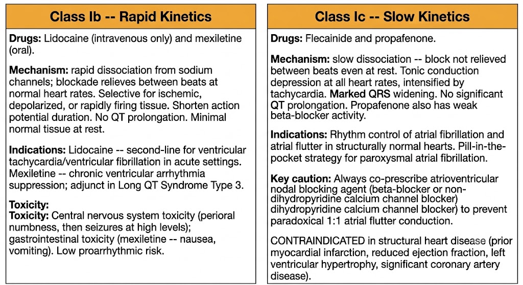
Two-panel comparison diagram contrasting Class Ib agents on the left showing rapid kinetics with lidocaine and mexiletine, and Class Ic agents on the right showing slow kinetics with flecainide and propafenone, including mechanisms, indications, and the structural heart disease contraindication.
Source: AI-generated figure (Gemini). Educational use.

Section 4

Class Ic Agents

Flecainide and propafenone — potent conduction slowing, effective in structurally normal hearts only

Class Ic agents are the most potent sodium channel blockers among the antiarrhythmics. Their slow recovery kinetics produce profound, rate-independent conduction slowing that is highly effective for atrial arrhythmias in patients without structural heart disease — and potentially fatal in those who have it.

Flecainide

Flecainide produces marked slowing of conduction in all cardiac tissue — atria, ventricles, and the His-Purkinje system — resulting in QRS widening on the electrocardiogram without significant change in QT interval. Its sodium channel blockade intensifies with increasing heart rate (use-dependence), making it particularly effective at terminating and preventing re-entrant atrial arrhythmias.

Flecainide is a first-line option for rhythm control of atrial fibrillation and atrial flutter in patients with no structural heart disease, no left ventricular hypertrophy, and no ischemic heart disease. It can be used as a "pill-in-the-pocket" strategy, in which a patient takes a single oral dose at the onset of palpitations to self-cardiovert a paroxysmal atrial fibrillation episode at home. This approach requires that the patient have had prior successful cardioversion with the drug in a monitored setting and that they have no contraindications.

A critical safety rule: when flecainide is used for atrial fibrillation or atrial flutter, an atrioventricular nodal blocking agent (a beta-blocker or non-dihydropyridine calcium channel blocker) must always be co-prescribed. Flecainide slows atrial flutter rate from approximately 300 beats per minute to approximately 200 beats per minute. At the slower flutter rate, the atrioventricular node may conduct every impulse (1:1 atrioventricular conduction), producing a paradoxical and dangerous acceleration of the ventricular rate. Co-administration of an atrioventricular nodal blocking agent prevents this complication.

Propafenone

Propafenone shares flecainide's Class Ic sodium channel blocking profile and is used for the same indications: rhythm control of atrial fibrillation and supraventricular tachycardias in structurally normal hearts, including the pill-in-the-pocket strategy. The same structural heart disease contraindication applies, and the same requirement for atrioventricular nodal agent co-prescription when used for atrial flutter.

What distinguishes propafenone from flecainide is its additional weak beta-adrenergic receptor blocking activity. This beta-blocking effect is clinically meaningful and can cause bradycardia, atrioventricular block, and bronchospasm — the last being particularly relevant in patients with reactive airway disease or asthma, who may not tolerate propafenone even if flecainide would be acceptable. In patients who are poor metabolizers of the hepatic enzyme responsible for propafenone's primary metabolic pathway, the parent compound accumulates and beta-blocking effects become more pronounced.

Class Ic Agents: The Absolute Structural Heart Disease Contraindication

Flecainide and propafenone are contraindicated in any patient with structural heart disease — including prior myocardial infarction, reduced left ventricular ejection fraction, significant left ventricular hypertrophy, or significant coronary artery disease. This contraindication applies regardless of the arrhythmia being treated. A patient with a history of myocardial infarction who develops atrial fibrillation cannot receive flecainide, even though flecainide would be appropriate for the same atrial fibrillation in a structurally normal heart.


Section 5

The Cardiac Arrhythmia Suppression Trial and Clinical Prescribing Rules

Why suppressing ectopy does not equal reducing mortality — and how this shapes drug selection

The Cardiac Arrhythmia Suppression Trial is among the most consequential trials in cardiovascular pharmacology. It permanently changed antiarrhythmic prescribing and established the most important safety boundary in this drug class: Class Ic agents are contraindicated in structural heart disease.

The Trial and Its Finding

The Cardiac Arrhythmia Suppression Trial enrolled patients with prior myocardial infarction and asymptomatic or mildly symptomatic ventricular premature beats. The hypothesis — widely accepted at the time — was that suppressing ventricular ectopy after myocardial infarction would reduce sudden cardiac death. Patients who achieved ventricular premature beat suppression with encainide, flecainide, or placebo were enrolled and followed.

The trial was stopped early when encainide and flecainide were found to increase arrhythmic death and cardiac arrest compared to placebo, despite effectively suppressing ventricular premature beats. The drugs did exactly what they were supposed to do pharmacologically — and patients died at a higher rate than those who received sugar pills.

The mechanism: use-dependent sodium channel blockade in peri-infarct scar tissue converts minor, non-sustained re-entrant circuits into sustained, fatal ventricular tachycardia or ventricular fibrillation. Ventricular premature beat suppression is a surrogate endpoint, not a clinical benefit. This lesson — that surrogate endpoints can mislead — applies far beyond antiarrhythmic pharmacology.

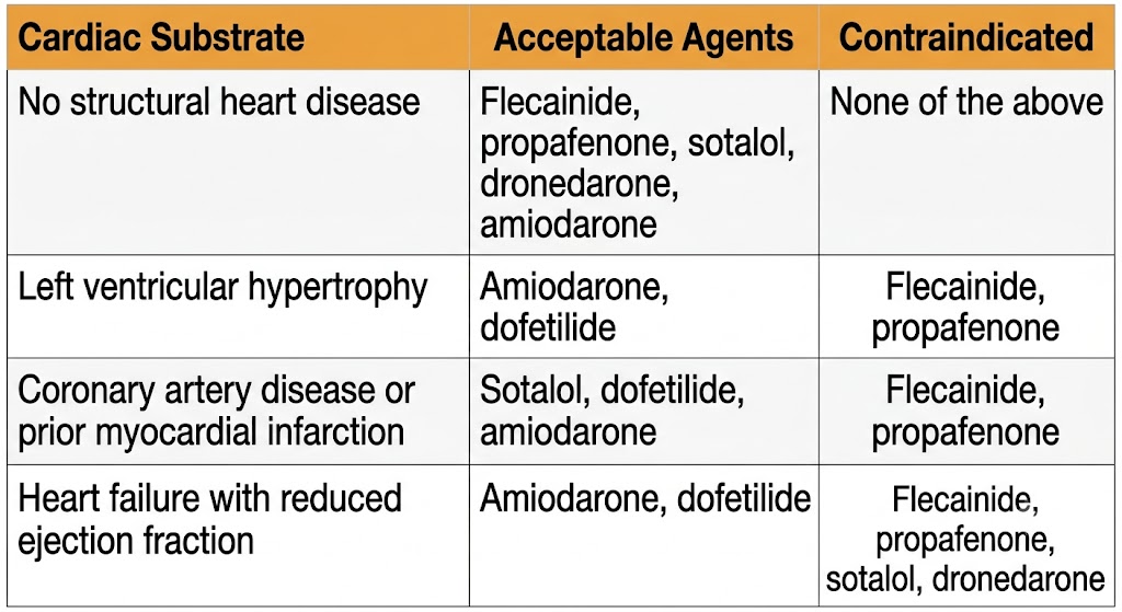
Reference table showing antiarrhythmic drug selection for atrial fibrillation rhythm control by cardiac substrate, with four rows covering no structural heart disease, left ventricular hypertrophy, coronary artery disease or prior myocardial infarction, and heart failure with reduced ejection fraction, listing acceptable and contraindicated agents for each.
Source: AI-generated figure (Gemini). Educational use.

Drug Selection by Cardiac Substrate

The Cardiac Arrhythmia Suppression Trial findings drive a critical decision branch whenever rhythm control is considered for atrial fibrillation. The patient's cardiac substrate — not just the arrhythmia — determines which agents are safe.

No Structural Heart Disease

Broadest Options Available

  • Flecainide — acceptable
  • Propafenone — acceptable
  • Sotalol — acceptable (preserved ejection fraction)
  • Dronedarone — acceptable
  • Amiodarone — acceptable (usually reserved if others fail)

Left Ventricular Hypertrophy

Class Ic Contraindicated

  • Flecainide — contraindicated
  • Propafenone — contraindicated
  • Amiodarone — preferred
  • Dofetilide — acceptable
  • Sotalol — use with caution

Coronary Artery Disease / Prior Myocardial Infarction

Class Ic Contraindicated

  • Flecainide — contraindicated
  • Propafenone — contraindicated
  • Sotalol — acceptable (preserved ejection fraction)
  • Dofetilide — acceptable
  • Amiodarone — acceptable

Heart Failure with Reduced Ejection Fraction

Most Agents Contraindicated

  • Flecainide — contraindicated
  • Propafenone — contraindicated
  • Sotalol — contraindicated
  • Dronedarone — contraindicated
  • Amiodarone — acceptable
  • Dofetilide — acceptable

Class-Specific Proarrhythmia Patterns

Two proarrhythmic patterns are high-yield for second-year pharmacology and deserve explicit recognition alongside the Cardiac Arrhythmia Suppression Trial finding.

Torsades de pointes from Class Ia agents arises from QT interval prolongation due to potassium channel blockade, which enables early afterdepolarizations during a prolonged phase 3. Risk is highest at slow heart rates, with concurrent hypokalemia or hypomagnesemia, and when other QT-prolonging drugs are co-administered. Treatment is intravenous magnesium, electrolyte correction, and withdrawal of the offending agent. If episodes are pause-dependent, heart rate must be increased with isoproterenol or temporary cardiac pacing.

Paradoxical ventricular rate acceleration from Class Ic agents occurs when flecainide or propafenone slows the atrial flutter circuit rate from approximately 300 beats per minute to approximately 200 beats per minute. At the slower atrial rate, the atrioventricular node may conduct every impulse rather than blocking alternate ones, producing 1:1 atrioventricular conduction and a ventricular rate of 200 or more beats per minute. Co-prescription of a beta-blocker or non-dihydropyridine calcium channel blocker is mandatory whenever Class Ic agents are used for atrial arrhythmias.


Suggested References
Author / Organization Title Source
Hille B Local anesthetics: hydrophilic and hydrophobic pathways for the drug-receptor reaction J Gen Physiol. 1977;69(4):497–515
Harrison DC Antiarrhythmic drug classification: new science and practical applications Am J Cardiol. 1985;56(4):185–187
Roden DM Antiarrhythmic drugs. In: Brunton LL, Hilal-Dandan R, Knollmann BC, eds. Goodman & Gilman's: The Pharmacological Basis of Therapeutics. 13th ed. McGraw-Hill; 2018:547–571
Roden DM Risks and benefits of antiarrhythmic therapy N Engl J Med. 1994;331(12):785–791
Chinn K, Brown BS, Carlsson L Lidocaine. In: Singh BN, Wellens HJJ, Hiraoka M, eds. Electropharmacological Control of Cardiac Arrhythmias Futura; 1994:191–210
Aliot E, Capucci A, Crijns HJ, Goette A, Tamargo J Twenty-five years in the making: flecainide is safe and effective in the long-term treatment of atrial fibrillation Europace. 2011;13(2):161–173
Echt DS, Liebson PR, Mitchell LB, et al Mortality and morbidity in patients receiving encainide, flecainide, or placebo: the Cardiac Arrhythmia Suppression Trial N Engl J Med. 1991;324(12):781–788
January CT, Wann LS, Calkins H, et al 2019 AHA/ACC/HRS focused update of the 2014 AHA/ACC/HRS guideline for the management of patients with atrial fibrillation J Am Coll Cardiol. 2019;74(1):104–132
Brugada J, Katritsis DG, Arbelo E, et al 2019 ESC Guidelines for the management of patients with supraventricular tachycardia Eur Heart J. 2020;41(5):655–720
Connolly SJ, Dorian P, Roberts RS, et al Comparison of beta-blockers, amiodarone plus beta-blockers, or sotalol for prevention of shocks from implantable cardioverter defibrillators: the OPTIC study JAMA. 2006;295(2):165–171