Abbreviations used in this module: glucocorticoid (GC), glucocorticoid-induced osteoporosis (GIOP), avascular necrosis (AVN), hypothalamic-pituitary-adrenal (HPA), adrenocorticotropic hormone (ACTH), nonsteroidal anti-inflammatory drug (NSAID), proton pump inhibitor (PPI), interferon-gamma release assay (IGRA), tuberculosis (TB), hepatitis B virus (HBV), Pneumocystis jirovecii pneumonia (PJP), trimethoprim-sulfamethoxazole (TMP-SMX), monosodium urate (MSU), NLRP3 inflammasome (NOD-like receptor family pyrin domain-containing protein 3), interleukin-1 beta (IL-1β), xanthine oxidase (XO), urate-lowering therapy (ULT), estimated glomerular filtration rate (eGFR).
Corticosteroid adverse effects reflect the ubiquitous distribution of the GC receptor throughout the body. Every organ that participates in the therapeutic response also participates in the toxicity profile. Adverse effects are dose-dependent and duration-dependent — the risk of clinically significant toxicity rises substantially above prednisone 5 mg per day chronically and with courses extending beyond four weeks.
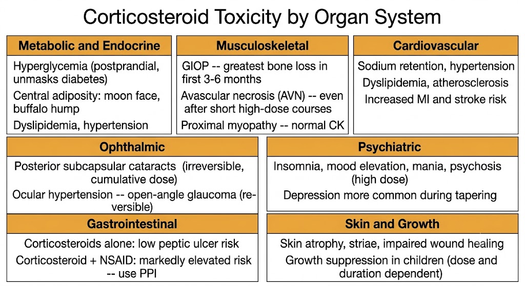
GC excess drives hyperglycemia through two converging mechanisms: increased hepatic glucose output via upregulation of gluconeogenic enzymes, and peripheral insulin resistance at the level of glucose transporter type 4 in muscle and adipose tissue. The hyperglycemia is characteristically postprandial and most pronounced in the afternoon and evening following morning dosing — fasting glucose measurements underestimate its severity. Corticosteroids may unmask latent type 2 diabetes or convert well-controlled diabetes into insulin-requiring disease. Central redistribution of adipose tissue (moon face, buffalo hump, truncal obesity with relative limb wasting) is driven by differential effects of GC excess on adipocyte populations in different anatomical depots.
GIOP is the most clinically consequential long-term musculoskeletal adverse effect. Corticosteroids suppress osteoblast activity and promote osteoclast-mediated bone resorption through multiple transcriptional mechanisms. The greatest rate of bone loss occurs in the first three to six months of therapy — early prophylaxis is therefore more effective than late intervention. Guidelines recommend bisphosphonate prophylaxis for patients prescribed prednisone equivalent to 2.5 mg per day or higher for three months or longer. Calcium and vitamin D supplementation is required in all patients on systemic corticosteroids.
AVN is a distinct and serious musculoskeletal complication: corticosteroids cause fat embolism and endothelial injury in subchondral bone vasculature, leading to ischemic necrosis at the femoral head, humeral head, and femoral condyles. AVN can follow even short high-dose courses — including pulse methylprednisolone — and may present months after treatment ends. Any patient who has received high-dose corticosteroids and develops hip, knee, or shoulder pain should be evaluated with MRI, which detects AVN before plain radiographs become abnormal. Steroid myopathy causes insidious proximal muscle weakness (hip flexors, shoulder girdle) from type II muscle fiber atrophy; it is distinguished from inflammatory myopathy by normal creatine kinase levels and improves with dose reduction.
Even low-dose chronic corticosteroids increase cardiovascular risk through sodium retention, hypertension, dyslipidemia (elevated low-density lipoprotein and triglycerides), endothelial dysfunction, and acceleration of atherosclerosis. Blood pressure monitoring and lipid management are components of long-term corticosteroid management.
Two distinct ophthalmic adverse effects occur with corticosteroid use. Posterior subcapsular cataracts form through direct GC receptor-mediated effects on lens epithelial cells; they correlate with cumulative dose and duration and are largely irreversible. Ocular hypertension and open-angle glaucoma develop through corticosteroid-induced impairment of trabecular meshwork drainage of aqueous humor; this effect is more pronounced with topical ophthalmic corticosteroids but occurs with systemic use as well. Unlike cataracts, pressure elevation is generally reversible on drug discontinuation. Annual ophthalmologic review is appropriate for patients on long-term therapy.
Psychiatric effects span a wide spectrum: insomnia and mild mood elevation are common at moderate doses, while severe mania or frank psychosis occur predominantly at high doses (prednisone equivalent 40 mg per day or higher) and typically resolve with dose reduction. Depression is more common than mania during tapering phases. A prior history of psychiatric illness does not absolutely contraindicate corticosteroid use but mandates close monitoring.
Corticosteroids alone are not a major cause of peptic ulcer disease, but the combination of a corticosteroid with an NSAID multiplies the gastrointestinal bleeding risk markedly — PPI co-prescription is mandatory when these two drug classes are used together. Dermatologic effects include skin atrophy and striae from inhibition of fibroblast collagen synthesis, easy bruising from capillary fragility, and impaired wound healing. In children, growth suppression from inhibition of growth hormone and insulin-like growth factor-1 signaling is a dose- and duration-dependent adverse effect that is substantially attenuated by alternate-day dosing regimens.
AVN of the femoral head can occur after pulse methylprednisolone and may present months after treatment ends. There is no safe minimum dose below which AVN risk is zero. Any patient who has received high-dose corticosteroids and develops hip, knee, or shoulder pain requires MRI evaluation — plain radiographs are often normal in early AVN. Early diagnosis allows core decompression before structural collapse. This is a class effect of corticosteroid therapy.
Immunosuppression from corticosteroids increases susceptibility to opportunistic pathogens and reactivation of latent infections. Risk is dose-dependent, duration-dependent, and synergistic with other immunosuppressive agents. Three mandatory pre-therapy screens and a prophylaxis decision must be completed before initiating chronic immunosuppressive corticosteroid doses.
Prednisone below 10 mg per day for less than two weeks carries low risk for opportunistic infections in otherwise immunocompetent patients. Prednisone at or above 20 mg per day for four weeks or longer substantially increases the risk of opportunistic infections, particularly PJP — formerly called Pneumocystis carinii pneumonia — which carries mortality rates of 30 to 50% in immunosuppressed patients. TMP-SMX is the preferred prophylactic agent, with a single-strength tablet daily (80 mg trimethoprim plus 400 mg sulfamethoxazole) providing nearly complete protection. TMP-SMX also provides prophylaxis against toxoplasmosis and nocardiosis. Current guidance recommends PJP prophylaxis for patients receiving prednisone at or above 20 mg per day for four weeks or longer in combination with other immunosuppressive agents. For patients intolerant of TMP-SMX, alternatives include dapsone, atovaquone, or inhaled pentamidine.
Latent TB can reactivate with immunosuppressive corticosteroid doses. All patients about to begin prolonged moderate-to-high dose corticosteroid therapy should be screened with an IGRA (such as QuantiFERON-TB Gold) or tuberculin skin test; IGRA is preferred in patients who have received BCG vaccination, as it does not produce false-positive results from that exposure. Active TB must be excluded clinically and radiologically. Patients with confirmed latent TB without active disease should receive isoniazid preventive therapy for nine months, ideally initiated at least four weeks before immunosuppression in non-urgent settings. In urgent clinical situations, corticosteroids and isoniazid can be started simultaneously.
HBV reactivation can cause fulminant hepatitis in immunosuppressed patients. All patients initiating corticosteroid therapy at or above prednisone 10 mg per day for four weeks or longer, or who will receive concurrent immunosuppressants, should be screened with hepatitis B surface antigen, total antibody to hepatitis B core antigen, and antibody to hepatitis B surface antigen. Patients who are hepatitis B surface antigen-positive (chronic HBV infection) require antiviral prophylaxis with a high-barrier nucleotide analogue (entecavir or tenofovir) continued for six to twelve months after immunosuppression ends. Patients with past infection (positive core antibody, negative surface antigen) have lower reactivation risk but warrant monitoring with HBV DNA levels during high-level immunosuppression.
Live attenuated vaccines — including measles-mumps-rubella, varicella-zoster, yellow fever, and intranasal influenza — are contraindicated in patients receiving prednisone at or above 20 mg per day for two weeks or longer, because corticosteroid-mediated immunosuppression can allow vaccine-strain replication and dissemination. Inactivated vaccines (injectable influenza, pneumococcal, recombinant zoster, tetanus-diphtheria-pertussis) are safe and should be given whenever feasible, ideally before immunosuppression begins. Steroid-sparing strategies — disease-modifying antirheumatic drugs, conventional immunosuppressants such as methotrexate and azathioprine, and targeted biologics — allow corticosteroid dose reduction and should be employed whenever the underlying condition permits, to minimize cumulative corticosteroid exposure and toxicity.
Three clinical entities can present with overlapping symptoms during corticosteroid tapering: adrenal insufficiency, corticosteroid withdrawal syndrome, and relapse of the underlying disease. Distinguishing them is clinically essential because their management differs completely.
Adrenal insufficiency is confirmed by a low morning serum cortisol (below 3 micrograms per deciliter) or subnormal cortisol response on ACTH stimulation testing. It may be accompanied by hyponatremia and hypoglycemia. Management requires slowing the taper and potentially temporarily increasing the dose; patients with persistent adrenal insufficiency may need stress dosing during physiological challenges.
Corticosteroid withdrawal syndrome is a distinct entity from adrenal insufficiency. It results from physiological dependence on supraphysiological GC concentrations: tissues that have adapted to chronically elevated GC receptor activation experience relative GC deficiency when drug levels decline, even when plasma cortisol is within the normal range. Symptoms include arthralgia, myalgia, fatigue, headache, and nausea — overlapping with adrenal insufficiency — but morning cortisol and ACTH stimulation testing are normal. There are no electrolyte abnormalities. Management requires patient education, reassurance, and a slower taper over a longer time course; dose escalation is not required and can perpetuate dependence.
Relapse of the underlying inflammatory disease is identified by recurrence of disease-specific symptoms alongside rising inflammatory markers. Management requires increased immunosuppressive therapy rather than simply adjusting the steroid taper pace.
Before prednisone ≥10 mg per day for anticipated four weeks or longer: screen for latent TB (IGRA preferred), screen for HBV (hepatitis B surface antigen, total core antibody, surface antibody), update vaccinations (live vaccines at least four weeks before if possible; inactivated vaccines at any time), assess cardiovascular risk factors, obtain baseline bone density if course expected to exceed three months, start calcium and vitamin D, plan bisphosphonate if ≥2.5 mg per day for ≥three months, consider PJP prophylaxis with TMP-SMX if ≥20 mg per day for ≥four weeks with concurrent immunosuppression, counsel on sick day rules.
Gout is the most common form of inflammatory arthritis in adults. It results from deposition of MSU crystals in joints and periarticular tissues when serum urate concentrations persistently exceed the physiological solubility threshold of approximately 6.8 mg per deciliter. The acute attack is driven by a defined innate immune pathway centered on the NLRP3 inflammasome and IL-1β — knowledge of this pathway directly explains the mechanisms of all drugs used to treat acute gout.
Uric acid is the end product of purine catabolism in humans. Unlike most mammals, humans lack functional uricase — the enzyme that converts uric acid to the more soluble allantoin — because the gene encoding it was inactivated during primate evolution. Human serum urate therefore sits near the upper limit of solubility, placing the entire human species at baseline risk for crystal deposition when additional hyperuricemic factors are present.
Approximately 90% of patients with primary gout have decreased renal urate excretion as the dominant mechanism. The kidney handles urate through glomerular filtration followed by extensive proximal tubular reabsorption mediated by specific transporters (urate anion transporter 1 and glucose transporter 9), with net excretion representing only about 10% of filtered load. Drug-induced hyperuricemia occurs with thiazide and loop diuretics (reduced renal excretion), low-dose aspirin (reduced tubular secretion), cyclosporine, niacin, pyrazinamide, and ethambutol.
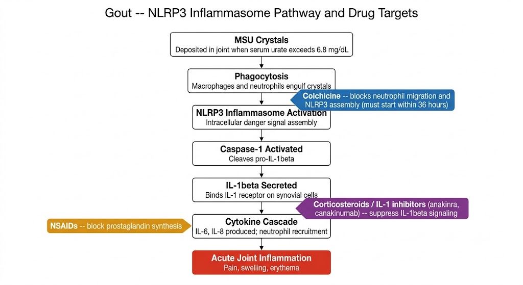
When serum urate exceeds 6.8 mg per deciliter, MSU crystals precipitate preferentially in cooler peripheral joints — the first metatarsophalangeal joint, ankles, and knees — where lower temperatures favor crystallization. Phagocytosis of MSU crystals by synovial macrophages and neutrophils triggers activation of the NLRP3 inflammasome, a multiprotein intracellular complex that assembles in response to cellular danger signals. NLRP3 activation drives autocleavage of procaspase-1 to its active form, which in turn cleaves pro-IL-1β to the mature secreted cytokine. IL-1β then binds the interleukin-1 receptor on synovial endothelial cells and fibroblasts, triggering a cascade of downstream cytokines (including interleukin-6 and interleukin-8) that recruits neutrophils and drives the intense joint inflammation characteristic of an acute attack. The neutrophil influx amplifies the response: phagocytosis of more crystals causes further NLRP3 activation, creating a self-amplifying loop that explains the rapid escalation of acute gout pain.
NSAIDs are first-line treatment for acute gouty attacks in the absence of contraindications. Indomethacin, naproxen, and diclofenac are the most commonly used agents. Full anti-inflammatory doses should be maintained for the full treatment course (typically five to seven days) rather than tapered; early discontinuation is the most common cause of treatment failure. PPI co-prescription is appropriate for patients with gastrointestinal risk factors.
Colchicine is a plant alkaloid derived from the autumn crocus with a mechanism distinct from NSAIDs. It binds tubulin and inhibits microtubule polymerization, impairing neutrophil chemotaxis, phagocytosis, and degranulation. It also inhibits NLRP3 inflammasome assembly by disrupting the microtubule-dependent processes required for inflammasome formation. Low-dose colchicine — 1.2 mg at onset followed by 0.6 mg one hour later — is as effective as historical high-dose regimens and substantially less toxic. Colchicine must be initiated within 36 hours of attack onset for maximal efficacy; it is significantly less effective when started later.
Colchicine is renally cleared and requires dose reduction when the eGFR falls below 30 mL per minute per 1.73 m². A critical drug interaction: colchicine is a substrate of both cytochrome P450 3A4 and P-glycoprotein. Co-administration with potent inhibitors of either pathway — including clarithromycin, cyclosporine, and ritonavir — can raise colchicine plasma levels to life-threatening concentrations, causing myopathy, neuromuscular toxicity, and cytopenias.
Systemic corticosteroids are highly effective for acute gout and are first-line when both NSAIDs and colchicine are contraindicated — as in severe renal impairment, anticoagulation, or intolerance. Oral prednisone for five to ten days is equivalent to indomethacin in efficacy in randomized trials. Intraarticular triamcinolone is effective for monoarticular attacks when systemic therapy is not preferred. A practical hazard is rebound attack after a short steroid course; a longer, more gradual taper reduces this risk.
IL-1 inhibitors target the central cytokine mediator of gout inflammation directly. Anakinra, an IL-1 receptor antagonist, is used off-label for refractory acute gout and polyarticular attacks. Canakinumab, a monoclonal antibody against IL-1β, is approved in some countries for frequent refractory attacks. These agents are reserved for patients in whom first-line therapies are contraindicated or ineffective.
Initiating or adjusting ULT during an active gout attack — or within two to four weeks of resolution — can trigger or prolong attacks through crystal shedding: rapid changes in serum urate in either direction mobilize crystals from tissue depots, intensifying synovial inflammation. Standard practice is to wait until the acute attack has fully resolved before starting or adjusting ULT. However, patients already established on ULT who experience a flare should continue their ULT without interruption; stopping would cause serum urate fluctuation and worsen crystal instability.
ULT is the cornerstone of long-term gout management. Its goals are to dissolve existing MSU crystal deposits, prevent new crystal formation, reduce attack frequency, and prevent tophaceous gout and urate nephropathy. ULT is indicated in patients with two or more attacks per year, visible tophi, radiographic joint damage, or chronic kidney disease with gout. The serum urate target is below 6 mg per deciliter for most patients, and below 5 mg per deciliter in patients with tophi or frequent attacks, to accelerate crystal dissolution.
Allopurinol is the most widely prescribed ULT and the first-line agent in most guidelines. It is a structural analogue of hypoxanthine that inhibits XO, the enzyme that catalyzes the final two steps of uric acid biosynthesis — conversion of hypoxanthine to xanthine and xanthine to uric acid. Allopurinol itself is rapidly metabolized by XO to oxypurinol, which is the primary pharmacologically active species. Oxypurinol has a long half-life and is renally cleared; dose adjustment is mandatory in chronic kidney disease to prevent oxypurinol accumulation, which is the principal pharmacokinetic cause of allopurinol hypersensitivity reactions.
The starting dose should be low — 50 to 100 mg per day — and titrated upward in increments every two to four weeks to achieve the serum urate target; doses well above 300 mg per day are frequently required and are safe with appropriate renal function monitoring. The allopurinol hypersensitivity syndrome is a rare but potentially fatal reaction characterized by fever, rash (ranging from maculopapular to Stevens-Johnson syndrome and toxic epidermal necrolysis), hepatitis, eosinophilia, and renal failure. Risk is substantially increased by starting at doses disproportionately high for the patient's renal function and by the HLA-B*5801 allele, which is more prevalent in East Asian populations. Initiating at a low dose and titrating slowly substantially reduces hypersensitivity risk regardless of genetic background.
Febuxostat is a non-purine selective XO inhibitor that is structurally unrelated to allopurinol and does not require dose adjustment for mild-to-moderate chronic kidney disease (eGFR at or above 30 mL per minute per 1.73 m²). This makes it useful in the subset of gout patients with renal insufficiency who cannot tolerate the required allopurinol dose reduction. Febuxostat is predominantly hepatically metabolized and excreted via bile, with limited renal dependence. A large clinical trial raised a cardiovascular safety concern by finding higher all-cause and cardiovascular mortality with febuxostat compared to allopurinol in patients with established cardiovascular disease. Current US guidance recommends allopurinol as first-line, with febuxostat reserved for patients who have failed or cannot tolerate allopurinol.
Uricosuric agents reduce serum urate by inhibiting the proximal tubular transporters (urate anion transporter 1 and glucose transporter 9) responsible for reabsorbing filtered urate, thereby increasing renal urate excretion. Probenecid is effective as monotherapy in gout patients with normal renal function and without a history of urolithiasis. Contraindications include eGFR below 30 mL per minute per 1.73 m² (insufficient urine flow for uricosuric effect) and urate overproduction with high urinary uric acid (increased urolithiasis risk). Low-dose aspirin at or below 325 mg per day blocks the uricosuric effect of probenecid by competing for tubular secretion transporters; this combination should be avoided when probenecid is used for gout management.
Pegloticase is a pegylated recombinant porcine uricase that converts uric acid to allantoin — the highly soluble metabolite that most mammals excrete — providing an enzymatic activity that humans lack. It is reserved for refractory tophaceous gout in patients who have failed or cannot tolerate conventional ULT, administered as an intravenous infusion every two weeks. The principal clinical challenge is immunogenicity: approximately 40 to 50% of patients develop antibodies against pegloticase that neutralize its hypouricemic effect and dramatically increase the risk of anaphylaxis and serious infusion reactions. Loss of the serum urate response — urate rising above 6 mg per deciliter during treatment — signals antibody formation and requires immediate drug discontinuation before the next infusion to prevent anaphylaxis. Co-administration of methotrexate (typically 15 mg per week) substantially reduces anti-drug antibody formation and is now endorsed in guidelines as a strategy to improve the proportion of patients who maintain a durable response.
Any ULT — whether XO inhibitor or uricosuric — reduces serum urate and mobilizes MSU crystals from tissue depots, triggering paradoxical gout flares in the first three to six months of therapy. This crystal shedding phenomenon is the primary reason patients discontinue ULT before reaching the target serum urate. Co-prescribing prophylactic low-dose colchicine (0.5 to 0.6 mg once or twice daily) for the first three to six months of ULT initiation substantially reduces flare frequency and improves ULT persistence. Low-dose NSAIDs or low-dose prednisone are alternatives when colchicine is contraindicated. Prophylaxis should continue for at least six months after achieving the serum urate target, and longer in patients with tophi.
Acute attack: NSAIDs full dose for five to seven days, or colchicine 1.2 mg then 0.6 mg one hour later (start within 36 hours), or prednisone (if first-line agents contraindicated). Do not start ULT during the attack.
After resolution (two to four weeks): Start allopurinol at 50 to 100 mg per day with colchicine prophylaxis. Titrate every two to four weeks to serum urate below 6 mg per deciliter. Continue prophylaxis for three to six months after target is achieved.
Allopurinol failure or intolerance: Febuxostat (caution in established cardiovascular disease), or add probenecid (if renal function adequate). Refractory tophaceous gout: pegloticase plus methotrexate co-administration.
| Author / Organization | Title | Source |
|---|---|---|
| Liu D, Ahmet A, Ward L, et al | A practical guide to the monitoring and management of the complications of systemic corticosteroid therapy | Allergy Asthma Clin Immunol. 2013;9(1):30 |
| Strehl C, Bijlsma JW, de Wit M, et al | Defining conditions where long-term glucocorticoid treatment has an acceptably low level of harm | Ann Rheum Dis. 2016;75(6):952-957 |
| Huscher D, Thiele K, Gromnica-Ihle E, et al | Dose-related patterns of glucocorticoid-induced side effects | Ann Rheum Dis. 2009;68(7):1119-1124 |
| Green H, Paul M, Vidal L, Leibovici L | Prophylaxis of Pneumocystis pneumonia in immunocompromised non-HIV-infected patients | Mayo Clin Proc. 2007;82(9):1052-1059 |
| Winthrop KL, Weinblatt ME, Bathon J, et al | Unmet need in rheumatology: reports from the Targeted Therapies meeting 2019 | Ann Rheum Dis. 2020;79(1):88-93 |
| Czock D, Keller F, Rasche FM, Haussler U | Pharmacokinetics and pharmacodynamics of systemically administered glucocorticoids | Clin Pharmacokinet. 2005;44(1):61-98 |
| Dinsen S, Baslund B, Klose M, et al | Why glucocorticoid withdrawal may sometimes be as dangerous as the treatment itself | Eur J Intern Med. 2013;24(8):714-720 |
| Dalbeth N, Merriman TR, Stamp LK | Gout | Lancet. 2016;388(10055):2039-2052 |
| Martinon F, Petrilli V, Mayor A, Tardivel A, Tschopp J | Gout-associated uric acid crystals activate the NALP3 inflammasome | Nature. 2006;440(7081):237-241 |
| FitzGerald JD, Dalbeth N, Mikuls T, et al | 2020 American College of Rheumatology Guideline for the Management of Gout | Arthritis Care Res. 2020;72(6):744-760 |
| Richette P, Doherty M, Pascual E, et al | 2016 updated EULAR evidence-based recommendations for the management of gout | Ann Rheum Dis. 2017;76(1):29-42 |
| Coutinho AE, Chapman KE | The anti-inflammatory and immunosuppressive effects of glucocorticoids, recent developments and mechanistic insights | Mol Cell Endocrinol. 2011;335(1):2-13 |