Antimalarial pharmacology is organized around the parasite life cycle. Each drug class acts at a specific stage, and that stage-specificity determines whether a drug can treat acute infection, prevent relapse, or provide prophylaxis. The single most important conceptual distinction is between blood-stage activity and liver-stage activity, because drugs that clear blood-stage parasites do not eliminate the latent liver reservoir.
An infected female Anopheles mosquito injects sporozoites into the human bloodstream. Sporozoites travel to the liver and develop into hepatic schizonts, which rupture to release thousands of merozoites into the blood. Merozoites invade red blood cells and cycle through ring, trophozoite, and schizont stages every 48 hours for most species (72 hours for Plasmodium malariae). Erythrocytic schizont rupture releases new merozoites plus malarial toxins, triggering the cyclical fever and rigors of clinical malaria.
In Plasmodium vivax and Plasmodium ovale, some hepatic sporozoites do not develop immediately but instead become dormant hypnozoites that persist in liver cells for months to years. Hypnozoites are the source of delayed relapse. No blood-stage drug eliminates them. Only the 8-aminoquinolines primaquine and tafenoquine reach and kill this liver reservoir.
Causal prophylactics kill hepatic schizonts before they release merozoites, preventing blood-stage infection entirely. Atovaquone-proguanil and doxycycline have this property, which allows them to be stopped shortly after leaving an endemic area. Suppressive prophylactics such as chloroquine and mefloquine do not kill liver-stage parasites; they suppress the erythrocytic cycle and must be continued for four weeks after return to ensure all liver-stage parasites have matured and entered the blood where the drug can act.
Blood schizonticides form the largest drug category and include chloroquine, quinine, mefloquine, and the artemisinins. These clear erythrocytic parasites but do not touch hypnozoites, so Plasmodium vivax and Plasmodium ovale infections treated with blood schizonticides alone will relapse unless an 8-aminoquinoline is added for radical cure.
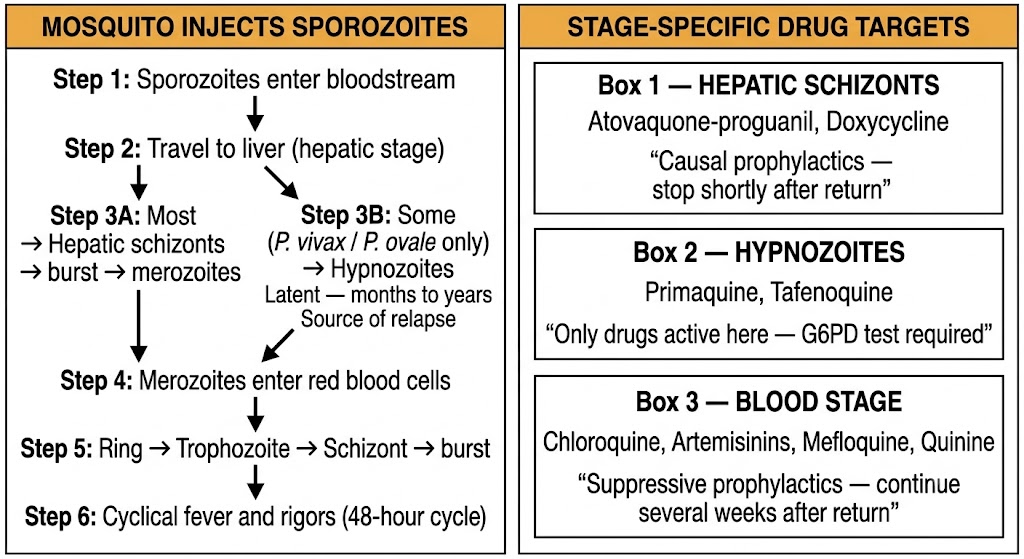
Causal prophylactics (kill hepatic schizonts): atovaquone-proguanil, doxycycline — can be stopped shortly after leaving endemic area. Suppressive prophylactics (kill blood-stage only): chloroquine, mefloquine — continue 4 weeks after return. Antirelapse agents (kill hypnozoites): primaquine, tafenoquine — require glucose-6-phosphate dehydrogenase testing before use; essential for radical cure of Plasmodium vivax and Plasmodium ovale. Blood schizonticides (treatment): chloroquine, quinine, artemisinins, mefloquine.
The quinoline class spans several structurally related subgroups with distinct mechanisms and clinical roles. Chloroquine and hydroxychloroquine are 4-aminoquinolines; quinine and mefloquine are arylaminoalcohols; primaquine and tafenoquine are 8-aminoquinolines. All except the 8-aminoquinolines act against blood-stage parasites.
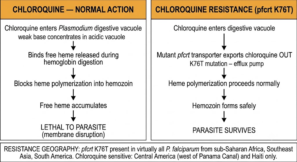
Chloroquine is a weak base that concentrates in the acidic digestive vacuole of the Plasmodium trophozoite. There it binds free heme (released during hemoglobin digestion) and prevents heme polymerization into the inert storage form hemozoin. Free heme accumulates to toxic levels, disrupting the parasite membrane and killing the trophozoite. The drug is active against blood-stage parasites of all four Plasmodium species but has no activity against liver stages or hypnozoites.
Chloroquine resistance in Plasmodium falciparum is mediated by mutations in the pfcrt gene (the Plasmodium falciparum chloroquine resistance transporter), particularly the K76T substitution. The mutant transporter pumps chloroquine out of the digestive vacuole, preventing lethal heme accumulation. Resistance is now present in virtually all Plasmodium falciparum-endemic regions outside Central America west of the Panama Canal and Haiti. Chloroquine remains effective against Plasmodium vivax in most areas, though resistance is established in parts of Southeast Asia and the Pacific.
Quinine and its stereoisomer quinidine share the heme polymerization inhibition mechanism with chloroquine. Quinine is used to treat chloroquine-resistant Plasmodium falciparum, always combined with a second agent (doxycycline or clindamycin) to prevent recrudescence and slow resistance selection. Intravenous quinidine was historically used for severe malaria in the United States, but intravenous artesunate has replaced it.
Mefloquine also inhibits heme polymerization and has a long half-life of several weeks, making once-weekly dosing feasible for prophylaxis. Its use is limited by neuropsychiatric adverse effects including vivid dreams, anxiety, dizziness, and, less commonly, psychosis and seizures. Mefloquine is contraindicated in patients with a personal history of psychiatric illness or seizure disorder.
Primaquine and tafenoquine are the only drugs that eliminate hypnozoites, making them essential for radical cure of Plasmodium vivax and Plasmodium ovale infections. They are also active against hepatic schizonts (causal prophylactic activity) and against mature Plasmodium falciparum gametocytes, giving them a role in transmission interruption.
Both drugs generate reactive oxygen species through metabolic activation. These oxidative metabolites overwhelm the antioxidant defenses of glucose-6-phosphate dehydrogenase (G6PD)-deficient red blood cells, causing hemolytic anemia. Glucose-6-phosphate dehydrogenase deficiency is common (approximately 400 million people worldwide) and follows the geographic distribution of malaria endemicity. Glucose-6-phosphate dehydrogenase testing is mandatory before prescribing either drug. Tafenoquine requires a quantitative test rather than a qualitative screen because even intermediate deficiency levels cause clinically significant hemolysis with its single high dose.
Never use chloroquine alone to treat Plasmodium falciparum from chloroquine-resistant areas (virtually all of sub-Saharan Africa, Southeast Asia, South America). Quinine monotherapy selects resistance — always combine with doxycycline or clindamycin. Mefloquine: contraindicated with psychiatric history or seizure disorder. Primaquine and tafenoquine: glucose-6-phosphate dehydrogenase test required — qualitative test insufficient for tafenoquine.
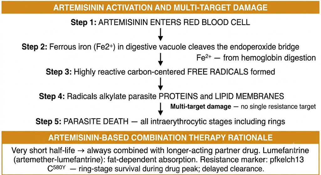
Artemisinins are the most rapidly acting antimalarials known, reducing parasite biomass by orders of magnitude per 48-hour cycle. Their very short half-life makes monotherapy impractical and combination with a longer-acting partner drug pharmacologically essential. Artemisinin-based combination therapies are the current first-line treatment for uncomplicated Plasmodium falciparum malaria worldwide.
Artemisinins contain an endoperoxide bridge that is cleaved by ferrous iron within the parasite digestive vacuole, generating highly reactive carbon-centered free radicals. These radicals alkylate and damage a broad range of parasite proteins and membrane lipids. The multi-target mechanism — distinct from heme polymerization inhibition — explains why artemisinins retain activity against chloroquine-resistant parasites and why resistance has been slower to develop than with single-target drugs.
Artemisinins act against all intraerythrocytic stages including ring-stage parasites, which are largely resistant to older blood schizonticides. This ring-stage activity accounts for their rapid parasite clearance rate. Artesunate is the intravenous form used for severe malaria; artemether-lumefantrine is the most widely used oral artemisinin-based combination therapy globally.
The very short half-life of artemisinins means that a standard course clears most parasites but cannot sustain drug exposure long enough to eliminate the last surviving organisms. A longer-acting partner drug with a half-life of days covers the period after artemisinin levels fall. Lumefantrine in artemether-lumefantrine and piperaquine in dihydroartemisinin-piperaquine are both long-acting partner drugs that fill this pharmacokinetic gap, maintaining drug exposure after the artemisinin component is cleared. Lumefantrine absorption depends critically on dietary fat — artemether-lumefantrine must always be taken with a fatty meal; absorption falls dramatically in the fasted state.
Intravenous artesunate is the treatment of choice for severe Plasmodium falciparum malaria, having demonstrated superiority over intravenous quinine in large randomized trials across Africa and Southeast Asia. Once the patient can take oral therapy and shows clinical improvement, a full oral artemisinin-based combination therapy course completes treatment.
Partial resistance to artemisinins, mediated by mutations in the pfkelch13 (Plasmodium falciparum kelch 13 propeller) gene, was first confirmed on the Thailand-Cambodia border and has since spread across the Greater Mekong Subregion and has been detected in East Africa. The key mutation is C580Y (cysteine to tyrosine at position 580). Resistant ring-stage parasites reduce their metabolic activity when drug levels peak, surviving the artemisinin exposure window and resuming development when drug levels fall — producing delayed parasite clearance. Artemisinin partial resistance alone does not reliably cause clinical treatment failure when the partner drug remains effective; the clinical problem arises when partner drug resistance (for example, piperaquine resistance in Cambodia) co-exists with kelch13 mutations, eliminating the backup component of the combination.
Artemether-lumefantrine: take with fatty food — lumefantrine absorption is fat-dependent. Intravenous artesunate: first-line for severe malaria; superior to intravenous quinine. Kelch13 C580Y mutation: marker of artemisinin partial resistance; endemic in Southeast Asia; now detected in Africa. Treatment failure within 28 days of artemisinin-based combination therapy requires polymerase chain reaction (PCR) genotyping to distinguish recrudescence from reinfection and to guide switch to alternative regimen.
Antimalarial prophylaxis selection integrates destination resistance patterns, drug mechanism type (causal versus suppressive), patient contraindications, and practical factors including dosing frequency and duration after return. No single agent is appropriate for all travelers.
Causal prophylactics (atovaquone-proguanil, doxycycline) kill hepatic schizonts before blood-stage infection is established. Because the liver stage is eliminated, these drugs can be stopped shortly after leaving the endemic area. Suppressive prophylactics (chloroquine, mefloquine) act only on blood-stage parasites and must be continued for several weeks after return, long enough for any remaining liver-stage parasites to mature into the bloodstream where the drug can act.
Chloroquine weekly is appropriate only for destinations where Plasmodium falciparum remains chloroquine-sensitive — essentially limited to Central America west of the Panama Canal, Haiti, and parts of the Middle East. For all other malaria-endemic destinations, chloroquine alone is inadequate for Plasmodium falciparum.
Atovaquone-proguanil combines two mechanisms: atovaquone inhibits the Plasmodium mitochondrial electron transport chain (cytochrome bc1 complex), collapsing mitochondrial membrane potential, while proguanil (via its active metabolite cycloguanil) inhibits dihydrofolate reductase. The causal mechanism and early discontinuation make it a preferred agent for most travelers. It is contraindicated in severe renal impairment.
Doxycycline acts as a causal prophylactic by inhibiting protein synthesis in the Plasmodium apicoplast — a non-photosynthetic organelle with prokaryotic-type ribosomes essential for the parasite. Photosensitivity is a significant adverse effect requiring sun protection. Esophageal irritation is a risk if the drug is taken without adequate fluid or while supine. Doxycycline is contraindicated in pregnancy and children under 8 years.
Mefloquine weekly is effective for chloroquine-resistant areas but carries neuropsychiatric risks. Starting mefloquine well before departure — earlier than other agents — allows time to detect neuropsychiatric intolerance before reaching a remote destination.
| Agent | Type | Continuation After Return | Key Contraindications |
|---|---|---|---|
| Chloroquine | Suppressive | Several weeks (suppressive) | Resistant areas (most of world) |
| Atovaquone-proguanil | Causal | Short (causal) | Severe renal impairment; pregnancy (limited data) |
| Doxycycline | Causal/suppressive | Several weeks (suppressive activity) | Pregnancy; age under 8 years |
| Mefloquine | Suppressive | Several weeks (suppressive) | Psychiatric history; seizure disorder; cardiac conduction abnormality |
| Primaquine | Causal | Short (causal) | Glucose-6-phosphate dehydrogenase deficiency; pregnancy |
Central America (west of Panama Canal), Haiti: chloroquine acceptable. Sub-Saharan Africa, South Asia: atovaquone-proguanil or doxycycline preferred; mefloquine acceptable if no psychiatric history. Southeast Asia (Mekong region): doxycycline or atovaquone-proguanil preferred. Plasmodium vivax-endemic areas: add primaquine or tafenoquine terminal prophylaxis after glucose-6-phosphate dehydrogenase testing. Pregnancy: chloroquine for sensitive areas; mefloquine from second trimester for resistant areas; doxycycline and atovaquone-proguanil generally avoided.
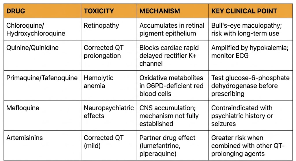
The antimalarial drug class carries four mechanistically distinct toxicity patterns, each linked to the drug's pharmacological properties. Understanding these mechanisms is more useful than memorizing monitoring schedules: the mechanism predicts which patients are at risk and which monitoring approach is appropriate.
Chloroquine and hydroxychloroquine accumulate in melanin-containing tissues including the retinal pigment epithelium, where they progressively damage photoreceptors. The damage is dose-dependent, largely irreversible once established, and produces a characteristic bull's-eye maculopathy pattern. Chloroquine is more retinotoxic than hydroxychloroquine at equivalent doses. Retinopathy is not a concern for short-term travel prophylaxis but becomes clinically relevant for patients on long-term therapy for rheumatologic conditions.
Multiple quinoline antimalarials block the cardiac rapid delayed rectifier potassium channel, prolonging cardiac repolarization (the corrected QT interval, or QTc). Quinine and quinidine carry the highest risk; mefloquine, lumefantrine, and piperaquine also prolong the corrected QT interval to varying degrees. The risk is amplified by electrolyte abnormalities (hypokalemia, hypomagnesemia) that are common in severe falciparum malaria. Patients with congenital long QT syndrome, structural heart disease, or concurrent corrected QT-prolonging medications are at elevated risk and warrant monitoring or drug selection adjustment.
Glucose-6-phosphate dehydrogenase catalyzes the rate-limiting step of the pentose phosphate pathway in red blood cells, generating reduced nicotinamide adenine dinucleotide phosphate, which maintains glutathione in its reduced antioxidant form. In glucose-6-phosphate dehydrogenase-deficient cells, oxidative stress from primaquine or tafenoquine metabolites cannot be neutralized. Hemoglobin denatures, Heinz bodies form, and red blood cells are destroyed. The degree of hemolysis depends on both the severity of glucose-6-phosphate dehydrogenase deficiency and the dose of the oxidant drug. Because glucose-6-phosphate dehydrogenase deficiency is most prevalent in populations from malaria-endemic regions — precisely the populations who need these drugs — testing before prescribing is non-negotiable.
Mefloquine crosses the blood-brain barrier and accumulates in central nervous system tissue. The neuropsychiatric spectrum ranges from mild — vivid dreams, sleep disturbance, dizziness, anxiety — to severe — acute psychosis, depression, seizures, and encephalopathy. Mild symptoms occur in a substantial minority of prophylaxis recipients. Severe neuropsychiatric effects can persist for months after discontinuation. The United States Food and Drug Administration black box warning mandates disclosure of neuropsychiatric risks before prescribing. Mefloquine is absolutely contraindicated with a current or prior history of psychiatric illness or seizure disorder.
Chloroquine resistance: the K76T mutation in pfcrt (Plasmodium falciparum chloroquine resistance transporter) is the primary marker. The mutant protein exports chloroquine from the digestive vacuole before lethal heme accumulation can occur. This mutation is present in Plasmodium falciparum from essentially all sub-Saharan Africa, Southeast Asia, and South America.
Artemisinin partial resistance: mutations in the pfkelch13 propeller domain, particularly C580Y, cause ring-stage parasites to temporarily reduce metabolic activity during drug exposure, surviving the peak artemisinin concentration and resuming development afterward. The clinical consequence is delayed parasite clearance. When partner drug resistance is present simultaneously — as in parts of Southeast Asia — clinical artemisinin-based combination therapy failure results. Clinical treatment failure within 28 days of artemisinin-based combination therapy treatment should be evaluated with genotyping to distinguish recrudescence from reinfection.
Chloroquine resistance (pfcrt K76T): present in nearly all Plasmodium falciparum outside Central America and Haiti. Artemisinin partial resistance (pfkelch13 C580Y): established in Mekong region; detected in East Africa. Partner drug resistance + kelch13 mutation = artemisinin-based combination therapy clinical failure. Sulfadoxine-pyrimethamine resistance (pfdhfr [dihydrofolate reductase gene] and pfdhps [dihydropteroate synthase gene] mutations): widely prevalent in sub-Saharan Africa; limits use as treatment but retained in intermittent preventive therapy in pregnancy in select settings.
| Author / Organization | Title | Source |
|---|---|---|
| Baird JK, Hoffman SL | Primaquine therapy for malaria | Clin Infect Dis. 2004;39(9):1336-1345 |
| Miller LH, Baruch DI, Marsh K, Doumbo OK | The pathogenic basis of malaria | Nature. 2002;415(6872):673-679 |
| World Health Organization | Guidelines for the Treatment of Malaria. 3rd ed. | WHO; 2015 |
| Lacerda MVG, Llanos-Cuentas A, Krudsood S, et al. | Single-dose tafenoquine to prevent relapse of Plasmodium vivax malaria | N Engl J Med. 2019;380(3):215-228 |
| Fidock DA, Nomura T, Talley AK, et al. | Mutations in the P. falciparum digestive vacuole transmembrane protein PfCRT and evidence for their role in chloroquine resistance | Mol Cell. 2000;6(4):861-871 |
| Marmor MF, Kellner U, Lai TY, et al. | Recommendations on screening for chloroquine and hydroxychloroquine retinopathy (2016 revision) | Ophthalmology. 2016;123(6):1386-1394 |
| Nosten F, White NJ | Artemisinin-based combination treatment of falciparum malaria | Am J Trop Med Hyg. 2007;77(6 Suppl):181-192 |
| Brunton L, Knollmann B, Hilal-Dandan R, eds. | Goodman & Gilman's The Pharmacological Basis of Therapeutics. 14th ed. Chapter 49: Chemotherapy of Malaria | McGraw-Hill; 2023 |
| Toovey S | Mefloquine neurotoxicity: a literature review | Travel Med Infect Dis. 2009;7(1):2-6 |
| Ariey F, Witkowski B, Amaratunga C, et al. | A molecular marker of artemisinin-resistant Plasmodium falciparum malaria | Nature. 2014;505(7481):50-55 |
| Dondorp AM, Fanello CI, Hendriksen ICE, et al. | Artesunate versus quinine in the treatment of severe falciparum malaria in African children (AQUAMAT) | Lancet. 2010;376(9753):1647-1657 |
| Schlagenhauf P, Petersen E | Malaria chemoprophylaxis: strategies for risk groups | Clin Microbiol Rev. 2008;21(3):466-472 |
| Ashley EA, Dhorda M, Fairhurst RM, et al. | Spread of artemisinin resistance in Plasmodium falciparum malaria | N Engl J Med. 2014;371(5):411-423 |