Introduction to Medical Pharmacology
Coagulation Physiology and Pharmacological Targets
Chapter 39 · Module 1 of 6Section 1
Vascular response, platelet adhesion, activation, and aggregation
Hemostasis arrests bleeding from an injured vessel through two overlapping phases. Primary hemostasis produces an initial platelet plug within seconds to minutes of injury. Secondary hemostasis then reinforces that plug with a cross-linked fibrin meshwork. The cellular events of primary hemostasis also define the pharmacological targets of the antiplatelet drug class.
The immediate vascular response to endothelial disruption is vasoconstriction, mediated by local reflex mechanisms and endothelin-1 released from injured endothelial cells. This reduces blood flow through the injured segment while the platelet plug forms. Disruption of the endothelial monolayer simultaneously exposes subendothelial collagen and von Willebrand factor, the two primary adhesive substrates that initiate platelet recruitment.
Platelet adhesion occurs in two steps. At high shear rates in small arteries and arterioles, the platelet surface receptor glycoprotein Ib-IX-V tethers transiently to von Willebrand factor immobilized on exposed collagen. Firm adhesion then follows through direct platelet collagen receptors, glycoprotein VI and integrin alpha-2 beta-1. Glycoprotein VI binding to collagen is the primary activating signal that transitions platelets from passive adhesion to active plug formation.
Following adhesion, platelets undergo activation: a dramatic morphological and biochemical transformation. The glycoprotein VI-collagen interaction activates phospholipase C, triggering calcium release from intracellular stores. Rising cytosolic calcium drives shape change, granule mobilization, and activation of the arachidonic acid pathway. Cyclooxygenase-1 converts arachidonic acid to thromboxane A2, which is secreted and acts on adjacent platelets and vascular smooth muscle, amplifying both platelet activation and vasoconstriction. This autocrine and paracrine amplification loop is the pharmacological target of aspirin, which irreversibly acetylates cyclooxygenase-1 and permanently blocks thromboxane A2 synthesis for the platelet lifetime of eight to ten days.
Dense granule release contributes adenosine diphosphate and serotonin to the local environment. Adenosine diphosphate binds the P2Y12 receptor (a Gi-coupled purinergic receptor) on adjacent platelets, sustaining platelet activation by suppressing cyclic adenosine monophosphate levels. P2Y12 is the target of clopidogrel, prasugrel, ticagrelor, and cangrelor. Alpha-granule release contributes fibrinogen, von Willebrand factor, and factor V, further enriching the prothrombotic environment at the injury site.
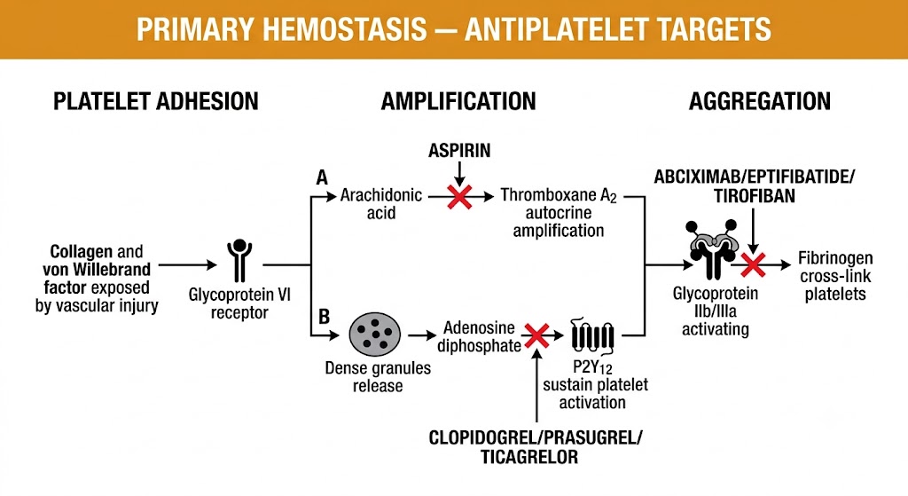
The culminating event of primary hemostasis is aggregation, the cross-linking of activated platelets into a plug. Aggregation is mediated almost entirely through the glycoprotein IIb/IIIa receptor (integrin alpha-IIb beta-3), which undergoes a conformational change from low- to high-affinity state following platelet activation. Activated glycoprotein IIb/IIIa binds fibrinogen and von Willebrand factor, cross-linking adjacent platelets into a three-dimensional plug. Intravenous glycoprotein IIb/IIIa antagonists (abciximab, eptifibatide, tirofiban) block this receptor pharmacologically and are used in high-risk percutaneous coronary intervention to prevent platelet-mediated coronary thrombosis.
Intact endothelium adjacent to the injury site continuously suppresses platelet activation through prostacyclin and nitric oxide. Prostacyclin elevates cyclic adenosine monophosphate in platelets, inhibiting the calcium release that sustains activation. Nitric oxide raises cyclic guanosine monophosphate, activating protein kinase G, which phosphorylates and inactivates platelet activation signaling proteins. These mechanisms confine platelet aggregation to the injury site and prevent pathological extension of the thrombus into adjacent intact vessels.
Primary Hemostasis — Pharmacological Targets
Thromboxane A2 synthesis via cyclooxygenase-1: target of aspirin (irreversible inhibition, permanent for platelet lifetime). P2Y12 receptor: target of clopidogrel, prasugrel, ticagrelor, and cangrelor. Glycoprotein IIb/IIIa receptor: target of abciximab, eptifibatide, and tirofiban.
Section 2
Extrinsic and intrinsic pathways, thrombin generation, and fibrin clot formation
Secondary hemostasis activates the plasma coagulation cascade, generating thrombin and converting soluble fibrinogen to insoluble fibrin polymer. Although the cascade is classically divided into intrinsic and extrinsic pathways based on laboratory coagulation tests, in vivo coagulation is initiated almost exclusively through the extrinsic pathway, with the intrinsic pathway amplifying thrombin generation rather than initiating it.
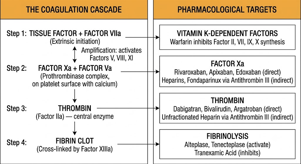
Coagulation initiates when tissue factor, a transmembrane glycoprotein expressed by subendothelial fibroblasts and smooth muscle cells, is exposed to circulating blood following vascular injury. Tissue factor forms a high-affinity complex with circulating factor VIIa (present in plasma at low concentrations in its active form). The tissue factor-factor VIIa complex activates both factor X and factor IX through proteolytic cleavage.
Activated factor X assembles with activated factor V on a negatively charged phospholipid surface (provided by activated platelets that flip phosphatidylserine to their outer leaflet) in the presence of calcium to form the prothrombinase complex. The prothrombinase complex cleaves prothrombin (factor II) to generate thrombin at rates far exceeding what factor Xa alone can achieve. This initial burst of thrombin is insufficient to produce a stable clot but is adequate to activate the amplification phase.
The small amount of thrombin generated by the initial tissue factor-factor VIIa complex activates multiple cascade components in a positive feedback loop. Thrombin activates factor V to factor Va and factor VIII to factor VIIIa, both of which serve as non-enzymatic cofactors that dramatically accelerate their respective enzymatic complexes. Thrombin also activates factor XI on the platelet surface, which in turn activates factor IX, allowing the intrinsic pathway to contribute substantially to the amplification phase.
Factor IXa assembles with its cofactor factor VIIIa on the platelet phospholipid surface (the intrinsic tenase complex), which activates factor X with far greater efficiency than the tissue factor-factor VIIa complex. This intrinsic tenase amplification explains why hemophilia A (factor VIII deficiency) and hemophilia B (factor IX deficiency) produce severe bleeding despite an intact extrinsic pathway: the amplification limb of thrombin generation is absent, leaving only the small initial extrinsic burst insufficient to sustain hemostasis.
The culminating steps of the common pathway convert fibrinogen to fibrin. Thrombin cleaves fibrinopeptides A and B from fibrinogen, exposing polymerization sites that allow fibrin monomers to self-assemble into long protofibrils. These protofibrils aggregate laterally to form fibrin fibers, creating the initial fibrin network. Factor XIII, activated by thrombin, cross-links fibrin polymers through covalent isopeptide bonds between adjacent fibrin molecules, creating a mechanically stable clot resistant to fibrinolytic dissolution.
Thrombin is the central effector molecule of the entire coagulation system and is directly or indirectly the target of every major anticoagulant drug class. Beyond cleaving fibrinogen, thrombin activates factors V, VIII, and XIII and stimulates platelet activation through protease-activated receptor 1 cleavage. Anticoagulants that inhibit thrombin generation (heparins, vitamin K antagonists, direct factor Xa inhibitors) or directly block its active site (direct thrombin inhibitors) have broad effects on both clot formation and platelet function.
Coagulation Test Correlations
Prothrombin time (international normalized ratio): reflects extrinsic and common pathway function (factors VII, X, V, II, fibrinogen). Prolonged by warfarin and factor VII deficiency. Activated partial thromboplastin time: reflects intrinsic and common pathway function (factors XII, XI, IX, VIII, X, V, II, fibrinogen). Prolonged by heparins, direct thrombin inhibitors, and hemophilia A and B. Thrombin time: measures fibrinogen-to-fibrin conversion directly; prolonged by direct thrombin inhibitors and hypofibrinogenemia. Anti-factor Xa assay: measures inhibition of factor Xa; used to monitor low-molecular-weight heparins, fondaparinux, and direct factor Xa inhibitors.
Section 3
Antithrombin III, the protein C and S pathway, and tissue factor pathway inhibitor
Under normal physiological conditions, coagulation is tightly regulated by endogenous anticoagulant systems that prevent pathological clot extension beyond the site of injury. These systems are clinically critical because they are the molecular targets through which major anticoagulant drugs act, and their inherited or acquired deficiency produces the hypercoagulable states encountered in clinical thrombosis management.
Antithrombin III is a serine protease inhibitor synthesized by the liver and circulating in plasma. It inhibits thrombin, factor Xa, factor IXa, factor XIa, and factor XIIa by forming stable, irreversible covalent complexes with these serine proteases at their active sites. In the absence of heparin, inhibition is slow. In the presence of heparin, which binds antithrombin III and induces a conformational change that exposes its reactive site loop, the rate of thrombin and factor Xa inhibition is accelerated approximately 1,000-fold. This is the fundamental mechanism of action of all heparin-based anticoagulants.
Inherited antithrombin III deficiency is a cause of familial venous thromboembolism. Acquired deficiency occurs in nephrotic syndrome (urinary protein losses), liver failure (reduced synthesis), and disseminated intravascular coagulation (consumption). Patients with severe antithrombin III deficiency may require antithrombin III concentrate to achieve adequate anticoagulation with heparin, because the drug has no antithrombin III to bind and accelerate.
Protein C is a vitamin K-dependent serine protease synthesized by the liver. When thrombin binds to thrombomodulin on the endothelial surface, the resulting complex loses procoagulant activity and instead activates protein C. Activated protein C, in complex with its cofactor protein S (also vitamin K-dependent), proteolytically cleaves and inactivates factor Va and factor VIIIa, the two non-enzymatic cofactors that are rate-limiting for the prothrombinase and intrinsic tenase complexes. By eliminating factor Va and factor VIIIa, the protein C pathway powerfully suppresses thrombin generation.
The factor V Leiden mutation, the most common inherited thrombophilia in populations of European ancestry, creates a form of factor Va that is resistant to cleavage by activated protein C. This results in sustained factor Va activity, amplified thrombin generation, and a three- to eightfold increased risk of venous thromboembolism in heterozygotes.
A particularly important clinical scenario is the initiation of warfarin in a protein C-deficient patient without adequate heparin bridging. Warfarin inhibits all vitamin K-dependent factor synthesis, including protein C, which has a shorter half-life than the procoagulant factors II, IX, and X. During the first one to three days of warfarin initiation, protein C levels fall before the procoagulant factors are sufficiently reduced, creating a transient procoagulant state that can precipitate warfarin-induced skin necrosis. This is the primary reason adequate heparin anticoagulation must overlap with warfarin initiation for at least four to five days.
Tissue factor pathway inhibitor is a serine protease inhibitor produced by endothelial cells. It exerts its anticoagulant effect in two steps: first binding and inhibiting factor Xa, and then the resulting complex binding and inhibiting the tissue factor-factor VIIa complex. This feedback mechanism limits the duration of extrinsic pathway activation and explains why the amplification and propagation phases of coagulation, dependent on the intrinsic pathway, are necessary to sustain clot formation. Intravenous heparin releases tissue factor pathway inhibitor from the endothelial surface, contributing an additional mechanism to heparin's anticoagulant effect beyond antithrombin III enhancement.
Inherited Thrombophilia
Prothrombotic States
Natural Anticoagulants
Endogenous Control Systems
Section 4
The plasminogen-plasmin system and pharmacological manipulation
Fibrinolysis is the physiological process by which fibrin clots are dissolved once vascular repair is complete, preventing permanent vessel occlusion. The fibrinolytic system is also the target of pharmacological intervention in two opposing directions: thrombolytic drugs activate fibrinolysis to dissolve pathological thrombi, while antifibrinolytic agents inhibit fibrinolysis to control excessive bleeding.
Plasmin is the principal fibrinolytic protease, capable of cleaving fibrin at multiple sites to generate soluble fibrin degradation products including D-dimer. Plasmin circulates as its inactive zymogen, plasminogen, which is synthesized by the liver. Plasminogen binds fibrin through lysine-binding sites on its kringle domains, positioning it for activation at the clot surface. The most physiologically important plasminogen activators are tissue-type plasminogen activator, released from endothelial cells in response to thrombin and shear stress, and urokinase-type plasminogen activator.
In the absence of fibrin, tissue-type plasminogen activator has low catalytic activity toward plasminogen. When tissue-type plasminogen activator binds fibrin, a ternary complex forms that dramatically accelerates plasminogen activation, concentrating fibrinolytic activity at the clot surface. This is the basis for the fibrin specificity of recombinant tissue-type plasminogen activator (alteplase) and its modified forms (reteplase, tenecteplase): at therapeutic doses, plasminogen activation is largely confined to the fibrin surface, minimizing systemic fibrinogenolysis.
In contrast, streptokinase forms an equimolar complex with plasminogen that nonspecifically converts both clot-bound and circulating plasminogen to plasmin, producing a systemic lytic state with marked fibrinogen depletion. This mechanistic distinction explains the superior bleeding safety profile of fibrin-specific thrombolytics compared to streptokinase, though all thrombolytics carry significant hemorrhagic risk, particularly intracranial hemorrhage.
Plasminogen activator inhibitor-1 is the principal physiological inhibitor of fibrinolysis, rapidly and irreversibly inactivating tissue-type plasminogen activator and urokinase-type plasminogen activator. Elevated plasminogen activator inhibitor-1 levels are associated with impaired fibrinolysis and increased thrombotic risk in metabolic syndrome, type 2 diabetes mellitus, and sepsis. Plasmin itself is inhibited by alpha-2 antiplasmin, the primary circulating plasmin inhibitor, which prevents indiscriminate proteolysis of plasma proteins.
D-dimer is a specific fibrin degradation product generated when cross-linked fibrin is cleaved by plasmin. Because D-dimer formation requires both thrombin (to cross-link fibrin via factor XIII activation) and plasmin (to degrade that cross-linked fibrin), an elevated D-dimer indicates active fibrin formation and dissolution. D-dimer assays have high sensitivity but low specificity for venous thromboembolism: a negative D-dimer in a patient with low-to-intermediate pretest probability effectively excludes acute deep vein thrombosis or pulmonary embolism, while a positive result requires imaging because D-dimer is elevated in many conditions including surgery, trauma, malignancy, and pregnancy.
Tranexamic acid and epsilon-aminocaproic acid are lysine analogues that competitively inhibit the lysine-binding sites on plasminogen, preventing its binding to fibrin and blocking plasminogen activation at the clot surface. These agents reduce surgical blood loss and traumatic hemorrhage and are addressed further in Module 6 of this series.
Section 5
Mapping approved drug classes to their specific molecular targets within the hemostatic system
The clinical pharmacology of anticoagulation is best understood as a map of interventions at defined nodes within the coagulation and platelet activation systems. Each drug class exploits a specific vulnerability in the hemostatic architecture. This target-to-drug mapping provides the framework developed in mechanistic detail across Modules 2 through 6 of this series.
The heparin family — unfractionated heparin, low-molecular-weight heparins (enoxaparin, dalteparin, tinzaparin), and fondaparinux (a synthetic pentasaccharide) — all exert their anticoagulant effects by binding antithrombin III and accelerating its inhibitory activity. Unfractionated heparin accelerates antithrombin III inhibition of both thrombin and factor Xa in approximately equal proportion. Low-molecular-weight heparins preferentially accelerate antithrombin III inhibition of factor Xa over thrombin due to their shorter chain lengths. Fondaparinux accelerates antithrombin III inhibition of factor Xa exclusively, with no direct anti-thrombin activity. Because all three drug types depend on antithrombin III, patients with antithrombin III deficiency may have reduced or absent heparin effect.
The direct oral anticoagulants targeting factor Xa — rivaroxaban, apixaban, edoxaban, and betrixaban — bind directly to the active site of factor Xa without requiring antithrombin III as an intermediary. This antithrombin III independence means they work equally well in antithrombin III-deficient patients. Because factor Xa sits at the convergence of the extrinsic and intrinsic pathways, inhibiting it blocks thrombin generation from both upstream pathways simultaneously. Andexanet alfa, a recombinant modified factor Xa decoy molecule, is the reversal agent for this drug class.
Direct thrombin inhibitors bind directly to the thrombin active site, blocking thrombin's ability to cleave fibrinogen, activate coagulation factors V, VIII, and XIII, and stimulate platelet protease-activated receptor 1. Parenteral direct thrombin inhibitors include bivalirudin and argatroban. The oral direct thrombin inhibitor dabigatran etexilate is a prodrug converted to dabigatran after intestinal absorption. Dabigatran binds both free and clot-bound thrombin, distinguishing it from heparins, which cannot inhibit thrombin already incorporated into a fibrin clot due to steric protection. Because direct thrombin inhibitors do not require antithrombin III, they are used when heparin therapy is contraindicated due to heparin-induced thrombocytopenia. Idarucizumab, a monoclonal antibody fragment with extremely high affinity for dabigatran, is the specific reversal agent.
Warfarin acts at the vitamin K epoxide reductase complex subunit 1 enzyme, inhibiting regeneration of the reduced form of vitamin K required for post-translational modification of the vitamin K-dependent coagulation factors: factor II (prothrombin), factor VII, factor IX, and factor X, as well as the anticoagulant proteins C and S. Without this modification, these proteins cannot bind calcium and are unable to assemble on phospholipid surfaces, rendering them functionally inactive. Because warfarin effects are mediated through protein synthesis, onset and offset are governed by factor half-lives, creating the characteristic delayed onset and prolonged offset that necessitate heparin bridging strategies.
Indirect Inhibitors
Antithrombin III-Dependent
Direct Oral Anticoagulants
Factor Xa Inhibitors
Direct Thrombin Inhibitors
Factor IIa Direct Inhibitors
Synthesis Inhibitor
Vitamin K Antagonist
Section 6
Matching pathophysiology to drug class across the major thrombotic syndromes
Rational anticoagulant prescribing begins with matching the pathophysiology of the thrombotic process to the pharmacological target most relevant to that process. Arterial thrombosis is primarily platelet-driven and occurs at sites of atherosclerotic plaque rupture under high shear stress. Venous thrombosis is primarily coagulation cascade-driven and occurs under low shear stress with relative stasis. This distinction has direct implications for drug selection.
Venous thromboembolism, encompassing deep vein thrombosis and pulmonary embolism, is a coagulation cascade-driven process occurring under conditions of stasis, endothelial injury, and hypercoagulability (Virchow's triad). Because the initial thrombus is fibrin-rich with relatively few platelets, anticoagulant therapy targeting thrombin generation or thrombin itself is the mainstay of treatment. Direct oral anticoagulants (rivaroxaban, apixaban, edoxaban, dabigatran) have become first-line therapy for venous thromboembolism treatment and secondary prevention in most patients without contraindications. Low-molecular-weight heparins are preferred for venous thromboembolism in active malignancy.
In atrial fibrillation, loss of organized atrial contraction creates stasis within the left atrial appendage, where slow and turbulent blood flow promotes thrombus formation. These thrombi are predominantly fibrin-based and can embolize to the cerebral circulation, producing cardioembolic stroke. Anticoagulation with direct oral anticoagulants or warfarin substantially reduces stroke risk. Warfarin remains the drug of choice for atrial fibrillation patients with mechanical heart valves, where direct oral anticoagulants have not demonstrated equivalent efficacy; dabigatran was found inferior to warfarin in patients with mechanical valves in the relevant randomized trial, leading to contraindication of all direct oral anticoagulants in this indication.
In acute coronary syndromes, the triggering event is rupture or erosion of an atherosclerotic plaque with exposure of subendothelial collagen and tissue factor to circulating blood. The resulting thrombus is platelet-rich, forming under high arterial shear stress, explaining why antiplatelet therapy is the cornerstone of management. Dual antiplatelet therapy with aspirin plus a P2Y12 inhibitor (ticagrelor or prasugrel preferred over clopidogrel in high-risk acute coronary syndromes based on randomized trial data) is mandatory following coronary stenting to prevent stent thrombosis. Short-term anticoagulation during the acute phase targets coagulation cascade amplification accompanying platelet activation.
Arterial vs. Venous Thrombosis — Drug Class Matching
Arterial thrombosis (platelet-rich thrombus, high shear): antiplatelet therapy is primary — aspirin, P2Y12 inhibitors, glycoprotein IIb/IIIa antagonists. Anticoagulants are adjunctive during acute phase. Venous thrombosis (fibrin-rich thrombus, low shear): anticoagulant therapy is primary — direct oral anticoagulants, heparins, or warfarin. Antiplatelet agents have minimal efficacy in venous thromboembolism treatment.
| Author / Organization | Title | Source |
|---|---|---|
| Nieswandt B, Watson SP | Platelet-collagen interaction: is GPVI the central receptor? | Blood. 2003;102(2):449-461. |
| Brass LF | Thrombin and platelet activation | Chest. 2003;124(3 Suppl):18S-25S. |
| Bhatt DL, Topol EJ | Current role of platelet glycoprotein IIb/IIIa inhibitors in acute coronary syndromes | JAMA. 2000;284(12):1549-1558. |
| Hoffman M, Monroe DM | A cell-based model of hemostasis | Thromb Haemost. 2001;85(6):958-965. |
| Monroe DM, Hoffman M | What does it take to make the perfect clot? | Arterioscler Thromb Vasc Biol. 2006;26(1):41-48. |
| Hirsh J, Anand SS, Halperin JL, Fuster V | Guide to anticoagulant therapy: heparin | Circulation. 2001;103(24):2994-3018. |
| Crowther MA, Kelton JG | Congenital thrombophilic states associated with venous thrombosis: a qualitative overview and proposed classification system | Ann Intern Med. 2003;138(2):128-134. |
| Esmon CT | The protein C pathway | Chest. 2003;124(3 Suppl):26S-32S. |
| Cesarman-Maus G, Hajjar KA | Molecular mechanisms of fibrinolysis | Br J Haematol. 2005;129(3):307-321. |
| Mannucci PM | Desmopressin (DDAVP) in the treatment of bleeding disorders: the first 20 years | Blood. 1997;90(7):2515-2521. |
| Weitz JI, Harenberg J | New developments in anticoagulants: past, present and future | Thromb Haemost. 2017;117(7):1283-1288. |
| Kearon C, Akl EA, Ornelas J, et al. | Antithrombotic therapy for VTE disease: CHEST guideline and expert panel report | Chest. 2016;149(2):315-352. |
| January CT, Wann LS, Calkins H, et al. | 2019 AHA/ACC/HRS focused update of the 2014 AHA/ACC/HRS guideline for the management of patients with atrial fibrillation | J Am Coll Cardiol. 2019;74(1):104-132. |
| Valgimigli M, Bueno H, Byrne RA, et al. | 2017 ESC focused update on dual antiplatelet therapy in coronary artery disease | Eur Heart J. 2018;39(3):213-260. |