CHAPTER 23  ·  ERGOT ALKALOID PHARMACOLOGY
Section 1

Chemistry, History, and Overview

The origin of ergot alkaloids in the fungus Claviceps purpurea, the historical consequences of ergotism, and the structural feature that explains the class’s broad receptor activity

Ergot alkaloids are derived from a fungus that grows on grain, and their pharmacological history stretches from medieval epidemics to modern obstetric wards and neurology clinics. Understanding where these drugs come from and how their shared chemistry produces wide receptor activity sets the foundation for every clinical application in this chapter.

Biological Origin: Claviceps purpurea

Ergot alkaloids originate from Claviceps purpurea, a fungus that infects rye and other cereal grains. The fungus replaces the grain kernel with a dark, hard structure called a sclerotium, which concentrates a mixture of alkaloid compounds. When contaminated grain was milled into flour and consumed, the alkaloids entered the human body in quantities sufficient to produce dramatic physiological effects.

This contamination was the cause of epidemic ergotism, a disease that swept through grain-dependent populations in medieval Europe and continued to appear sporadically into the early twentieth century. Modern grain inspection and storage practices have made epidemic ergotism a historical phenomenon in developed countries, but the pharmacology of the alkaloids responsible for it became the foundation of an important drug class.

Historical Ergotism: Two Clinical Forms

Epidemic ergotism appeared in two distinct clinical forms, each reflecting different pharmacological consequences of alkaloid exposure. Gangrenous ergotism, historically called St. Anthony’s Fire, was characterized by intense burning pain in the extremities followed by dry gangrene of the fingers, toes, and limbs. The mechanism was sustained peripheral vasoconstriction severe enough to produce ischemia and tissue death. Victims described the sensation of fire in their limbs as circulation was progressively cut off.

Convulsive ergotism, the second form, was characterized by seizures, muscle spasms, and neurological symptoms reflecting direct effects on the central nervous system. Both forms could affect the same population, and the predominant form in any given outbreak appeared to depend on the specific alkaloid mixture in the contaminated grain as well as nutritional factors in the affected population.
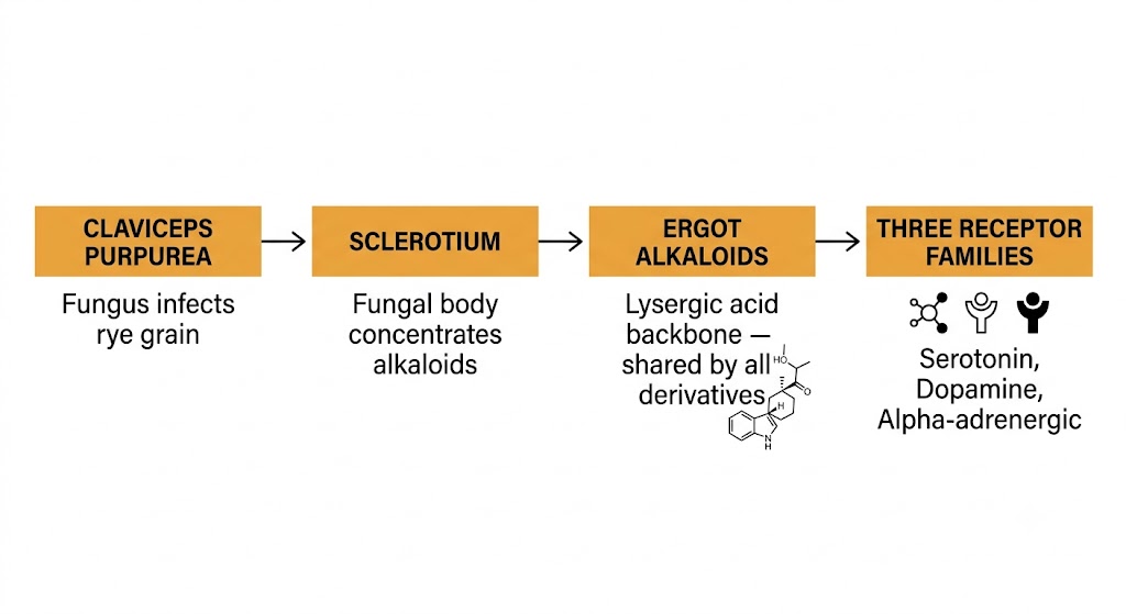
Flow diagram showing the origin of ergot alkaloids from Claviceps purpurea fungus through sclerotium formation to the lysergic acid backbone and its three receptor targets.
Origin and pharmacological basis of ergot alkaloids. Figure generated by Gemini AI.
The Lysergic Acid Backbone: Why Ergots Bind Everywhere

All clinically active ergot alkaloids share a common structural core — the lysergic acid backbone. This tetracyclic ring system resembles the molecular shapes of multiple neurotransmitters simultaneously, which is why ergot alkaloids can bind to serotonin receptors, dopamine receptors, and alpha-adrenergic receptors within the same molecule. No other clinical drug class achieves this breadth of receptor engagement from a single structural template.

The clinical derivatives differ from one another primarily in the substituents attached to this core. These modifications shift the balance of receptor binding affinities — one derivative may be predominantly dopaminergic, another predominantly vasoconstrictor, another primarily uterotonic — but all retain the capacity for activity at multiple receptor families to some degree. The lysergic acid backbone is the pharmacological reason this class produces such diverse effects across organ systems.

High-Yield Structural Concept

The lysergic acid backbone structurally mimics serotonin, dopamine, and norepinephrine simultaneously. This single chemical feature explains why ergot alkaloids are not selective drugs — they engage multiple receptor families at once. Every clinical effect and adverse effect in this chapter traces back to this structural reality.

Section 2

Receptor Pharmacology of Ergot Alkaloids

Simultaneous activity at serotonin, dopamine, and alpha-adrenergic receptors — and why the same drug can act as an agonist in one tissue and an antagonist in another

Ergot alkaloids are pharmacologically unusual because they act at three different receptor families simultaneously and behave as partial agonists at most of them. This partial agonist behavior is not a curiosity — it is the central pharmacological concept that explains why ergot effects vary dramatically depending on which tissue is being examined and what the baseline level of neurotransmitter activity is in that tissue.

Three Receptor Families, One Drug Class

Ergot alkaloids bind to serotonin receptors (primarily serotonin type 1 and type 2 subtypes), dopamine receptors (primarily the dopamine type 2 receptor), and alpha-adrenergic receptors. The degree of activity at each receptor family varies considerably across the class. Ergotamine and dihydroergotamine are predominantly vasoconstrictor drugs whose activity is driven mainly by serotonin type 2 and alpha-adrenergic receptor engagement. Bromocriptine is predominantly dopaminergic, with its therapeutic effects arising almost entirely from dopamine type 2 receptor agonism. Methylergonovine acts primarily on uterine smooth muscle through alpha-adrenergic and serotonin type 2 receptor mechanisms.

Despite these differences in emphasis, every ergot alkaloid retains some activity across all three receptor families. This broad engagement is what produces the complex adverse effect profiles that characterize the class — a drug given primarily for its dopaminergic effect may still produce vasoconstriction through its residual alpha-adrenergic activity, and a drug given for vasoconstriction may still influence prolactin secretion through its residual dopaminergic activity.

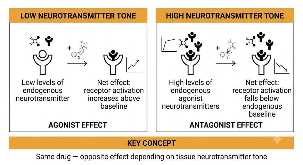
Two-panel diagram showing ergot partial agonism producing an agonist effect in low-tone tissue and an antagonist effect in high-tone tissue.
Ergot alkaloid partial agonism: tissue-dependent agonist versus antagonist behavior. Figure generated by Gemini AI.
Partial Agonism: The Core Pharmacological Concept

A partial agonist binds a receptor and activates it, but produces less than the maximum response that a full agonist would produce at the same receptor. This has an important consequence: in tissues where endogenous neurotransmitter activity is high, a partial agonist will compete with the endogenous agonist and reduce the overall receptor response — it acts as a functional antagonist. In tissues where endogenous neurotransmitter activity is low, the same partial agonist will increase receptor activation above baseline — it acts as an agonist.

Ergot alkaloids are partial agonists at most of their target receptors. This means that the same ergot alkaloid may produce opposite effects depending on the tissue being examined. In a blood vessel where serotonin tone is high, an ergot may block serotonin-mediated vasodilation and cause vasoconstriction. In a different vascular bed where serotonin tone is low, the same drug may produce minimal vascular effect. This tissue dependence of ergot action is a frequently tested concept on the United States Medical Licensing Examination and is essential for understanding why ergot effects are not predictable from simple receptor binding data alone.

Receptor Action
When Ergots Act as Agonists
  • Endogenous neurotransmitter tone is low in the target tissue
  • Ergot occupies unoccupied receptors and activates them
  • Net effect: receptor activation exceeds baseline
  • Example: uterine contraction via alpha-adrenergic and serotonin type 2 agonism
Receptor Action
When Ergots Act as Antagonists
  • Endogenous neurotransmitter tone is high in the target tissue
  • Ergot competes with endogenous agonist and limits full receptor activation
  • Net effect: receptor activation falls below endogenous baseline
  • Example: blockade of serotonin-mediated vasodilation during migraine
Ergotamine Versus Dihydroergotamine: Receptor Selectivity Differences

Ergotamine is the prototype natural ergot alkaloid used in migraine treatment. It is a potent partial agonist at serotonin type 1 and type 2 receptors, with significant alpha-adrenergic activity as well. Its serotonin type 2 and alpha-adrenergic receptor activity produces pronounced peripheral vasoconstriction, which is responsible for both its therapeutic effect on meningeal vessels and its risk of peripheral ischemia with excessive use.

Dihydroergotamine is a semi-synthetic derivative produced by hydrogenation of ergotamine. This structural modification reduces the potency of vasoconstriction at peripheral blood vessels while preserving activity at serotonin type 1B and 1D receptors on meningeal vessels. The result is a compound with a better ratio of therapeutic cranial vascular effect to peripheral vasoconstrictive risk. Dihydroergotamine also produces considerably less nausea than ergotamine and is better suited to parenteral administration, which matters for patients with severe migraine who cannot tolerate oral medications.


Section 3

Vasoconstriction: Mechanism and Clinical Consequences

How alpha-adrenergic and serotonin type 2 receptor activation by ergot alkaloids produces vasoconstriction in peripheral and cranial vascular beds, and what happens when this effect becomes pathological

Vasoconstriction is the most clinically consequential pharmacological property of the non-dopaminergic ergot alkaloids. In migraine treatment, carefully controlled cranial vasoconstriction is therapeutic. In peripheral vessels, excessive or sustained vasoconstriction is the mechanism of the most dangerous ergot complication — ergotism.

Dual Receptor Mechanism

Ergot alkaloid-induced vasoconstriction results from simultaneous activation of two receptor types on vascular smooth muscle: alpha-adrenergic receptors and serotonin type 2 receptors. Both receptor types, when activated, increase intracellular calcium in smooth muscle cells and promote contraction. Ergot alkaloids activate both simultaneously, producing a degree of vasoconstriction that exceeds what would be achieved through either receptor alone.

This dual mechanism is clinically important because it means that standard vasodilator drugs that work through a single pathway may be insufficient to reverse severe ergot-induced vasoconstriction. Management of ergotism typically requires multiple vasodilator approaches and may include anticoagulation to address the secondary thrombotic complications that can develop when blood flow through severely constricted vessels becomes turbulent or ceases.

Cranial Versus Peripheral Vascular Beds

Ergot alkaloids constrict blood vessels throughout the body, but the therapeutic relevance of this effect is specific to the cranial circulation. During migraine, abnormal dilation of meningeal blood vessels contributes to the activation of trigeminovascular pain fibers. Ergotamine and dihydroergotamine constrict these vessels by acting as serotonin type 1B and 1D receptor agonists on meningeal blood vessel walls, which reduces vessel diameter and decreases the mechanical activation of pain fibers wrapped around them.

The same vasoconstriction that is therapeutic in meningeal vessels is potentially harmful in peripheral vessels, where there is no therapeutic intent. Peripheral vasoconstriction from ergots reduces blood flow to the extremities, and with repeated or high-dose use, this can progress from cold extremities and paresthesias to frank ischemia. Coronary vasoconstriction is also a risk, which is why coronary artery disease is an absolute contraindication to ergot use in migraine treatment.

Coronary Vasospasm Risk

Ergot alkaloids can produce coronary artery vasospasm through the same alpha-adrenergic and serotonin type 2 receptor mechanisms that cause peripheral vasoconstriction. This is an absolute contraindication to ergot use in any patient with known coronary artery disease, Prinzmetal angina, or any condition associated with impaired coronary blood flow. The coronary vasospasm risk is shared by both ergotamine and dihydroergotamine, though the risk is somewhat lower with dihydroergotamine at therapeutic doses.


Section 4

Ergotism: Clinical Toxicology

Acute and chronic consequences of excessive ergot alkaloid exposure, the cytochrome P450 3A4 drug interaction that most commonly precipitates modern ergotism, and the principles of management

Ergotism in the modern clinical setting is almost always iatrogenic — it results from therapeutic ergot use combined with a drug interaction that dramatically elevates ergot plasma concentrations. Recognizing the trigger for this complication, identifying it clinically, and understanding why it is difficult to reverse are all high-yield concepts for the United States Medical Licensing Examination.

Acute Ergotism

Acute ergotism presents with nausea, vomiting, and vasoconstriction. The nausea and vomiting reflect dopaminergic activity at the chemoreceptor trigger zone. The vasoconstriction manifests as cold, pale, or cyanotic extremities with diminished or absent peripheral pulses. Patients may report paresthesias and pain in the extremities that reflects ischemia from reduced blood flow.

In severe acute cases, vasoconstriction may extend to mesenteric vessels, causing abdominal pain, and to coronary arteries, causing angina or myocardial ischemia. Central nervous system effects including confusion and seizures can occur in the most severe cases, particularly when very high plasma concentrations are reached through a drug interaction.

Chronic Ergotism and Gangrene

Chronic ergotism reflects the cumulative vascular consequences of sustained peripheral vasoconstriction. When blood flow through peripheral vessels is consistently reduced over days to weeks — either from repeated high-dose use or from a drug interaction that maintains elevated ergot concentrations — the ischemia progresses from reversible functional impairment to irreversible tissue death. Gangrene of the fingers, toes, and distal limbs results from this sustained ischemia and is the most severe consequence of ergotism.

The gangrenous form is dry gangrene because the mechanism is arterial occlusion from vasospasm rather than infection. Treatment requires discontinuation of the ergot alkaloid, aggressive vasodilator therapy, and anticoagulation if thrombosis has developed in the constricted vessels. In established gangrene, the tissue loss is permanent, and amputation may be required.

The Cytochrome P450 3A4 Drug Interaction

Ergot alkaloids are metabolized almost entirely by the hepatic enzyme cytochrome P450 3A4. When this enzyme is inhibited by another drug, ergot plasma concentrations rise dramatically — sometimes by tenfold or more. The elevation in plasma concentration converts a therapeutic dose into a toxic dose, and severe ergotism can result within hours of starting the inhibitor.

The drug classes that inhibit cytochrome P450 3A4 and most commonly precipitate this interaction include macrolide antibiotics (particularly erythromycin and clarithromycin), azole antifungal agents (particularly itraconazole and ketoconazole), and human immunodeficiency virus protease inhibitors (particularly ritonavir). This is a classic and frequently tested Step 1 drug interaction: the combination of an ergot alkaloid with any potent cytochrome P450 3A4 inhibitor is contraindicated. Azithromycin is a macrolide that does not meaningfully inhibit cytochrome P450 3A4 and does not carry this risk.

Classic Step 1 Drug Interaction

Ergot alkaloid + macrolide antibiotic (erythromycin, clarithromycin) = risk of severe ergotism from cytochrome P450 3A4 inhibition. The same risk applies with azole antifungal agents and protease inhibitors. This combination is absolutely contraindicated. The mechanism: inhibition of cytochrome P450 3A4 dramatically raises ergot plasma concentrations, converting a therapeutic dose into a toxic dose.

Management of Ergotism

Treatment of ergotism begins with immediate discontinuation of the ergot alkaloid and, if applicable, discontinuation or substitution of the cytochrome P450 3A4 inhibitor that triggered the interaction. Vasodilator therapy — using agents such as nitroprusside, calcium channel blockers, or prostaglandin analogs — is used to reverse peripheral vasoconstriction. Anticoagulation is added when there is concern for thrombosis in the vasospastic vessels.

The difficulty in treating ergotism reflects the pharmacokinetics of these drugs: ergot alkaloids have long half-lives and produce vasoconstriction that persists well after plasma concentrations begin to fall. Recovery from severe vasoconstriction may take days even with aggressive vasodilator therapy. This is why prevention through contraindication enforcement and drug interaction screening is essential before starting any ergot alkaloid.


Suggested References
Author / Organization Title Source
Schiff PL Ergot and its alkaloids Am J Pharm Educ. 2006;70(5):98
Boissier JR General pharmacology of ergot alkaloids Pharmacology. 1978;16(Suppl 1):12–26
Missale C, Nash SR, Robinson SW, Jaber M, Caron MG Dopamine receptors: from structure to function Physiol Rev. 1998;78(1):189–225
Silberstein SD, McCrory DC Ergotamine and dihydroergotamine: history, pharmacology, and efficacy Headache. 2003;43(2):144–166
Tfelt-Hansen P, Saxena PR, Dahlof C, et al Ergotamine in the acute treatment of migraine: a review and European consensus Brain. 2000;123(1):9–18
de Groot AN, van Dongen PW, Vree TB, Hekster YA, van Roosmalen J Ergot alkaloids: current status and review of clinical pharmacology and therapeutic use compared with other oxytocics in obstetrics and gynaecology Drugs. 1998;56(4):523–535
Matossian MK Poisons of the Past: Molds, Epidemics, and History Yale University Press; 1989
Dresser GK, Spence JD, Bailey DG Pharmacokinetic-pharmacodynamic consequences and clinical relevance of cytochrome P450 3A4 inhibition Clin Pharmacokinet. 2000;38(1):41–57
Diener HC, Limmroth V Medication-overuse headache: a worldwide problem Lancet Neurol. 2004;3(8):475–483
Aellig WH, Nuesch E Comparative pharmacokinetic investigations with tritium-labelled ergot alkaloids after oral and intravenous administration in man Int J Clin Pharmacol Biopharm. 1977;15(3):106–112