Neuromuscular Junction Physiology and Classification of Block
NMBL-01 · Module 1 of 4
Section 1
Anatomy of the Neuromuscular Junction
The structural basis for neuromuscular transmission and pharmacological blockade
The neuromuscular junction is a specialized chemical synapse between a motor nerve terminal and the membrane of a skeletal muscle fiber. Its architecture directly determines where neuromuscular blocking drugs act, how quickly they reach their target, and how reversal agents restore transmission.
Three-Compartment Structure
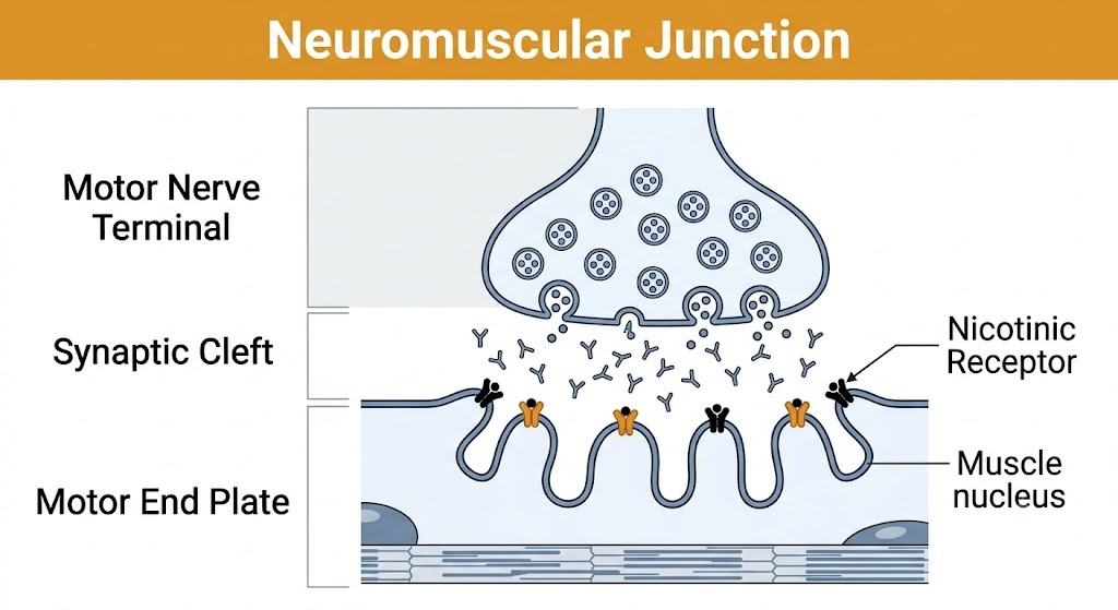
The neuromuscular junction consists of three anatomically distinct zones. The presynaptic motor nerve terminal is the expanded, unmyelinated ending of a lower motor neuron. It is packed with synaptic vesicles, each containing several thousand molecules of acetylcholine. The synaptic cleft is the narrow gap between the nerve terminal and the muscle membrane, roughly 20 to 50 nanometers wide. Within the cleft, acetylcholinesterase is anchored at high density and terminates acetylcholine signaling within milliseconds of receptor activation. The postsynaptic membrane — the motor end plate — is thrown into deep folds that increase its surface area and concentrate nicotinic acetylcholine receptors at the peaks of those folds, directly opposite the nerve terminal active zones.
This spatial organization is pharmacologically relevant. Nondepolarizing neuromuscular blocking drugs compete with acetylcholine at the postsynaptic receptor. Reversal agents that inhibit acetylcholinesterase increase acetylcholine concentration in the cleft, allowing it to displace the blocking drug from the receptor. The geometry of the junction — with acetylcholinesterase positioned to intercept acetylcholine molecules and receptors clustered at the end plate — sets the competitive playing field that determines drug effect.
Junctional and Extrajunctional Receptors
Under normal conditions, nicotinic acetylcholine receptors are concentrated almost exclusively at the motor end plate and are not expressed across the broader surface of the muscle fiber. This distribution changes under pathological conditions — particularly denervation, prolonged immobilization, burns, and crush injury. In these states, the muscle upregulates a fetal isoform of the nicotinic receptor across the extrajunctional membrane surface. These extrajunctional receptors have altered channel properties: they open more readily in response to depolarizing stimuli and close more slowly, allowing prolonged potassium efflux. The clinical consequence is exaggerated hyperkalemia when succinylcholine is administered to patients with these conditions, a phenomenon detailed in Module 2.
Anatomy of the neuromuscular junction showing the three-compartment structure: motor nerve terminal (vesicles), synaptic cleft (acetylcholinesterase), and motor end plate (nicotinic receptors at fold peaks). Figure generated by Gemini AI.
Section 2
Acetylcholine at the Junction
Synthesis, vesicular storage, calcium-triggered release, and enzymatic termination
Every aspect of normal neuromuscular transmission depends on the cycle of acetylcholine synthesis, release, receptor activation, and rapid hydrolysis. Neuromuscular blocking drugs intervene at one step in this cycle — receptor activation — and reversal agents intervene at another — hydrolysis. Understanding the full cycle clarifies both the pharmacological mechanism and the logic of reversal.
Synthesis and Storage
Acetylcholine is synthesized in the nerve terminal from two precursors: choline, which is actively transported into the terminal from the extracellular space, and acetyl coenzyme A, which is derived from mitochondrial metabolism. The enzyme choline acetyltransferase catalyzes the condensation of these two substrates to form acetylcholine. Once synthesized, acetylcholine is packaged into synaptic vesicles by a dedicated vesicular transporter and stored until a nerve impulse arrives.
Calcium-Triggered Release
When an action potential reaches the nerve terminal, voltage-gated calcium channels in the presynaptic membrane open, allowing calcium to flow into the terminal down its concentration gradient. The rise in intracellular calcium triggers fusion of acetylcholine-containing vesicles with the presynaptic membrane — a process of exocytosis — releasing acetylcholine into the synaptic cleft in discrete quantal packets. The calcium dependence of this release step explains why drugs that reduce presynaptic calcium entry, such as aminoglycoside antibiotics and magnesium, can impair neuromuscular transmission and potentiate the effect of nondepolarizing blocking drugs.
Termination by Acetylcholinesterase
Once acetylcholine diffuses across the cleft and binds to nicotinic receptors, it is released and rapidly hydrolyzed by acetylcholinesterase into choline and acetate. The choline is taken back up into the nerve terminal and recycled into new acetylcholine synthesis. Acetylcholinesterase operates at extraordinary speed — it can hydrolyze tens of millions of acetylcholine molecules per minute — ensuring that each nerve impulse produces a brief, precisely timed end-plate potential rather than sustained depolarization.
Drugs that inhibit acetylcholinesterase — the anticholinesterase agents neostigmine and pyridostigmine — extend the residence time of acetylcholine in the cleft by preventing its hydrolysis. This is the pharmacological basis of reversal for nondepolarizing neuromuscular block: the elevated acetylcholine concentration competes with the blocking drug at the nicotinic receptor, gradually restoring transmission. This reversal strategy is examined in detail in Module 4.
Section 3
The Nicotinic Receptor and the Safety Margin of Transmission
Ligand-gated ion channel activation, end-plate potential generation, and the physiological buffer that neuromuscular blocking drugs must overcome
The nicotinic acetylcholine receptor at the motor end plate is the molecular target for all neuromuscular blocking drugs. Its structural requirements for activation and the physiological safety margin built into normal transmission determine both why blocking drugs work and why a substantial fraction of receptors must be occupied before clinical weakness appears.
Receptor Structure and Activation
The neuromuscular nicotinic acetylcholine receptor is a pentameric ligand-gated ion channel — five protein subunits arranged around a central ion-conducting pore. Critically, the receptor has two acetylcholine binding sites, and both must be occupied simultaneously for the channel to open. This dual-site requirement has pharmacological significance: a single molecule of a competitive antagonist occupying one of the two sites is sufficient to prevent channel opening, even if the other site is occupied by acetylcholine.
When both sites are occupied by acetylcholine, the channel opens and allows cation flow: sodium moves inward, potassium moves outward, and the net effect is a rapid depolarization of the end-plate membrane called the end-plate potential. This local depolarization then activates voltage-gated sodium channels in the surrounding muscle membrane, generating an action potential that propagates along the fiber and triggers contraction.
The Safety Margin
A normal neuromuscular junction operates with a substantial safety margin. Under physiological conditions, the end-plate potential is roughly three to four times larger than the minimum amplitude needed to trigger a muscle action potential. This excess amplitude means that a large fraction of nicotinic acetylcholine receptors can be blocked without producing any measurable weakness. Experimental studies suggest that approximately 70 to 80 percent of receptors must be occupied by a nondepolarizing blocking drug before peak twitch strength begins to fall, and 90 to 95 percent must be occupied before the patient cannot sustain a tetanic contraction.
This safety margin has important clinical implications. Train-of-four monitoring — the standard clinical tool for assessing depth of block — detects progressive loss of this margin. The safety margin is also eroded by disease. In myasthenia gravis, autoantibodies destroy nicotinic acetylcholine receptors, reducing receptor density by 70 to 80 percent in some patients. These patients begin with a much smaller safety margin and are therefore exquisitely sensitive to nondepolarizing neuromuscular blocking drugs; even small doses can produce prolonged and profound block. The reverse logic applies to succinylcholine: fewer available receptors means succinylcholine must reach a greater proportion of them to produce depolarization, so myasthenia gravis patients may show partial resistance to the depolarizing agent.
Myasthenia Gravis and Neuromuscular Blocking Drug Sensitivity
Myasthenia gravis destroys nicotinic acetylcholine receptors through autoantibodies directed against the receptor or against proteins that anchor it. The reduced receptor density narrows the safety margin of transmission — these patients are already close to the threshold for transmission failure. Nondepolarizing neuromuscular blocking drugs are therefore used in dramatically reduced doses with intensive monitoring. Conversely, the depleted receptor pool may confer relative resistance to succinylcholine, since fewer receptors are available to be depolarized.
Section 4
Two Classes of Neuromuscular Block
Depolarizing and nondepolarizing mechanisms — how they differ and why it matters for reversal
All neuromuscular blocking drugs produce skeletal muscle paralysis by preventing the nicotinic acetylcholine receptor from generating a normal end-plate potential. They accomplish this through two fundamentally different mechanisms, and that mechanistic difference determines which reversal agents work and which do not.
Nondepolarizing Block — Competitive Antagonism
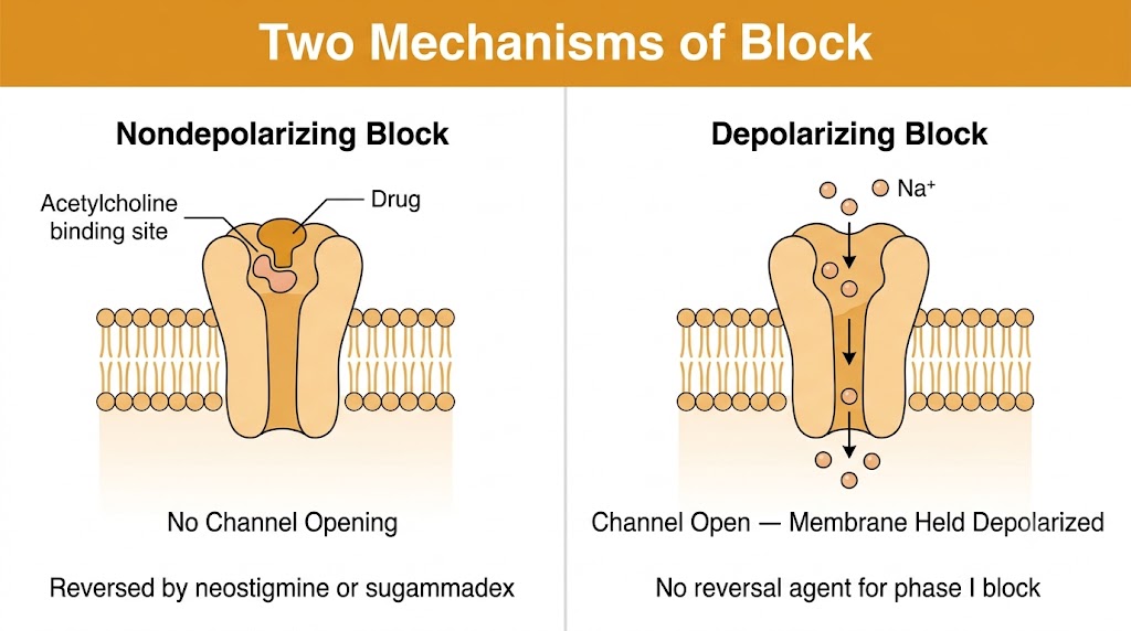
Nondepolarizing neuromuscular blocking drugs — which include the large majority of agents in clinical use — act as competitive antagonists at the nicotinic acetylcholine receptor. They bind to one or both of the acetylcholine binding sites on the receptor without activating it, physically preventing acetylcholine from binding and opening the channel. The muscle membrane remains at its resting potential: no depolarization occurs, no action potential is generated, and the muscle does not contract. Because the block is competitive, it can be overcome by increasing the concentration of acetylcholine in the cleft — which is exactly what anticholinesterase reversal agents do.
Nondepolarizing block produces no fasciculations before the onset of paralysis. The patient transitions smoothly from normal muscle tone to flaccid paralysis. On train-of-four stimulation, nondepolarizing block produces a characteristic fade pattern: the fourth twitch in the sequence is weaker than the first, because each successive stimulus depletes the releasable pool of acetylcholine and reduces the receptor-occupying competition for the blocking drug. The depth of block is graded by the magnitude of this fade.
Depolarizing Block — Persistent Activation
Succinylcholine, the sole depolarizing neuromuscular blocking drug in clinical use, works by a completely different mechanism. Rather than blocking the receptor, succinylcholine activates it — it is an acetylcholine receptor agonist. Unlike acetylcholine, however, succinylcholine is not rapidly hydrolyzed by acetylcholinesterase. It persists in the cleft, maintaining continuous receptor activation and holding the end-plate membrane in a depolarized state.
The initial binding of succinylcholine to the receptor opens ion channels, producing a brief depolarization of the end-plate membrane. This depolarization propagates as action potentials along the muscle fiber, causing the visible and palpable muscle fasciculations that characterize succinylcholine administration. After the initial fasciculations subside, the membrane remains persistently depolarized and cannot respond to subsequent nerve impulses. The voltage-gated sodium channels adjacent to the end plate, which require repolarization before they can reopen, remain inactivated — and the muscle falls into a sustained, flaccid paralysis despite the continued presence of acetylcholine receptor activation at the end plate. This is called phase I block.
With prolonged or repeated succinylcholine dosing, the character of the block can shift — the end-plate membrane partially repolarizes and the receptor undergoes a conformational change to a desensitized state. This phase II block resembles nondepolarizing block in its train-of-four characteristics and may be partially reversible with anticholinesterase agents, though this is unpredictable.
Mechanism
Nondepolarizing Block
Competitive antagonism at nicotinic receptor
No depolarization — no fasciculations
Smooth onset of flaccid paralysis
Train-of-four: fade pattern
Reversed by anticholinesterases or sugammadex
Mechanism
Depolarizing Block (Phase I)
Agonist at nicotinic receptor — persistent depolarization
Fasciculations precede paralysis
Flaccid paralysis due to sodium channel inactivation
Train-of-four: no fade (uniform depression)
No reversal agent for phase I block
Why Phase I Block Cannot Be Reversed with Anticholinesterases
The principle behind anticholinesterase reversal is straightforward: inhibit acetylcholinesterase, increase acetylcholine in the cleft, and allow acetylcholine to compete the nondepolarizing drug off the receptor. This strategy fails completely in phase I depolarizing block. Succinylcholine has already activated the receptor and depolarized the membrane. Adding more acetylcholine to the cleft — by inhibiting acetylcholinesterase — cannot repolarize a membrane that is already depolarized. In fact, anticholinesterases would prolong and deepen phase I block by slowing hydrolysis of succinylcholine. If a patient has an unexpectedly prolonged response to succinylcholine, the only management is continued ventilatory support until succinylcholine is metabolized.
Nondepolarizing block (left): competitive antagonism at the nicotinic receptor prevents channel opening and is reversible. Depolarizing block (right): persistent agonism holds the channel open and the membrane depolarized; phase I block has no reversal agent. Figure generated by Gemini AI.
Suggested References
Author / Organization
Title
Source
Brunton LL, Knollmann BC (eds)
Goodman and Gilman's The Pharmacological Basis of Therapeutics, 14th ed. Chapter 12: Neuromuscular Blocking Agents