Dihydrofolate reductase inhibition, polyglutamation, leucovorin rescue, and critical drug interactions
The folate antagonists are among the most widely used cytotoxic agents in clinical oncology and represent a paradigm for understanding how enzyme inhibition kinetics, cellular retention mechanisms, and renal pharmacokinetics interact to determine both efficacy and toxicity.
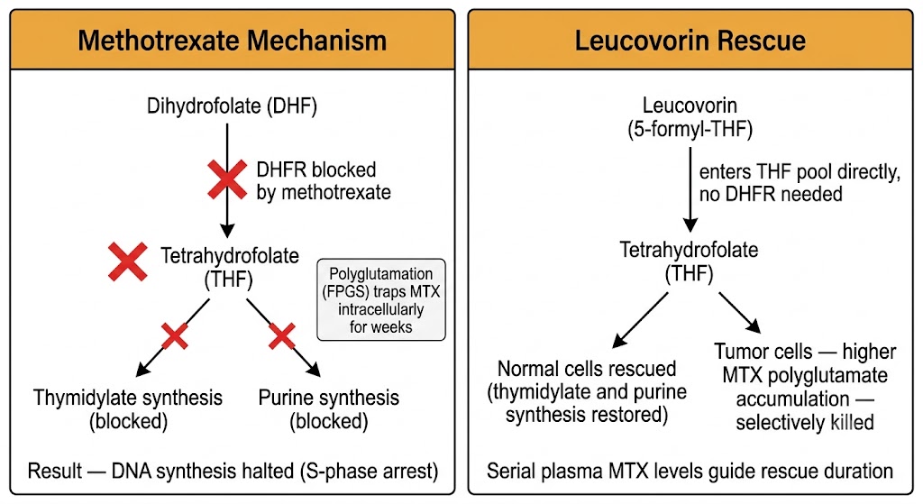
Methotrexate inhibits dihydrofolate reductase, the enzyme that reduces dihydrofolate to tetrahydrofolate, the active reduced folate cofactor required for one-carbon transfer reactions in de novo purine synthesis and thymidylate synthesis. By blocking dihydrofolate reductase, methotrexate depletes the intracellular tetrahydrofolate pool, halting synthesis of thymidylate and de novo purines. The cytotoxic effect is predominantly expressed in S phase, making methotrexate cycle-specific with activity dependent on sustained drug exposure.
Once inside the cell, methotrexate undergoes polyglutamation by folylpolyglutamate synthetase, an enzyme that adds multiple glutamate residues to methotrexate. Methotrexate polyglutamates are retained intracellularly for weeks because they cannot exit through the reduced folate carrier. They also directly inhibit thymidylate synthase and an enzyme in de novo purine synthesis, compounding the antiproliferative effect beyond dihydrofolate reductase inhibition alone.
Methotrexate is excreted approximately 80 to 90% unchanged in urine via glomerular filtration and active tubular secretion. Any drug that reduces renal blood flow, glomerular filtration rate, or organic anion tubular secretion prolongs methotrexate plasma half-life and dramatically increases toxicity. The most clinically important interactions are with nonsteroidal anti-inflammatory drugs, which reduce renal prostaglandin synthesis and decrease glomerular filtration rate, and with proton pump inhibitors, which compete for organic anion transporter 3-mediated tubular secretion. Both interactions can increase methotrexate plasma concentrations two- to tenfold, causing life-threatening mucositis, nephrotoxicity, myelosuppression, and neurotoxicity. Third-space fluid accumulations (ascites, pleural effusions) sequester methotrexate and release it slowly back into plasma after the infusion ends, prolonging exposure.
High-dose methotrexate with leucovorin rescue is used in osteosarcoma, acute lymphoblastic leukemia consolidation and central nervous system prophylaxis, diffuse large B-cell lymphoma, and primary central nervous system lymphoma. Leucovorin (5-formyltetrahydrofolate) is a reduced folate that directly enters the tetrahydrofolate pool without requiring dihydrofolate reductase, bypassing the dihydrofolate reductase block. Given 24 to 42 hours after methotrexate administration, leucovorin rescues normal cells while tumor cells, which accumulate more methotrexate polyglutamates, are more selectively killed. Rescue is timed by serial plasma methotrexate level monitoring and is continued until plasma methotrexate falls below 0.05 to 0.1 micromolar. Glucarpidase (carboxypeptidase G2), an enzyme that cleaves methotrexate and rapidly reduces plasma concentrations by more than 98% within 15 minutes, is reserved for cases of severe toxicity or failure of enhanced leucovorin rescue.
Before any high-dose methotrexate infusion: (1) Confirm normal or near-normal renal function with creatinine clearance above the institutional threshold — obtain a 24-hour urine creatinine clearance in borderline cases rather than relying on estimated glomerular filtration rate alone. (2) Discontinue all nonsteroidal anti-inflammatory drugs and proton pump inhibitors for at least 48 to 72 hours, and avoid penicillin-class antibiotics which compete for organic anion transporter 3. (3) Drain large pleural effusions or ascites to minimize the third-space reservoir effect. Proceeding without these checks risks prolonged methotrexate exposure for which even aggressive leucovorin rescue may be insufficient.
Pemetrexed inhibits three folate-dependent enzymes: thymidylate synthase, dihydrofolate reductase, and an enzyme in de novo purine synthesis. It is approved specifically for malignant pleural mesothelioma and non-squamous non-small cell lung cancer. The dose-limiting toxicities of myelosuppression and mucositis are substantially ameliorated by folic acid supplementation (400 micrograms orally daily, starting at least 5 days before the first dose) and vitamin B12 (1,000 micrograms intramuscularly approximately 1 week before the first dose, then every 3 cycles). Without these supplements, severe and potentially life-threatening myelosuppression occurs in a disproportionate fraction of patients. Dexamethasone is given the day before, day of, and day after pemetrexed to reduce skin rash. Pemetrexed is renally cleared and requires dose modification when creatinine clearance falls below 45 mL per minute; nonsteroidal anti-inflammatory drugs should be held before and after pemetrexed administration.
Thymidylate synthase inhibition, leucovorin modulation, dihydropyrimidine dehydrogenase catabolism, and dihydropyrimidine dehydrogenase deficiency toxicity
5-Fluorouracil and its oral prodrug capecitabine are among the most widely administered cytotoxic agents globally, forming the pharmacological backbone of treatment for colorectal, gastric, esophageal, breast, and pancreatic cancers. Their pharmacokinetics are governed by a single enzyme, dihydropyrimidine dehydrogenase, which determines both the rate of systemic catabolism and the risk of life-threatening toxicity in individuals with genetic deficiency.
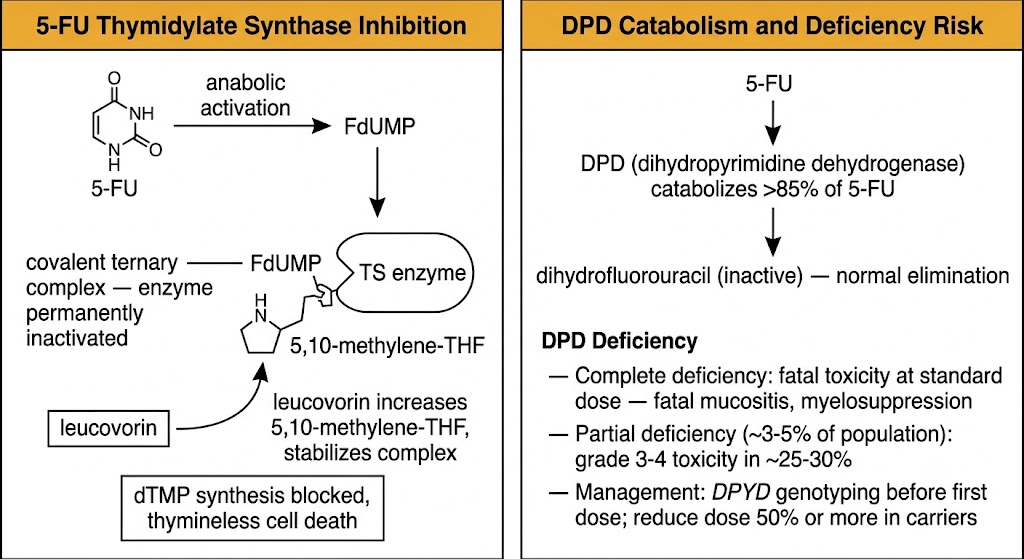
5-Fluorouracil exerts cytotoxicity through two distinct mechanisms that determine which toxicities predominate with different administration schedules. The first mechanism is thymidylate synthase inhibition via fluorodeoxyuridine monophosphate, the active metabolite that forms a covalent ternary complex with thymidylate synthase and the folate cofactor 5,10-methylenetetrahydrofolate, permanently inactivating the enzyme and blocking thymidylate synthesis. The second mechanism involves incorporation of fluorouridine triphosphate into ribonucleic acid, disrupting ribonucleic acid processing and ribosome assembly.
With bolus 5-fluorouracil administration, high peak concentrations favor ribonucleic acid disruption, producing predominantly myelosuppression. With continuous infusion, sustained lower concentrations favor thymidylate synthase inhibition, producing predominantly mucositis and hand-foot syndrome (palmar-plantar erythrodysesthesia). Leucovorin administered concurrently with 5-fluorouracil increases intracellular 5,10-methylenetetrahydrofolate, stabilizing the ternary complex and approximately doubling the response rate in metastatic colorectal cancer compared to 5-fluorouracil alone.
The primary catabolic pathway is hepatic oxidation by dihydropyrimidine dehydrogenase, which accounts for more than 85% of 5-fluorouracil elimination and is the dominant pharmacokinetic determinant of plasma half-life (approximately 10 to 20 minutes under normal dihydropyrimidine dehydrogenase activity). Dihydropyrimidine dehydrogenase activity varies approximately fivefold in the normal population, contributing to wide inter-patient variation in both toxicity and efficacy.
Dihydropyrimidine dehydrogenase deficiency is a pharmacogenomic variant affecting approximately 3 to 8% of the general population, caused by loss-of-function variants in the dihydropyrimidine dehydrogenase gene. The most clinically significant variant causes complete loss of dihydropyrimidine dehydrogenase activity in homozygotes. Patients with complete deficiency who receive standard 5-fluorouracil doses experience severe and potentially fatal toxicity: profound myelosuppression, life-threatening mucositis, neurotoxicity, and multi-organ failure occurring within days of the first dose. Patients with partial deficiency (heterozygotes, affecting approximately 3 to 5% of the population) experience severe grade 3 or 4 toxicity at standard doses in approximately 25 to 30% of cases. Dihydropyrimidine dehydrogenase gene testing before 5-fluorouracil or capecitabine administration is now recommended by the European Medicines Agency and is increasingly adopted in North America. Carriers require a starting dose reduction of at least 50%.
Capecitabine is an orally administered prodrug that undergoes three-step enzymatic conversion to 5-fluorouracil, with the final step catalyzed by thymidine phosphorylase, which is expressed at substantially higher levels in many tumor types than in surrounding normal tissue. This tumor-preferential final activation step produces predominantly a continuous infusion-like toxicity profile: hand-foot syndrome is the most frequent and dose-limiting adverse effect, occurring in 50 to 60% of patients, while myelosuppression is generally milder than with intravenous 5-fluorouracil regimens. Capecitabine is subject to the same dihydropyrimidine dehydrogenase deficiency risk as intravenous 5-fluorouracil. Dose reduction is required in renal impairment; capecitabine is contraindicated when creatinine clearance falls below 30 mL per minute.
A major drug interaction involves capecitabine and warfarin. Capecitabine inhibits cytochrome P450 2C9, the primary enzyme responsible for metabolizing the more potent S-warfarin enantiomer, and can increase the international normalized ratio dramatically within days to weeks of starting capecitabine. The international normalized ratio must be monitored weekly or more frequently in patients on warfarin receiving capecitabine. When possible, switching to a direct oral anticoagulant before capecitabine initiation avoids this interaction entirely.
Intracellular activation, deoxycytidine kinase dependence, self-potentiation, and schedule-dependent S-phase cytotoxicity
Cytarabine and gemcitabine are cytidine analogs that require intracellular phosphorylation to their active triphosphate forms and exert cytotoxicity predominantly through incorporation into deoxyribonucleic acid, where they terminate chain elongation and trigger apoptosis. Both are S-phase-specific agents whose efficacy is tightly linked to scheduling strategy and intracellular activation enzyme activity.
Cytarabine enters cells via nucleoside transporters and is phosphorylated sequentially to the active triphosphate form, ara-cytosine triphosphate. This triphosphate is incorporated into deoxyribonucleic acid by deoxyribonucleic acid polymerase in competition with the natural substrate deoxycytidine triphosphate. Once incorporated, it produces a steric block to further elongation, resulting in chain termination and apoptosis. Cytarabine is S-phase-specific, accounting for the clinical scheduling of cytarabine as a 7-day continuous infusion in acute myeloid leukemia induction — at any moment, only 20 to 40% of leukemic blasts are in S phase, and continuous infusion ensures every dividing cell is exposed as it enters S phase.
The rate-limiting step in cytarabine activation is phosphorylation by deoxycytidine kinase; loss of deoxycytidine kinase expression is the primary mechanism of cytarabine resistance. High-dose cytarabine (2 to 3 g per square meter every 12 hours for 3 to 6 days) achieves cytotoxic concentrations in the cerebrospinal fluid and is used in acute myeloid leukemia consolidation. Its serious specific toxicity is cerebellar toxicity (ataxia, dysarthria, nystagmus from Purkinje cell loss) occurring in approximately 10 to 25% of patients receiving high-dose cytarabine, requiring neurological examination before each dose cycle and immediate discontinuation if cerebellar signs appear.
Gemcitabine shares nucleoside transporter-mediated uptake and deoxycytidine kinase-dependent activation with cytarabine but has several additional mechanisms contributing to its broader clinical activity. After phosphorylation to the triphosphate, incorporation into deoxyribonucleic acid produces a masked chain termination that is relatively resistant to proofreading, enhancing its cytotoxic effect. Gemcitabine also inhibits ribonucleotide reductase, further impairing deoxyribonucleic acid synthesis.
Gemcitabine is used in pancreatic adenocarcinoma (gemcitabine plus nab-paclitaxel is a standard regimen), non-small cell lung cancer (gemcitabine plus platinum doublet), bladder cancer, and relapsed ovarian cancer. Its toxicity profile differs from cytarabine: myelosuppression (predominantly thrombocytopenia and neutropenia) is dose-limiting, and a unique toxicity is pulmonary toxicity manifesting as dyspnea and interstitial infiltrates in approximately 10 to 15% of patients. Gemcitabine-induced hemolytic uremic syndrome, a thrombotic microangiopathy characterized by microangiopathic hemolytic anemia, thrombocytopenia, and acute kidney injury, occurs rarely but requires immediate drug discontinuation.
Thiopurine methyltransferase pharmacogenomics, allopurinol interaction, fludarabine immunosuppression, and azacitidine mechanism
Purine analogs exploit the dependence of lymphoid and myeloid malignancies on purine synthesis pathways. The most clinically critical pharmacogenomic consideration in this class is thiopurine methyltransferase activity, which governs 6-mercaptopurine dosing safety and is the paradigm for pharmacogenomics-guided cancer therapy.
6-Mercaptopurine is a thiopurine prodrug used primarily in acute lymphoblastic leukemia maintenance therapy. It requires intracellular activation by hypoxanthine-guanine phosphoribosyltransferase to thioguanine nucleotides, which are incorporated into deoxyribonucleic acid and trigger mismatch repair-mediated apoptosis. The primary catabolic pathway is methylation by thiopurine methyltransferase, which converts 6-mercaptopurine to an inactive metabolite, and oxidation by xanthine oxidase to 6-thiouric acid.
Thiopurine methyltransferase activity is trimodally distributed: approximately 90% of patients have high (wild-type) activity, approximately 10% have intermediate activity (heterozygous for one low-activity allele), and approximately 0.3% have low or absent activity (homozygous). Patients with low thiopurine methyltransferase activity shunt virtually all 6-mercaptopurine through the activation pathway, accumulating very high thioguanine nucleotide concentrations and experiencing life-threatening myelosuppression at standard doses. Acute lymphoblastic leukemia maintenance protocols routinely include thiopurine methyltransferase genotyping or phenotyping before starting 6-mercaptopurine; patients with intermediate activity receive a 30 to 50% dose reduction and those with low activity receive 10 to 20% of the standard dose.
The interaction between 6-mercaptopurine and allopurinol is among the most dangerous drug interactions in clinical oncology. Allopurinol inhibits xanthine oxidase, a primary catabolic enzyme for 6-mercaptopurine, and co-administration blocks 6-mercaptopurine catabolism, increasing plasma concentrations approximately fourfold and causing potentially fatal aplasia. The standard management is to reduce the 6-mercaptopurine dose to 25% of the usual dose when allopurinol cannot be avoided. For tumor lysis syndrome prevention in patients who will receive 6-mercaptopurine, rasburicase rather than allopurinol should be used as the uric acid-lowering agent whenever possible.
Fludarabine is a fluorinated purine nucleoside analog active in chronic lymphocytic leukemia, typically as part of the fludarabine, cyclophosphamide, and rituximab regimen in fit patients. Its most clinically significant adverse effect is profound and sustained immunosuppression: fludarabine is selectively toxic to CD4-positive T lymphocytes, depleting them to very low levels for months to years after therapy. This creates risks for opportunistic infections including Pneumocystis jirovecii pneumonia, viral reactivations (cytomegalovirus, herpesvirus), and fungal infections. Pneumocystis jirovecii pneumonia prophylaxis with trimethoprim-sulfamethoxazole is mandatory during and for at least 6 to 12 months after fludarabine-containing regimens. Fludarabine can also trigger autoimmune hemolytic anemia in chronic lymphocytic leukemia patients; development of autoimmune hemolytic anemia during fludarabine therapy is an indication to discontinue the drug immediately.
Cladribine (2-chlorodeoxyadenosine) is the drug of choice for hairy cell leukemia, producing durable complete remissions in more than 90% of patients with a single 7-day continuous infusion course. Like fludarabine, cladribine causes profound immunosuppression requiring Pneumocystis jirovecii pneumonia and herpesvirus prophylaxis.
Azacitidine and decitabine are cytidine analogs that, after incorporation into deoxyribonucleic acid during S phase, form an irreversible bond with deoxyribonucleic acid methyltransferase 1, the maintenance methyltransferase responsible for replicating the parental methylation pattern on newly synthesized daughter strands. This traps and degrades deoxyribonucleic acid methyltransferase 1, causing progressive loss of methylation marks on CpG islands in promoters of epigenetically silenced tumor suppressor genes across successive cell divisions, a process called passive demethylation. Reactivation of silenced tumor suppressors — including cyclin-dependent kinase inhibitors and E-cadherin — restores differentiation, apoptosis responsiveness, and cell cycle checkpoint function in myelodysplastic syndrome and acute myeloid leukemia cells.
Azacitidine is administered subcutaneously or intravenously for 7 days every 28 days in myelodysplastic syndrome and acute myeloid leukemia unfit for intensive chemotherapy. Decitabine is administered intravenously for 5 days every 28 days in myelodysplastic syndrome and acute myeloid leukemia. The clinical benefit of hypomethylating agents often requires 3 to 6 treatment cycles before a response is apparent, reflecting the time required for passive demethylation to accumulate across multiple cell divisions. A minimum of 4 to 6 cycles should be administered before declaring treatment failure, provided hematological tolerance allows continuation.
| Author / Organization | Title | Source |
|---|---|---|
| Jolivet J, Cowan KH, Curt GA, Clendeninn NJ, Chabner BA | The pharmacology and clinical use of methotrexate | N Engl J Med. 1983;309(18):1094–1104 |
| Howard SC, McCormick J, Pui CH, Buddery RL, Henderson ES | Preventing and managing toxicities of high-dose methotrexate | Oncologist. 2016;21(12):1471–1482 |
| Adjei AA | Pemetrexed (Alimta): a novel multitargeted antineoplastic agent | Clin Cancer Res. 2004;10(12 Pt 2):4276s–4280s |
| Longley DB, Harkin DP, Johnston PG | 5-Fluorouracil: mechanisms of action and clinical strategies | Nat Rev Cancer. 2003;3(5):330–338 |
| Miura K, Kinouchi M, Ishida K, et al | 5-FU metabolism in cancer and orally-administrable 5-FU drugs | Cancers (Basel). 2010;2(3):1717–1730 |
| Deenen MJ, Meulendijks D, Cats A, et al | Upfront genotyping of DPYD*2A to individualize fluoropyrimidine therapy: a safety and cost analysis | J Clin Oncol. 2016;34(3):227–234 |
| Galmarini CM, Mackey JR, Dumontet C | Nucleoside analogues: mechanisms of drug resistance and reversal strategies | Leukemia. 2001;15(6):875–890 |
| Estey EH | Acute myeloid leukemia: 2021 update on risk-stratification and management | Am J Hematol. 2020;95(11):1368–1398 |
| Lennard L | TPMT in the treatment of Crohn's disease with azathioprine | Gut. 2002;51(2):143–146 |
| Keating MJ, O'Brien S, Albitar M, et al | Early results of a chemoimmunotherapy regimen of fludarabine, cyclophosphamide, and rituximab as initial therapy for chronic lymphocytic leukemia | J Clin Oncol. 2005;23(18):4079–4088 |
| Stresemann C, Lyko F | Modes of action of the DNA methyltransferase inhibitors azacytidine and decitabine | Int J Cancer. 2008;123(1):8–13 |
| Fenaux P, Mufti GJ, Hellstrom-Lindberg E, et al | Efficacy of azacitidine compared with that of conventional care regimens in the treatment of higher-risk myelodysplastic syndromes: a randomised, open-label, phase III study | Lancet Oncol. 2009;10(3):223–232 |