Ribonucleotide reductase inhibition, sickle cell disease applications, asparagine auxotrophy, and pegaspargase toxicities
Hydroxyurea and asparaginase occupy distinct pharmacological niches: hydroxyurea as the prototype ribonucleotide reductase inhibitor used in myeloproliferative disorders and sickle cell disease; asparaginase as a unique enzyme-based cytotoxic that exploits the asparagine auxotrophy of certain leukemic cell populations.
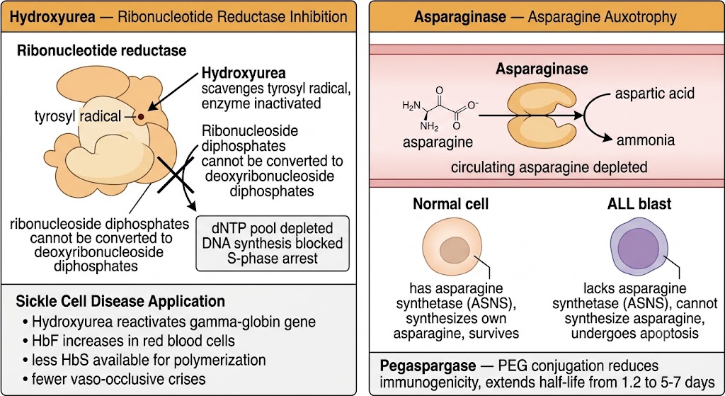
Hydroxyurea inhibits ribonucleotide reductase, the rate-limiting enzyme in de novo deoxyribonucleic acid synthesis that catalyzes the reduction of ribonucleoside diphosphates to deoxyribonucleoside diphosphates. Ribonucleotide reductase contains a stable tyrosyl radical essential for catalytic activity; hydroxyurea scavenges this radical, inactivating the enzyme and blocking deoxyribonucleic acid synthesis. The result is S-phase arrest. Hydroxyurea is administered orally and undergoes renal excretion; dose reduction is required when creatinine clearance falls below 60 mL per minute.
In oncology, hydroxyurea is used as cytoreductive therapy in essential thrombocythemia, polycythemia vera, primary myelofibrosis, and chronic myeloid leukemia when tyrosine kinase inhibitors are not immediately available or are contraindicated. In sickle cell disease, hydroxyurea increases fetal hemoglobin production in erythroid precursors through mechanisms that are not fully characterized. Elevated fetal hemoglobin reduces the proportion of hemoglobin S available for polymerization, decreasing the frequency of vaso-occlusive crises, acute chest syndrome, stroke, and the need for red blood cell transfusions. Hydroxyurea is the most widely used disease-modifying therapy in sickle cell disease. The most common toxicities are myelosuppression requiring periodic complete blood count monitoring, macrocytosis (a consistent finding that does not require dose reduction), and, with long-term use, lower extremity leg ulcers.
Asparaginase catalyzes the hydrolysis of L-asparagine to L-aspartic acid and ammonia, depleting circulating asparagine. Most normal cells synthesize asparagine endogenously and are relatively protected. Certain acute lymphoblastic leukemia blasts, particularly T-cell acute lymphoblastic leukemia, express very low levels of asparagine synthetase and are dependent on exogenous asparagine for protein synthesis; depletion starves these cells of an essential amino acid, inducing endoplasmic reticulum stress and apoptosis. Three formulations exist: native Escherichia coli-derived asparaginase (no longer widely available), pegaspargase (polyethylene glycol-asparaginase, the standard preparation), and Erwinia asparaginase (used as an alternative for patients allergic to Escherichia coli-based preparations).
Pegaspargase is standard in current acute lymphoblastic leukemia protocols. Polyethylene glycol conjugation reduces immunogenicity, extending the plasma half-life from approximately 1.2 days (native asparaginase) to approximately 5.5 to 7 days, allowing less frequent dosing. Hypersensitivity reactions (10 to 20% of patients) remain the most common clinically significant toxicity, ranging from local infusion reactions to systemic anaphylaxis. Silent inactivation of pegaspargase by neutralizing antibodies without clinical hypersensitivity is increasingly recognized and should be suspected when asparagine depletion is inadequate despite apparent clinical tolerance. Beyond hypersensitivity, asparaginase-related toxicities include pancreatitis (1 to 18% incidence), coagulopathy (depletion of fibrinogen and antithrombin III, increasing both thrombotic and hemorrhagic risks), hyperglycemia (from reduced insulin secretion), and hepatotoxicity. Central venous sinus thrombosis occurs in approximately 1 to 4% of patients and requires systemic anticoagulation.
Cereblon-mediated substrate degradation, anti-myeloma activity, teratogenicity, and Risk Evaluation and Mitigation Strategy requirements
The immunomodulatory drugs represent a class of thalidomide derivatives with pleiotropic anti-myeloma and immunomodulatory activity mediated primarily through their binding to the E3 ubiquitin ligase adaptor protein cereblon, which directs proteasomal degradation of specific transcription factors critical for myeloma cell survival.
Thalidomide was originally developed as a sedative-hypnotic in the 1950s and caused one of the most catastrophic teratogenic disasters in pharmaceutical history when administered to pregnant women for morning sickness, producing approximately 10,000 children with severe limb malformations (phocomelia) globally. It was withdrawn from clinical use in 1961. Its anti-myeloma activity was discovered serendipitously in 1999, and Food and Drug Administration approval for multiple myeloma followed in 2006. Thalidomide binds to cereblon, an intracellular regulatory protein, and redirects its activity to degrade transcription factors that myeloma cells require for survival and proliferation; without these factors, myeloma cells undergo apoptosis. Thalidomide also inhibits vascular endothelial growth factor and basic fibroblast growth factor-mediated angiogenesis and has direct anti-inflammatory properties.
Lenalidomide is a thalidomide analog with substantially greater potency for cereblon-mediated transcription factor degradation, greater immunostimulatory activity, and less neurotoxicity than thalidomide. It has remarkable activity in myelodysplastic syndrome with isolated deletion 5q (del[5q] myelodysplastic syndrome), where it produces red blood cell transfusion independence in approximately 67% of patients. In multiple myeloma, lenalidomide in combination with dexamethasone is a standard regimen for newly diagnosed and relapsed disease. Lenalidomide is primarily renally excreted and requires dose adjustment for renal impairment. The dose-limiting toxicity is myelosuppression. Lenalidomide significantly increases the risk of venous thromboembolism, particularly when combined with dexamethasone or doxorubicin; thromboprophylaxis with aspirin (low-risk patients), low molecular weight heparin, or warfarin (high-risk patients) is mandatory. Long-term lenalidomide maintenance after autologous stem cell transplantation in myeloma is associated with a small but statistically significant increased risk of second primary malignancies.
Pomalidomide is a third-generation immunomodulatory drug with the greatest potency for cereblon-mediated transcription factor degradation and retained anti-myeloma activity in lenalidomide-refractory disease. It is approved for relapsed/refractory multiple myeloma after at least two prior therapies including lenalidomide and a proteasome inhibitor. Unlike lenalidomide, pomalidomide undergoes extensive cytochrome P450 1A2 and cytochrome P450 3A4 metabolism, making it subject to drug interactions with cytochrome P450 1A2 inducers such as cigarette smoke and carbamazepine.
All three immunomodulatory drugs are absolutely contraindicated in pregnancy. Thalidomide carries the highest known human teratogenic risk of any pharmaceutical agent — a single dose at a critical developmental window is sufficient to cause severe limb malformations. Every prescriber must be registered in the applicable Risk Evaluation and Mitigation Strategy program (Thalomid REMS for thalidomide, Revlimid REMS for lenalidomide, Pomalyst REMS for pomalidomide). Requirements include: confirming a negative pregnancy test within 10 to 14 days before starting therapy for women of childbearing potential, ensuring two simultaneous forms of contraception throughout therapy and for 4 weeks after the last dose, and documenting monthly pregnancy testing in women of childbearing potential. Male patients must also use condoms throughout therapy because thalidomide is present in semen. These are federally mandated conditions, not institutional policies.
High-risk cytochrome P450 combinations, duloxetine evidence, and dose modification principles
Drug interactions mediated by the cytochrome P450 enzyme system are among the most clinically consequential pharmacological hazards in oncology. Most cytotoxic drugs have narrow therapeutic indices; modest changes in exposure from cytochrome P450-mediated interactions can precipitate life-threatening toxicity or unacceptable loss of efficacy.
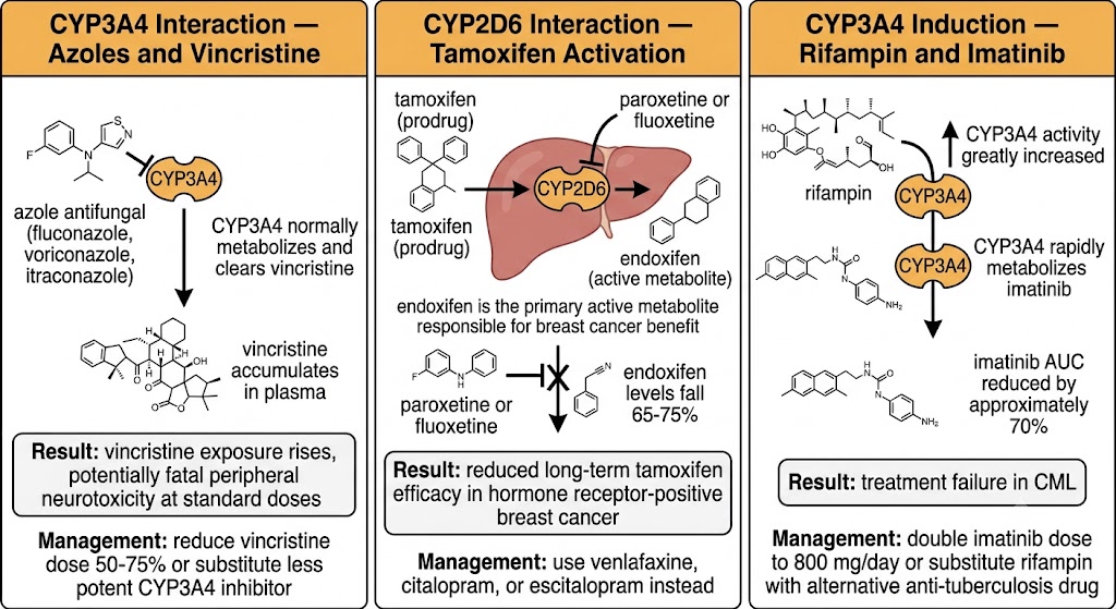
Cytochrome P450 3A4 is the most abundant hepatic cytochrome P450 isoform and metabolizes nearly all taxanes, vinca alkaloids, imatinib, erlotinib, gefitinib, cyclophosphamide, docetaxel, cabazitaxel, and many antiemetics. Major cytochrome P450 3A4 inhibitors encountered in oncology patients include azole antifungals (fluconazole, itraconazole, voriconazole, posaconazole), macrolide antibiotics (clarithromycin, erythromycin — azithromycin does not inhibit cytochrome P450 3A4), human immunodeficiency virus protease inhibitors, cobicistat, and grapefruit juice (which irreversibly inhibits intestinal cytochrome P450 3A4). Major inducers include rifampin, carbamazepine, phenytoin, phenobarbital, and St. John's wort — widely used as an over-the-counter supplement for depression that patients frequently do not disclose.
Cytochrome P450 2C8 is the primary metabolic pathway for paclitaxel; gemfibrozil (a fibrate used for hypertriglyceridemia) is a clinically significant cytochrome P450 2C8 inhibitor that increases paclitaxel exposure approximately twofold and should be discontinued before paclitaxel chemotherapy. Cytochrome P450 2D6 metabolizes tamoxifen to the active metabolite endoxifen; cytochrome P450 2D6 inhibitors (paroxetine, fluoxetine, bupropion) reduce endoxifen plasma concentrations by 65 to 75%, potentially reducing long-term tamoxifen efficacy in hormone receptor-positive breast cancer. Venlafaxine, citalopram, and escitalopram have low cytochrome P450 2D6 inhibitory activity and are preferred antidepressants for patients on tamoxifen. Aprepitant and fosaprepitant, the neurokinin-1 receptor antagonists used for prevention of chemotherapy-induced nausea and vomiting, are both moderate cytochrome P450 3A4 inhibitors and substrates, creating complex interactions with co-administered chemotherapy including docetaxel, vinorelbine, and cyclophosphamide.
Chemotherapy-induced peripheral neuropathy affects up to 40 to 60% of patients receiving neurotoxic chemotherapy, caused predominantly by taxanes, platinum compounds, vinca alkaloids, bortezomib, and thalidomide. Taxane-induced neuropathy results from axonal transport disruption caused by microtubule stabilization, affecting the longest axons first (distal lower extremities). Platinum-induced neuropathy targets dorsal root ganglia neurons directly; oxaliplatin produces both an acute cold-triggered sensory syndrome and a cumulative sensory neuropathy.
For prevention of chemotherapy-induced peripheral neuropathy, the evidence base for pharmacological neuroprotection is limited. Calcium and magnesium infusions before and after oxaliplatin failed to prevent cumulative neuropathy in a randomized trial and are no longer recommended. For treatment of established chemotherapy-induced peripheral neuropathy, duloxetine is the only agent with Level I evidence from a randomized controlled trial (the ACCRU Alliance trial) demonstrating significant reduction in chemotherapy-induced peripheral neuropathy pain and sensory symptoms; a 30 mg daily starting dose titrated to 60 mg daily is the standard approach. Dose reduction or schedule modification remains the most consistently effective neuropathy-sparing strategy where oncologically acceptable.
Alkylating agent versus topoisomerase II inhibitor secondary leukemia, chemotherapy in pregnancy, and organ impairment dose adjustment
Treatment-related secondary malignancies are a well-established long-term consequence of curative cytotoxic chemotherapy. The two most clinically important forms — treatment-related acute myeloid leukemia and treatment-related myelodysplastic syndrome — differ systematically in latency, cytogenetics, and prognosis according to the class of causative agent.
Alkylating agent-related treatment-related myeloid neoplasm follows a characteristic pattern: latency of 5 to 10 years from exposure to diagnosis, typical presentation as treatment-related myelodysplastic syndrome with a hypocellular marrow and a prolonged myelodysplastic phase before transformation to frank acute myeloid leukemia, and cytogenetic abnormalities of monosomy 5 or deletion 5q and monosomy 7 or deletion 7q. Prognosis is poor: median survival from diagnosis is approximately 8 to 12 months, complete remission rates with standard acute myeloid leukemia induction are 20 to 40%, and the only potentially curative approach is allogeneic hematopoietic stem cell transplantation in eligible patients.
Topoisomerase II inhibitor-related treatment-related myeloid neoplasm (caused by anthracyclines and etoposide) has a strikingly different pattern: latency is short (1 to 5 years, most cases within 3 years), the disease presents de novo as frank acute myeloid leukemia without a preceding myelodysplastic phase, and the cytogenetic hallmark is a balanced chromosomal translocation rather than the chromosomal losses seen with alkylating agents. Despite their de novo presentation, these treatment-related acute myeloid leukemia cases can respond to intensive induction chemotherapy, with allogeneic hematopoietic stem cell transplantation in first remission recommended for most patients.
No chemotherapy is safe in the first trimester. Organogenesis (weeks 3 to 8 post-conception) is the critical window for major structural teratogenicity, and essentially all cytotoxic chemotherapy is contraindicated during this period. After the first trimester, many cytotoxic agents have been administered in the second and third trimesters with acceptable maternal and neonatal outcomes, particularly ABVD (doxorubicin, bleomycin, vinblastine, dacarbazine) for Hodgkin lymphoma and CHOP (cyclophosphamide, doxorubicin, vincristine, prednisone) with rituximab for diffuse large B-cell lymphoma. Antimetabolites, particularly methotrexate and 5-fluorouracil, remain relatively contraindicated throughout pregnancy because of their anti-folate mechanisms. All chemotherapy should be stopped at least 3 to 4 weeks before anticipated delivery to allow maternal and fetal drug clearance and reduce the risk of neonatal bone marrow suppression.
Dose adjustment for organ impairment requires knowing the primary elimination pathway of each drug. Renally cleared drugs (carboplatin, bleomycin, topotecan, methotrexate, etoposide, capecitabine, hydroxyurea) require dose reduction proportional to renal dysfunction. Hepatically metabolized drugs requiring biliary excretion (doxorubicin, vincristine, vinblastine, docetaxel, paclitaxel, irinotecan) require dose reduction in hepatic impairment; the bilirubin-based dose reduction guidelines for anthracyclines (50% dose reduction for bilirubin 1.2 to 3.0 mg per deciliter; 25% dose for bilirubin above 3.0 mg per deciliter) are the most widely applied.
| Author / Organization | Title | Source |
|---|---|---|
| Platt OS, Brambilla DJ, Rosse WF, et al | Mortality in sickle cell disease: life expectancy and risk factors for early death | N Engl J Med. 1994;330(23):1639–1644 |
| Charache S, Terrin ML, Moore RD, et al | Effect of hydroxyurea on the frequency of painful crises in sickle cell anemia | N Engl J Med. 1995;332(20):1317–1322 |
| Tram Nguyen T, Mattingly M, Ghassemieh B, Bhatt DL, Kim BK | Asparaginase in pediatric and adult acute lymphoblastic leukemia: pharmacology and clinical relevance | Ann Pharmacother. 2016;50(5):374–387 |
| Vrooman LM, Stevenson KE, Supko JG, et al | Postinduction dexamethasone and individualized dosing of Escherichia coli L-asparaginase each improve outcome of children and adolescents with newly diagnosed acute lymphoblastic leukemia | J Clin Oncol. 2013;31(9):1202–1210 |
| Kronke J, Udeshi ND, Narla A, et al | Lenalidomide causes selective degradation of IKZF1 and IKZF3 in multiple myeloma cells | Science. 2014;343(6168):301–305 |
| List A, Kurtin S, Roe DJ, et al | Efficacy of lenalidomide in myelodysplastic syndromes | N Engl J Med. 2005;352(6):549–557 |
| Scripture CD, Figg WD | Drug interactions in cancer therapy | Nat Rev Cancer. 2006;6(7):546–558 |
| Beijnen JH, Schellens JH | Drug interactions in oncology | Lancet Oncol. 2004;5(8):489–496 |
| Hershman DL, Lacchetti C, Dworkin RH, et al | Prevention and management of chemotherapy-induced peripheral neuropathy in survivors of adult cancers: American Society of Clinical Oncology clinical practice guideline | J Clin Oncol. 2014;32(18):1941–1967 |
| Staff NP, Grisold A, Grisold W, Windebank AJ | Chemotherapy-induced peripheral neuropathy: a current review | Ann Neurol. 2017;81(6):772–781 |
| Smith EM, Pang H, Cirrincione C, et al | Effect of duloxetine on pain, function, and quality of life among patients with chemotherapy-induced painful peripheral neuropathy: a randomized clinical trial | JAMA. 2013;309(13):1359–1367 |
| Godley LA, Larson RA | Therapy-related myeloid leukemia | Semin Oncol. 2008;35(4):418–429 |