CHAPTER 30  ·  THYROID PHARMACOLOGY
Section 1

The Hypothalamic-Pituitary-Thyroid Axis

Feedback architecture, thyroid-stimulating hormone as a monitoring tool, and drug-induced disruptions

The hypothalamic-pituitary-thyroid axis operates as a negative-feedback endocrine loop. Understanding its components is essential because drugs can disrupt it at multiple points, and because thyroid-stimulating hormone serves as the primary clinical tool for monitoring thyroid hormone therapy.

Axis Overview

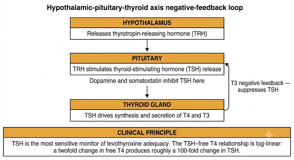
Thyrotropin-releasing hormone is secreted from the hypothalamus and stimulates the pituitary to release thyroid-stimulating hormone. Thyroid-stimulating hormone then acts on thyroid follicular cells to drive iodide trapping, thyroid hormone synthesis, and hormone secretion. When thyroid hormone levels rise sufficiently, they feed back on both the pituitary and hypothalamus to suppress further thyroid-stimulating hormone release. This closed-loop architecture is what makes thyroid-stimulating hormone such a sensitive monitor of thyroid hormone status.

Dopamine and somatostatin tonically inhibit thyroid-stimulating hormone secretion from the pituitary. This explains the mild secondary hypothyroidism occasionally seen with prolonged high-dose dopamine infusion and the transient thyroid-stimulating hormone suppression produced by somatostatin analogues such as octreotide.

The Thyroid-Stimulating Hormone Molecule

Thyroid-stimulating hormone shares an alpha subunit with luteinizing hormone, follicle-stimulating hormone, and human chorionic gonadotropin. The clinical relevance: in the first trimester of pregnancy and in gestational trophoblastic disease, human chorionic gonadotropin reaches very high concentrations and cross-reacts with the thyroid-stimulating hormone receptor, suppressing thyroid-stimulating hormone while stimulating thyroid hormone output enough to cause gestational hyperthyroidism.

Thyroid-Stimulating Hormone as a Clinical Monitor

The pituitary is uniquely rich in type 2 deiodinase, which locally converts thyroxine to triiodothyronine. This makes thyroid-stimulating hormone suppression highly sensitive to circulating thyroxine levels, and it is why thyroid-stimulating hormone is the most sensitive single marker of levothyroxine adequacy. A persistently elevated thyroid-stimulating hormone on a stable dose almost always signals inadequate dosing or an absorption problem.

The thyroid-stimulating hormone–free thyroxine relationship is log-linear: a twofold change in free thyroxine produces roughly a 100-fold change in thyroid-stimulating hormone. This amplification makes thyroid-stimulating hormone a sensitive detector of small changes in thyroid hormone status — but also means that a thyroid-stimulating hormone of 8 mIU/L represents a much smaller hormone deficit than a thyroid-stimulating hormone of 80 mIU/L.

Why Each Patient Has a Personal Thyroid-Stimulating Hormone Set Point

Population-based reference ranges (approximately 0.4–4.0 mIU/L) capture inter-individual variation, but each person's euthyroid thyroid-stimulating hormone clusters in a narrow personal range. Some patients remain symptomatic with a thyroid-stimulating hormone that is technically within the population normal range but outside their personal set point. This intra-individual stability supports titrating levothyroxine toward the lower half of the reference range in patients who remain symptomatic despite a "normal" thyroid-stimulating hormone.
Hypothalamic-pituitary-thyroid axis feedback loop showing TRH, TSH, and T3 negative feedback
The hypothalamic-pituitary-thyroid axis. Thyrotropin-releasing hormone (TRH) stimulates thyroid-stimulating hormone (TSH) release; T3 feeds back to suppress TSH. The TSH–free T4 relationship is log-linear, making TSH the most sensitive clinical monitor.

Section 2

Thyroid Hormone Biosynthesis

Sodium-iodide symporter, thyroid peroxidase, and each step as a drug target

Thyroid hormone biosynthesis is a sequential enzymatic process. Each enzymatic step is a target for pharmacological intervention, and understanding this sequence explains the mechanisms of thionamides, iodide preparations, and radioactive iodine.

Step 1: Iodide Capture — Sodium-Iodide Symporter

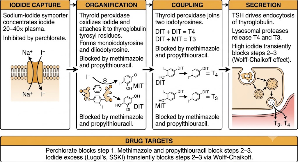
The sodium-iodide symporter on the basolateral membrane of thyroid follicular cells cotransports two sodium ions with each iodide ion, concentrating iodide within the thyrocyte to 20–40 times the plasma level. This transporter is the molecular basis for radioactive iodine uptake — iodine-131 enters the thyroid by exactly the same mechanism as stable iodide. Thyroid-stimulating hormone upregulates sodium-iodide symporter expression; high iodide concentrations and perchlorate downregulate or competitively inhibit it.

Step 2: Organification and Coupling — Thyroid Peroxidase

Once in the follicular lumen, thyroid peroxidase catalyzes two reactions using hydrogen peroxide generated by dual oxidase 2. First, organification: iodide is oxidized and covalently attached to tyrosyl residues on thyroglobulin, forming monoiodotyrosine and diiodotyrosine. Second, coupling: two iodotyrosyl residues join to form thyroxine (diiodotyrosine plus diiodotyrosine) or triiodothyronine (diiodotyrosine plus monoiodotyrosine). Thyroglobulin serves as both the scaffold for iodination and the storage matrix for thyroid hormones within follicular colloid.

Thionamide drugs — methimazole and propylthiouracil — inhibit thyroid peroxidase and block both organification and coupling. This is their primary mechanism of antithyroid action.

Step 3: Secretion and the Wolff-Chaikoff Effect

Hormone secretion requires thyroid-stimulating hormone-driven endocytosis of colloid back into the follicular cell, where lysosomal proteases cleave thyroxine and triiodothyronine from thyroglobulin. The thyroid secretes approximately 80–100 micrograms of thyroxine daily, but only 5–10 micrograms of triiodothyronine — approximately 80% of circulating triiodothyronine is produced by peripheral conversion of thyroxine.

Acute iodide loading transiently inhibits thyroid peroxidase-mediated organification through the Wolff-Chaikoff effect. This is exploited clinically in two situations: pre-operative preparation of the hyperthyroid gland (Lugol's solution for 7–10 days reduces gland vascularity), and thyroid storm (iodide given at least one hour after a thionamide to block ongoing hormone release). The thyroid escapes Wolff-Chaikoff inhibition after several days by downregulating the sodium-iodide symporter, which is why iodide alone is not a durable antithyroid therapy.

Mechanism
Sodium-Iodide Symporter Inhibitors
  • Perchlorate: competitive inhibitor; used in iodine-excess thyrotoxicosis
  • High iodide: downregulates symporter expression (Wolff-Chaikoff)
  • Radioactive iodine: enters via symporter to ablate thyroid tissue
Mechanism
Thyroid Peroxidase Inhibitors
  • Methimazole: blocks organification and coupling; preferred agent
  • Propylthiouracil: blocks organification and coupling plus peripheral conversion
  • Both: thionamide class, do not release preformed hormone
Four-step thyroid hormone biosynthesis pathway showing drug targets at each step
Thyroid hormone biosynthesis. Each enzymatic step is a pharmacological target. Thionamides (methimazole and propylthiouracil) block thyroid peroxidase-mediated organification and coupling. Iodide excess produces transient Wolff-Chaikoff inhibition of the same steps.

Section 3

Deiodinase Enzymes and Peripheral Metabolism

Type 1, 2, and 3 isoforms, sick euthyroid syndrome, and drug effects on peripheral conversion

Deiodinase enzymes determine whether thyroxine is converted to active triiodothyronine or inactivated to reverse triiodothyronine. Three isoforms with distinct tissue distribution and drug sensitivities govern this step.

Three Deiodinase Isoforms

Type 1 deiodinase is expressed in the liver, kidney, thyroid, and skeletal muscle. It converts thyroxine to triiodothyronine in peripheral tissues and clears reverse triiodothyronine from circulation. Type 1 deiodinase is inhibited by propylthiouracil, amiodarone, and propranolol at high doses — this explains the peripheral triiodothyronine-lowering effect of propylthiouracil beyond its thyroid peroxidase inhibition, and the rise in reverse triiodothyronine seen with amiodarone.

Type 2 deiodinase is expressed in the pituitary, brain, and heart, and is the primary source of intracellular triiodothyronine in the brain. It is upregulated in hypothyroid states and downregulated when thyroxine concentrations rise, serving as a local homeostatic buffer. Because the pituitary uses type 2 deiodinase to generate its triiodothyronine signal, thyroid-stimulating hormone suppression tracks circulating thyroxine levels closely.

Type 3 deiodinase is expressed at high levels in the placenta and fetal tissues. It inactivates both thyroxine and triiodothyronine, protecting the fetus from premature exposure to physiologically active thyroid hormone during neurodevelopment.

Sick Euthyroid Syndrome

In critically ill patients, type 3 deiodinase is upregulated in peripheral tissues while type 1 deiodinase activity falls. The result is low triiodothyronine, elevated reverse triiodothyronine, and low-normal thyroid-stimulating hormone — without any primary thyroid gland dysfunction. This pattern is called sick euthyroid syndrome or nonthyroidal illness syndrome.

Sick Euthyroid Syndrome: Do Not Treat the Numbers

The deiodinase shifts in critical illness reduce triiodothyronine and elevate reverse triiodothyronine as an adaptive response reducing metabolic demand. Levothyroxine administration in this setting has not been shown to reduce mortality and may be harmful. Treatment should target the underlying illness. Thyroid function tests should be rechecked 4–6 weeks after recovery before concluding that permanent thyroid disease is present.


Section 4

Thyroid Hormone Receptors and Mechanism of Action

Alpha and beta receptor isoforms, tissue-specific effects, and resistance syndrome

Thyroid hormones act primarily through nuclear receptors that regulate gene transcription. Two receptor genes produce distinct isoforms with different tissue distributions, which explains why excess or deficiency of thyroid hormone produces different effects in different organs.

Receptor Isoforms and Tissue Distribution

Thyroid hormone receptor alpha 1 predominates in the heart, bone, and gastrointestinal tract. Excess thyroid hormone at these sites produces tachycardia, atrial fibrillation, accelerated bone resorption, and increased gastrointestinal motility. Deficiency causes bradycardia, impaired cardiac output, and constipation.

Thyroid hormone receptor beta 1 predominates in the liver, where it regulates cholesterol metabolism — explaining the elevated low-density lipoprotein cholesterol seen in hypothyroidism. Thyroid hormone receptor beta 2 is expressed almost exclusively in the pituitary thyrotroph and hypothalamic neurons, mediating the negative feedback suppression of thyroid-stimulating hormone.

Genomic Mechanism of Action

Unliganded thyroid hormone receptors bind thyroid hormone response elements on deoxyribonucleic acid in association with corepressor proteins, actively suppressing gene transcription. When triiodothyronine binds, corepressors are exchanged for coactivator complexes, switching the receptor from a transcriptional repressor to an activator. This corepressor-to-coactivator switch is the central mechanism of thyroid hormone action and explains why hypothyroidism is not simply an absence of stimulation — it is the presence of active transcriptional repression at thyroid hormone response element-containing genes.

Resistance to Thyroid Hormone Syndrome

Mutations in the thyroid hormone receptor beta gene produce resistance to thyroid hormone syndrome, characterized by elevated thyroid hormone levels with a paradoxically non-suppressed or elevated thyroid-stimulating hormone. Because receptor beta 2 in the pituitary is dysfunctional, the pituitary cannot recognize the elevated thyroid hormone signal and continues to secrete thyroid-stimulating hormone. Tissues expressing normal receptor alpha 1 (heart, bone) remain sensitive, producing tachycardia and potential bone loss despite an apparently "normal" thyroid-stimulating hormone.


Section 5

Pharmacokinetics: Levothyroxine and Liothyronine

Absorption, half-lives, protein binding, and formulation selection

Levothyroxine and liothyronine are the two clinically used thyroid hormone preparations. Their pharmacokinetic differences govern when each is used and how they are monitored.

Levothyroxine

Levothyroxine (synthetic thyroxine) is the standard preparation for thyroid hormone replacement. Oral bioavailability averages 70–80% from standard tablets and is substantially affected by gastric pH — absorption requires an acidic environment and occurs primarily in the proximal small intestine. The elimination half-life is 6–7 days, meaning four to five half-lives are needed to reach steady state after any dose change. Thyroid-stimulating hormone should not be rechecked sooner than six weeks after initiation or dose adjustment, because earlier values do not reflect steady-state drug concentrations.

Approximately 99.97% of circulating thyroxine is bound to thyroid-binding globulin, transthyretin, and albumin; only the free fraction (0.03%) is biologically active. Standard assays measure free thyroxine to avoid the confounding effect of altered binding protein concentrations. Pregnancy and estrogen therapy increase thyroid-binding globulin, raising total thyroxine while free thyroxine and thyroid-stimulating hormone remain unchanged in euthyroid individuals.

Liothyronine

Liothyronine (synthetic triiodothyronine) has a much shorter half-life of approximately 1 day and higher bioavailability than levothyroxine, producing larger peak-to-trough fluctuations in serum triiodothyronine. Its primary clinical use is in hypothyroid preparation for radioactive iodine scanning — it can be stopped 2 weeks before scanning rather than the 4 weeks required for levothyroxine withdrawal, minimizing hypothyroid symptom burden. It is also used in myxedema coma for rapid triiodothyronine repletion and, controversially, in combination with levothyroxine for patients who remain symptomatic on levothyroxine monotherapy.

Pharmacokinetics
Levothyroxine
  • Half-life: 6–7 days; steady state at 6 weeks
  • Bioavailability: ~70–80% (tablets); higher with liquid or capsule
  • 99.97% protein-bound; free fraction is active
  • Monitor: thyroid-stimulating hormone (primary hypothyroidism)
Pharmacokinetics
Liothyronine
  • Half-life: ~1 day; faster onset and offset
  • Bioavailability: ~95%; absorption less food-dependent
  • Uses: radioactive iodine prep, myxedema coma, combination therapy
  • Monitor: free triiodothyronine levels

Section 6

Drug Interactions Affecting Thyroid Hormone

Absorption interference, binding protein effects, and drugs that alter thyroid hormone metabolism

Multiple drugs interfere with levothyroxine pharmacokinetics or thyroid hormone physiology. These interactions are clinically important because they can cause symptomatic hypothyroidism or hyperthyroidism in patients on stable thyroid regimens.

Absorption Interactions

Several drugs bind levothyroxine in the gastrointestinal tract and reduce absorption when taken simultaneously. Calcium carbonate, ferrous sulfate, antacids containing aluminum or magnesium, cholestyramine, and sucralfate all reduce levothyroxine absorption when co-administered. Proton pump inhibitors raise gastric pH and reduce dissolution of standard tablets. The management principle for all of these interactions is the same: take levothyroxine on an empty stomach, 30–60 minutes before breakfast, separated from any potentially interacting medications by at least 4 hours.

Drugs That Accelerate Levothyroxine Metabolism

Rifampin, phenytoin, carbamazepine, and phenobarbital induce hepatic cytochrome P450 enzymes and glucuronidation pathways that accelerate thyroxine metabolism. Patients on these anticonvulsants or antitubercular agents typically require levothyroxine dose increases of 20–50% to maintain target thyroid-stimulating hormone. Thyroid-stimulating hormone should be monitored after starting or stopping any of these agents.

Amiodarone

Amiodarone deserves specific attention because it affects thyroid hormone pharmacology through multiple mechanisms simultaneously. Containing approximately 37% iodine by weight, amiodarone delivers a massive iodine load that disrupts the normal Wolff-Chaikoff autoregulation. It inhibits type 1 deiodinase, reducing peripheral thyroxine-to-triiodothyronine conversion and raising reverse triiodothyronine levels. Amiodarone also directly inhibits thyroid hormone entry into cells and has a direct cytotoxic effect on thyroid follicular cells. The net result can be either hypothyroidism (most common, from iodine-induced thyroid suppression and type 1 deiodinase inhibition) or thyrotoxicosis (from iodine-induced autonomous synthesis in nodular thyroid or from destructive thyroiditis). Thyroid function testing is required before starting amiodarone and every 6 months during therapy.

Amiodarone-Induced Thyroid Dysfunction: Two Distinct Syndromes

Amiodarone-induced hypothyroidism: thyroid-stimulating hormone rises; treat with levothyroxine; amiodarone can often be continued. Amiodarone-induced thyrotoxicosis type 1: iodine excess drives autonomous thyroid hormone synthesis in a pre-existing goiter or nodular thyroid; treat with thionamides. Amiodarone-induced thyrotoxicosis type 2: destructive thyroiditis from direct thyroid toxicity; treat with glucocorticoids. The two thyrotoxicosis types often coexist and can be difficult to distinguish; combined therapy with thionamide plus glucocorticoid is used when the type is uncertain.


Suggested References
Author / Organization Title Source
Ortiga-Carvalho TM et al. Hypothalamus-pituitary-thyroid axis Compr Physiol. 2016;6(3):1387–1428
Portulano C et al. The Na+/I- symporter (NIS): mechanism and medical impact Endocr Rev. 2014;35(1):106–149
Peeters RP, Visser TJ Metabolism of thyroid hormone Endotext. MDText.com, 2017
Bianco AC et al. Biochemistry, cellular and molecular biology, and physiological roles of the iodothyronine selenodeiodinases Endocr Rev. 2002;23(1):38–89
Cheng SY et al. Molecular aspects of thyroid hormone actions Endocr Rev. 2010;31(2):139–170
Jonklaas J et al. Guidelines for the treatment of hypothyroidism: American Thyroid Association task force Thyroid. 2014;24(12):1670–1751
Ross DS et al. 2016 American Thyroid Association guidelines for diagnosis and management of hyperthyroidism Thyroid. 2016;26(10):1343–1421