Medical Pharmacology Chapter 44:  Clinical Correlation: Pain Management

Pain Management

Previous Page

 
  • Rationale for management of acute postoperative pain

    • Acute postoperative pain affects a number of organ systems- some of which are unrelated to the actual surgical intervention. within

    • For example: upper abdominal surgery may result in hypoventilation-which diminishes pain associated with the ventilatory process

      • Hypoventilation can cause atelectasis impairing ventilation/perfusion relationships, increasing the possibility of arterial hypoxemia and pneumonia

    • Another possibility is that postoperative pain limits mobility, which in turn increases the likelihood of deep vein thrombosis.  

    • Pain is also correlated with a stress-induced hypercoagulation state

    • Pain is associated with increased circulating catecholamine levels that not only favor hypertension but also arrhythmias secondary to increased  catecholamine-mediated automaticity.  

  • Development of new clinical methods for pain management: Rationale

    • Because of the number of adverse physiological effects associated with postoperative pain, the number of relatively new analgesic methods complement the traditional means of pain management.  These methods include:

      1. Patient-controlled analgesia (PCA pumps)

      2. Neuraxial analgesia

      3. Peripheral nerve blockade

Adverse Systems physiological Effects of postoperative pain

Pulmonary system (decrease lung volumes secondary to patient's pain associated with respiration)

Pneumonia

Hypercarbia

Atelectasis* (total/partial collapse of the lung)

Ventilation/perfusion mismatching

Arterial hypoxemia


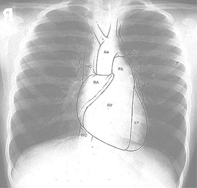
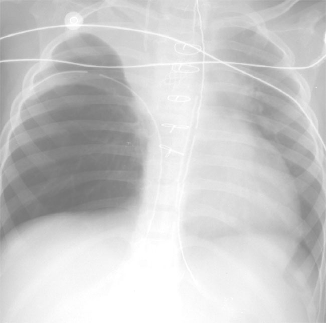
Normal Chest Radiograph (A, left)  and one illustrating Post-operative Atelectasis (B, right)


  • (B, above) Post-operative atelectasis may occur  following surgical correction of congenital heart disease. 

    • The right upper lobe is most commonly affected, and is possibly due to aspiration of secretions into this area of the lung.  (Atelectasis may develop due to low position of the endotrachial tube occluding one of the main stem bronchi)

  • Texas Children's Hospital, Houston, Texas; Texas Heart Institute; Edward B. Singleton Diagnostic Imaging Service;Colin McMahon, M.B.BCh.  Edward Singleton, M.D.

Atelectasis-Right Middle Lobe

Atelectasis-Right Middle Lobe

  • Atelectasis is the loss of lung volume and therefore a direct sign is the displacement of interlobular fissures. Generally this is accompanied by increased density and possibly elevation of the hemidiaphragm, mediastinal displacement, or compensatory over-inflation. If there has been resorption of air within the atelectatic segment, there is generally an absence of air bronchograms. The pattern of the specific lobar or segmental collapse produces relatively specific findings on the chest film, often requiring both PA and lateral films for clear and
    specific definition.

  • PA (posterior-anterior) radiograph of this female patient (note breast shadows bilaterally) showed obscuration of the lower right cardiac border merging with opacification of the lung field underlying the right breast. Because the right middle lobe is immediately adjacent to the cardiac silhouette in that position collapse or opacification of the right middle lobe will merge densities between the lung and the heart and thus, the normal sharp boundary between heart and lung is lost."

  • Yale University School of Medicine, used with permission

Medical Editor
C. Carl Jaffe, MD, FACC
Professor of Medicine
(Cardiology)
Section of
Cardiovascular Medicine

Site producer
Patrick J. Lynch, MS
Design Director
Web Design and Development
Information Technology
Services - Medicine


  • Example: thorax/abdominal surgical procedures: question: in this setting is how would pain lead to respiratory complications:

    • Pain causes in increase in skeletal muscle tension which in turn reduces total lung compliance, causes hypoventilation and splinting (splinting is defined as "rigidity of muscles occurring as a means of avoiding pain caused by movement of the part" ).  These changes cause subsequent effects including:

      • Additional ventilation/perfusion anomalies (some anomalies may occur is a result of increases in extracellular lung water) which promotes hypoxemia

      • This effect reduces significantly functional reserve capacity (25%-50% of preoperative levels)

      • Pain through its hypoxemia effect results tachypnea and hypocapnia initially; however, this increase in breathing work can result in hypercapnic respiratory failure

  • Hypoventilation, induced by pain, can promote pulmonary consolidation/pneumonitis which at the very least complicates clinical situation.   The significance of these adverse effects may ultimately depend on the clinical condition of the patient, e.g. presence of pre-existing pulmonary dysfunction, advance age, obesity, as well as the particular surgical procedure.

Possible Cardiovascular System Effects (secondary to excess sympathetic* outflow due to acute postoperative pain)

Tachycardia

Systemic hypertension

Ischemia

Arrhythmias

  • 2*-Increased sympathetic outflow secondary to painful stimulation can itself enhance, that is increase pain.  

    • The mechanism could involve (a) initial vasoconstriction which leads to (b) acidosis, tissue ischemia, and release those substances that themselves activate pain receptors.  

    • Increased pain therefore leads to increase sympathetic outflow in the initiation of the cycle, sometimes called reflex sympathetic dystrophy (complex regional pain syndrome(s) is now the preferred designation).

  • Following some types of nerve injury, pain may occur without requiring activation of pain receptors.  That is, spontaneous firing of injured peripheral nerves may occur and may be more likely to occur in response to sympathetic nervous system simulation.

    •  This increased likelihood may be caused by an increase in the number of alpha-adrenergic receptors localized at these injured sites.  (The phenomena of spontaneous firing also occur in dorsal root ganglia following interruption of their peripheral projections (nerve transection/limb amputation)

  • The cardiovascular effects of pain involve four major systems:(1) catecholamine release and sympathetic nerve terminals and adrenal medulla (2) aldosterone and cortisol release from the adrenal cortex and (3) antidiuretic hormone release from hypothalamus and (4) activation of the renin-angiotensin system.  Why should these changes influence the cardiovascular system?

    • Increased angiotensin II  promotes generalized vasoconstriction, causing hypertension and increasing cardiac work by increasing afterload

    • Catecholamines directly cause a positive chronotropic and inotropic effect and increase systemic vascular resistance, latter of which also increases afterload

    • Increase circulating catecholamines promote arrhythmias directly as well as  by exacerbating underlying myocardial ischemia.  (Recall that significant perioperative morbidity is associated with myocardial dysfunction)

    • Increased aldosterone, cortisol, and ADH (antidiuretic hormone) in concert with angiotensin II and catecholamine effects all work in the direction of promoting congestive heart failure, although this effect would be more likely patients with intrinsically limited cardiac reserve.  Accordingly, preoperative assessment for the purpose of identification of patients who may have limited cardiac reserve or some cardiac dysfunction is particularly important as it also underscores the necessity of effective clinical pain management.


Endocrine Effects associated with Postoperative Pain

Sodium and water retention

Increased protein catabolism

Hyperglycemia


Immunological Effects associated with Postoperative  Pain

Reduce Immune Function

  • Stress response to pain reduces immune function (cellular and humoral (humoral: refers to antibody-mediated immunity)).  Pain of the responsible for lymphopenia and leukocytosis with depression of the reticuloendothelial system. (definition: lymphopenia "decrease in the proportion lymphocytes in the blood"; definition: leukocytosis-- increase the number of leukocytes in the blood which may be associated with a pathological condition)

Coagulation System effects associated with Postoperative Pain

Hypercoagulation States

Deep vein thrombosis

 Increased platelet adhesiveness

Reduced fibrinolysis

  • *Note: Epidural anesthesia reduces postoperative thromboembolic complications; however, the underlying molecular mechanisms are not completely understood.  

    • In a recent study of major orthopedic surgical procedures, general anesthesia was compared with local anesthesia in terms of postoperative hypercoagulability states. 

    • With general anesthesia, orthopedic surgery induced a hypercoagulable state, as assessed by increased platelet-mediated hemostasis time, clotting time, and collagen-induced thrombus formation.  

    • By contrast, in the patient group receiving epidural anesthesia, these parameters were not altered. Apparently, epidural anesthesia reduces the likelihood of immediate post-operative hypercoagulability without influencing physiologic aggregation and the coagulation process. (Hollmann MW, Wieczorek KS, Smart M, Durieux ME.,Reg Anesth Pain Med 2001 May;26(3):215-222.)  3General anesthesia even with parenteral opioid administrationa has limited effect on postoperative hypercoagulability.

  • 3Coagulation effects are probably related to stress-associated changes in (a) blood viscosity, (b) platelet function, (c) fibrinolysis and (d) coagulation pathway effects.

    • The consequences of these changes are manifest as increased platelet adhesiveness,  reduced fibrinolysis and the production of a hypercoagulable state.

    • These coagulation effects, when taking in combination with microcirculatory effects of increased circulating catecholamines, are probably responsible for the increased incidence of thromboembolism.

Gastrointestinal system effects associated with Postoperative  Pain

Ileus

  • 3Reduction in gastrointestinal function would be expected based on sympathetic hyperactivity, recalling that the parasympathetic system promotes motility, whereas sympathetic action retards GI motility.  The clinical consequence of postoperative ileus include:

    • Increased nausea, vomiting, generalized discomfort as well as

    •  Delay in resumption of enteral diet

  • 3The delay in resumption of an enteral diet itself may have adverse clinical effects (postoperative morbidity) including an increase likely those septic complications and abnormal wound healing.

Genitourinary system effects associated with Postoperative Pain

Urinary retention

  • Sympathetic activity responses to pain can cause a reflex inhibition of the  visceral smooth muscle (e.g. urinary bladder smooth muscle) tone-therefore urinary retention. 

    • Urinary retention can promote urinary tract infection and associated complications.


Acute Post-Operative Pain Management

  • Objectives for the anesthesia provider in terms of management of acute pain:

    • Postoperative pain must be evaluated and treated

    • Side effects associated with pain management should be anticipated and managed.

  •  6Pain management approaches-routes of delivery:

    • 6Oral: disadvantages include relatively long onset times and difficulty in titrating to effect; furthermore, these agent require a functional gastrointestinal tract

      • In-patients may be started with systemically administered analgesics (typically opioids) and then transitionned to oral medication when the need for rapid adjustment decreases.

    • 6Transepithelial delivery is considered an alternative to oral administration.  One advantage is that the medication would not be subject to significant first-pass metabolic effects.  However onset times will be long relative to intravenous alternatives.  

      • Drug absorption variability is still a problem, as with oral administration and he would be difficult to titrate to effect.

      • Transepithelial dosing however may have a place in the outpatient setting particular

    • 6Parenteral routes of administration are the primary methods to manage moderate/severe postoperative pain. 

      • Patient controlled systems are now common.

      • Intramuscular: this route of administration results in a more rapid onset and time to maximal effect compared to oral administration; however, variability in absorption, injection site discomfort, and even delayed respiratory depression represent clinical concerns. 

        • Intermittent dosing results in sufficient variation in plasma concentration such that the patient may oscillate between sedation (a somewhat too high concentration of drug) and pain (the drug concentration has fallen below an analgesia threshold).

        • This oscillation between somewhat inadequate and somewhat excessive plasma drug concentration in terms of patient need follows from the observation that only a small change in plasma concentration (10%-20%) is sufficient to transition between these two undesirable states.

        • Not surprisingly the use of PCA pumps and continuous epidural infusions allow greater stability of the desired level of analgesia.

        • Finally, although many opioids may be administered by the intramuscular route only one nonsteroidal anti-inflammatory agent (NSAID), ketorolac (Toradol) is used for managing postoperative pain.  Clinical experience suggests that ketorolac (Toradol) efficacy is comparable to moderate opioid doses and therefore can be used to manage moderates-severe pain associated with a variety of surgeries.

      • 6Patient controlled analgesia (PCA): Contemporary PCA pumps allow a low-level of continuous, background, infusion of an opioid while still allowing the patient to administer additional small doses. 

        • The combination of background confusion with additional small boluses available have served to reduce drug blood level fluctuations.

        • Contrasting PCA with on-demand administration suggest that PCA is associated with reduced total drug use, less sedation, fewer sleep disturbances and more rapid return to activity.

        • Difficulty associated with PCA is in part related to available drugs and their intrinsic limitations.  

          • Other difficulties for correlated with operator or mechanical malfunctions -- note that since patients are responsible in part for their own dosing a certain level of patient understanding of the system is required.  Accordingly, PCA use may be more difficult in pediatric as well as geriatric settings.

        • Anesthesia providers in developing the PCA therapy approach would take into account the various parameters that may be set in the context of the particular patient's pathophysiological state and the particular surgery involved.

      • 6Central Neuraxial Analgesia:

        • Overview: For 50 years is in recognize that postoperative analgesia could be accomplished by continual catheter epidural infusion. 

          • Postoperative analgesia was maintained for days through use of intermittent local anesthetic injections (bolus doses). 

            • Sympathetic blockade associated with this approach resulted in the frequent need for at least one vasopressor dose.  

            • Furthermore, as described above, any "bolus" dosing approach is associated with fluctuations in analgesia levels which functionally required repeated injections every few hours.

          • An alternative to intermittent, bolus dosing was the use of continuous local anesthetic infusions; unfortunately, in order to maintain adequate analgesia, the local anesthetic doses were sufficient to cause sensory and sometimes motor block.  The significance of motor blockade is this setting is that more blockade prevented ambulation which extended postoperative recovery time.

          • The most important event responsible for the now common application of central neuraxial anesthesia was the substitution for local anesthetic agents by opioids.  

            • An additional factor is the avoidance of those limitations associated with intramuscular or oral analgesia; limitations of which have been described earlier. 

              • Also, interrupting the pain pathway more peripherally (in this case between the first-and second-order neurons) reuse the likelihood of CNS depression.

        • 6Intrathecal administration:

          • drawing by Lindsey Parker, (c) University of Kansas Medical Center

          • Advantage: single injections provide long-lasting analgesia.  Time to onset of action will depend on the lipophilic nature of the opioid.  Lipophilicity promotes penetration of the opioid to the target.

            • Specific example: morphine -- time course to effect: Following intrathecal administration of 0.25-4 mg morphine, peak analgesia occurred in about 20 -60 minutes and analgesia lasted from 2-12 hours.

            • For routine clinically used,  effective analgesia can be obtained in the 0.25-1 mg dose range while minimizing the likelihood of respiratory depression.

            • Note that this approach involves bolus administration and as such exhibits limitations including the need to carefully monitor, possibly re-inject and the intrinsic limitation of the difficulty to titrate to effect. 

              • Intrathecal administration appears less desirable than epidural administration because of expectable side effects, the more serious of which include respiratory depression secondary to rostral drug diffusion and infection risk.  Cauda equina syndrome has also been reported after continuous spinal anesthesia.

              • 7"The cauda equina (CE) is formed by nerve roots caudal to the level of spinal cord termination. Cauda equina syndrome (CES) has been defined as low back pain, unilateral or usually bilateral sciatica, saddle sensory disturbances, bladder and bowel dysfunction, and variable lower extremity motor and sensory loss." Spinal anesthesia is only one cause of cauda equina syndrome.  Other causes include trauma, lumbar disc disease,tumor, abscess, late stage ankylosing spondylitis (ankylosing spondylitis describes the condition by which some or all of the joints and bones of the spine fuse together) and idiopathic presentations. 

              • Side effect comparisons between spinal and epidural neuraxial  opioids:

                • Respiratory depression: spinal 5%-7% incidence; epidural 0.1-2% incidence.

                • Pruritus: spinal 60% incident; epidural 1-100% incidence

                • Urinary retention: spinal 50% incidence; epidural 15-25% incidence

                • Nausea and vomiting: spinal 20-30% incidence; epidural 20-30% incidence

              • Management of side effects:

                • Respiratory depression may require naloxone (Narcan) which will of course terminate the analgesia effect as well as reverse respiratory depression.  Ventilatory support may also be used.

                • Pruritus:  naloxone (Narcan) again can be used as yet will reverse essentially all opioid-induced effects, again with the loss of  analgesia.  Antihistamines are also helpful.

                • Nausea and vomiting: Naloxone (Narcan) as well as specific antiemetic agents including transdermal scopolamine

                • Urinary retention: naloxone (Narcan); catherization.

            • 6Intrathecal administration is relatively uncommon outside of labor analgesia; however, the combination of spinal anesthesia followed by epidural analgesia is used since it provides the advantages spinal anesthesia (rapid onset with significant role blockade) and allows ready transition to titratable, i.e. epidural analgesia following major surgery.


        • 6Epidural:

          • drawing by Lindsey Parker, (c) University of Kansas Medical Center


          • As noted above, one of the advantages of epidural administration is to reduce likelihood of serious CNS side effects, particularly respiratory depression.  

            • This reduced likelihood in the understood in terms of the pharmacokinetics associated with the absorption and distribution phases for epidurally administered opioids. 

            • Barrier is to CNS access include the dura itself as well as non-physical "barriers" which serve to promote drug redistribution to the systemic circulation.  For example, epidural space vascularity directly serves to transport the opioid away from the CNS.  Local drug reservoirs such as fat, connective tissue, lymphatic network all work to sequester drug away from direct CNS access.

          • 6Pharmacokinetic issues: The presence of various "barriers" to CNS penetration require that larger amounts of  epidural drug must be administered in order to ensure saturation of spinal cord opioid receptor sites, the target for pain management.  Despite the larger amounts of drug required by epidural administration compared intrathecal administration, CNS side effects are reduced.

            • The relative lipophilicity of the drug (e.g. fentanyl (Sublimaze)) or hydrophilicity of the drug (e.g. morphine) will influence the absorption and distribution characteristics in the expectable way.  For example, a fairly small proportion of an epidural morphine dose (about 2-10%) would actually diffuse across the dural barrier, buying to opioid receptors and produce analgesia.  The rate at which adequate analgesia will appear will be determined by Fick's Law or the law of mass action; therefore, if bolus administration is not used, continuous infusion will likely not achieve adequate analgesia for several hours, at least with hydrophilic agents.  One alternative, in this case, used to provide an initially high infusion rate (e.g. 5-10 ml/5-15 minutes) which would simulate a "bolus" and then transition to a slower, continuous flow. 

            • Analgesic titration is more readily available using continues infusion.  Effective titration is evident when fentanyl (Sublimaze) is used given its short onset of action (4-5 minutes) its short time to peak effect (within 20 minutes).  Morphine (less lipophilic) has own set time of about a half-hour with peak effects occurring within 60-90 minutes.

          • 6Safety issues: Concerns include accidental intrathecal drug delivery, respiratory depression, epidural hematoma, and infection related complications.

            • A protection against unnoticed, inadvertent subarachnoid catheter migration is the inclusion of a low concentration of local anesthetic (0.1%  bupivacaine (Marcaine)) which allows a progressive sensor blockade to be really identified, but otherwise will not produce a sensory/motor block upon epidural administration.  Infusions would be discontinued do sensory/motor deficits occurred because the possibility of intrathecal delivery and these sensory or motor deficits should regress within two-four hours.  If impairment persists for a longer period of time consideration should be given to a more serious basis for the dysfunction such as local hematoma.

            • Respiratory rate/sedation levels should be monitored typically every hour for the first day and subsequently every four hours.  Direct patient monitoring would be superior to apnea monitors.  Groups of patients and particularly  high risk for respiratory depression might include the elderly, patients with apnea or those with debilitating disorders.

            • 6Clotting issues:

              • Epidural catheter placement should occur at least one hour prior to IV heparinization in patients who do not have preoperative plotting abnormalities that will require intraoperative anticoagulants treatment with unfractionated.  Placement at least one hour before heparinization appears to guard against an increase in the incidence of clinically important epidural hemorrhage.

              • Hematoma formation and epidural catheters in patients receiving perioperative anticoagulation: can because those the increased use of low molecular weight heparins with possible increased associated risk for epidural hematoma, the current recommendation is that placement or removal of an epidural catheter should be performed at least 10-12 hours following a dose of low-molecular-weight heparin (LMWH) . 

                • LMWH should not be given to a patient within two hours following epidural catheter removal.

                • Risks of spinal hematoma will be increased when LMWH is used concurrently with other agents affecting hemostasis such as antiplatelet drugs, dextran, and standard heparin.

                • The general rule is that if perioperative anticoagulation is employed in patients whose pain is being managed by epidural analgesia, then anticoagulation and neurological status must be monitored carefully.

                • In patients receiving perioperative warfarin (Coumadin), epidural catheters may be placed providing coagulation status is carefully monitored.


Management of some pain syndromes (Chronic, subspecialty anesthesia practice)

  • Lumbosacral radiculopathy: symptoms may include low back pain (varying degrees) with radiation to lower extremity and sometimes sensory with motor changes.

    • 6Causes:

      • Originally presumed to a sole consequence of mechanical nerve root compression

        • an acute inflammatory process is associated with mechanical nerve root compression. 

          • The inflammatory process is associated with  intraneural accumulation of serum protein/fluid along with higher intraneural pressure, ischemia and axon degeneration.

        • Intervertebral contents also can cause significant spinal canal inflammation.

      • Other mechanisms are probably important because surgical decompression is always successful and there are many examples of asymptomatic patients with substantial disk protrusion. 

        • In human disk samples collected from patients with symptomatic radiculopathy, high levels (20-to 100-times higher than that obtained from human inflammatory synovial effusion, of phospholipase A2 have been noted.  These phospholipase A2  activities with subsequent activation of the arachidonic acid pathway may provide a basis for the inflammatory response.

      • Cytokines are probably also important in radiculopathy pain pathophysiology. 

        • The cytokines would include tumor necrosis factor (TNF alpha), nerve growth factor, interleukins, and interferons.

    • 4,5,6Intervention: epidural steroid injections may be particularly useful especially with new onset pain caused by mechanical/chemical nerve route injury. Pharmacology: triamcinolone diacetate (50 mg; range 50-100 mg) or methylprednisolone  (80 mg; range 80-120 mg) would be introduced into the epidural spaces close to the effective nerve roots. Special clinical considerations:

      1. Analgesia will be promoted by addition of lidocaine (Xylocaine) to the injected  steroid solution.  This method ensures proper location of steroid as well as breaking the cycle of pain and muscle spasms.

      2. If the first epidural administration was not effective, it is unlikely that a repeated, future ejection will be efficacious.

      3. Individuals with more chronic symptoms {patients who have had previous surgery or a more chronic back pain complaint} will probably not benefit from epidural steroids

    • The use of lidocaine (Xylocaine) alone in patients with lumbosacral radiculopathy is of benefit not only due to the immediate relief obtained with local anesthetic but also when compared to time to ambulation associated with bed-rest alone  (11 (LA) vs. 31 days (bed rest alone).  Moreover, in management of chronic pain patients combination of lidocaine (Xylocaine) with methylprednisolone for shown superior to both epidural saline or lidocaine (Xylocaine) injections alone.  In a separate study, patients treated with epidural methylprednisolone (Solu-Medrol) with or without the addition of local anesthetic were compared.  Either group was associated with a nearly 100% success rate, suggesting the steroid was providing the therapeutic efficacy.  

    • Despite the common usage of epidural steroids,  it has been difficult to conclusively demonstrate efficacy in highly controlled, randomized studies, although the results generally predict higher odds for a positive outcome with epidural steroids.

    • Epidural steroid injections are most likely to be effective in patients with evidence of nerve root inflammation associated with bulging disks, spondylolisthesis (forward displacement one vertebra over another, often the fourth lumbar over the fifth or the fifth lumbar over the body of the sacrum) or scoliosis with symptoms of radiculopathy:

      • Efficacy approximates 70%-100%

    • As suggested above, patients  whose symptoms have lasted > one-year: the success rate is about 30%-45%.

    • The underlying mechanism for those effect may be the significant suppression of the inflammatory response by the steroids

  • 5Epidural steroid injection -- injection administration site is at the lesion level; e.g.L4-5 in this case.  Drawing from Warfield, C.A.:Manual of Pain Management, p. 277, Philadelphia, Lippincott, 1991 obtained from Aberle, K.L and Warfield, C.A. "Basis of Contemporary Pain Management" in Principles and Procedures and Anesthesiology, Philip L. Liu, editor, J. B. Lippincott Company, Philadelphia, Chapter 23 p.368, 1992 (use by permission in secondary source)

  • 6Root compression is more commonly observed at the L5 and S1 levels due to anatomical factors (these nerves pass through a relatively narrow lateral bony recess in exiting the spinal canal. 

    • Lumbosacral radiculopathy symptoms include:

      • Low back pain with radiation to lower extremity (varying distance)

      • Consider disease, motor/sensory loss occurs

    • Although surgery may be indicated for large midline disk involvement that manifests as bowel and bladder dysfunction, initial treatment is usually limited to immobilization with mild analgesics along with slow resumption of activity (prolonged immobilization is unlikely to be helpful)

    • Therefore, only if conservative management (rest +analgesics) fails to resolve pain would intervention with epidural steroids be considered

      • As noted earlier, proper drug (steroid) localization is inferred from immediate analgesia due to the concurrently administered local anesthetic.

      • A concern mainly in patients with S1 radiculopathy is that the drug will not spread adequately.  Therefore caudal injection may be more appropriate, perhaps utilizing a radio-opaque catheter positioned using fluoroscopy.

    • Risk associated with epidural steroid injections: probably extremely low based on reports in which usually 1-3 injections have been used with 40-80 methylprednisolone acetate or 50 mg triamcinolone diacetate

      • Corticosteroid doses used for management of radiculopathy, however, are sufficient to suppress (acutely) the hypothalamic-pituitary-adrenal (HPA) axis as evidenced by reduced plasma cortisol and adrenocorticotropic hormone level response to provocation.

      • As an example, when the treatment protocol required three epidural triamcinolone injections  at one-week intervals,  all patients recovered from HPA suppression after three months.  

        • A more significant suppression of the HPA axis may be associated  when midazolam (Versed) was used for sedation. 

          • Altogether these findings suggest that exogenous steroids might be appropriate for patients undergoing significant stress during a "vulnerable" period following epidural steroid.  

        • Furthermore, epidural steroid treatment should be used with cautions in the diabetic patient because (a) of increased epidural infection risk and (b) since glucose control may be adversely affected.

        • Since immunosuppression will accompany epidural steroid use, aseptic technique must be emphasized during the procedure. 


Previous Page


  • Stoelting, R.K., and  Ronald D. Miller "Acute Post-Operative Pain Management", in Pharmacology and Physiology in Basics of Anesthesia, Churchill-Livingston, New York, 2000, 425-434.

  • 2Stoelting, R.K., "Pain", in Pharmacology and  Physiology in Anesthetic Practice, Lippincott-Raven, Philadelphia, third edition, pp. 628-633, 1999.

  • 3Lubenow, T.R., Ivankovich, A.D. and McCarthy, R.J.  "Management of Acute Postoperative  Pain" in Clinical Anesthesia, Barash, P.G., Cullen, B.F. and Stoelting, R.K.,eds, 4th edition, Lippincott-Williams and Wilkins, Philadelphia, Chapter 54, pp. 1403-1434,2001

  • 4Barash, P.G., Cullen, B.F. and Stoelting, R.K.,"Chronic Pain Management" in Handbook of Clinical Anesthesia, 4th edition, Lippincott-Williams and Wilkins, Philadelphia, Chapter 55, pp. 784-796, 2001

  • 5Aberle, K.L and Warfield, C.A. "Basis of Contemporary Pain Management" in Principles and Procedures and Anesthesiology, Philip L. Liu, editor, J. B. Lippincott Company, Philadelphia, Chapter 23 pp. 363-378, 1992

  • 6Abram, S.E. and Schlicht, C.R. "Chronic Pain Management" in  in Clinical Anesthesia, Barash, P.G., Cullen, B.F. and Stoelting, R.K.,eds, 4th edition, Lippincott-Williams and Wilkins, Philadelphia, Chapter 55, pp. 1435-1462, 2001

  • 7Michael S Beeson, MD, MBA,"Cauda Equina Syndrome" Program Director, Department of Emergency Medicine, Summa Health System; Associate Professor of Clinical Emergency Medicine, Northeastern Ohio Universities College


Disclaimer

This Web-based pharmacology and disease-based integrated teaching site is based on reference materials, that are believed reliable and consistent with standards accepted at the time of development. Possibility of human error and on-going research and development in medical sciences do not allow assurance that the information contained herein is in every respect accurate or complete. Users should confirm the information contained herein with other sources. This site should only be considered as a teaching aid for undergraduate and graduate biomedical education and is intended only as a teaching site. Information contained here should not be used for patient management and should not be used as a substitute for consultation with practicing medical professionals. Users of this website should check the product information sheet included in the package of any drug they plan to administer to be certain that the information contained in this site is accurate and that changes have not been made in the recommended dose or in the contraindications for administration.  Advertisements that appear on this site are not reviewed for content accuracy and it is the responsibility of users of this website to make individual assessments concerning this information.  Medical or other information  thus obtained should not be used as a substitute for consultation with practicing medical or scientific or other professionals.