Medical Pharmacology Chapter 43:  Adult Cardiac Procedures

Main Cardiovascular Section Menu

Site Table of Contents

Previous Page Next Page
Table of Contents


Chest Landmarks


  • Yale center for Advanced Instructional Media, Yale Tech University School of Medicine, Medical Editor: C. Carl Jaffe, MD; Site Producer: Patrick J. Lynch used with permission, copyright 2000,2001  Yale University School of Medicine


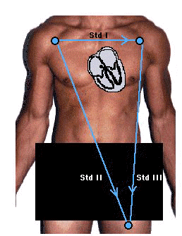
EKG Standard Leads
  • "There are three of these leads which are usually designated as I, II and III.

    • They are all bipolar (i.e., they detect a change in electric potential between two points) and detect an electrical potential change in the frontal plane. 

  • Lead I is between the right arm and left arm electrodes, the left arm being positive. 

  • Lead II is between the right arm and left leg electrodes, the left leg being positive. 

  • Lead III is between the left arm and left leg electrodes, the left leg again being positive

  • A diagrammatic representation of these three leads is termed Einthoven's triangle (shown in blue below), after the Dutch  doctor who first described the relationship. The central source of electrical potential in the triangle is the heart."

    • Image courtesy of New York University, Hippocrates Project NYU School of Medicine, Created by Steven Zweibel, Aaron Gindea, M.D. as Modified by Ambika Nayar. Animation and Art Coordinator: Kathleen Ruiz, Technical Coordination: Martin Nachbar, MD Program Director, Adam W. Hunger, BSE Systems Developer and Wei-Chen Chao, Multimedia / Virtual Reality Specialist, used with Permission


Precordial Lead Locations

  • "These six unipolar leads, each in a different position on the chest, record the electric potential changes in the heart in a cross sectional plane.

    • Each lead records the electrical variations that occur directly under the electrode."

    • Image courtesy of New York University, Hippocrates Project NYU School of Medicine, Created by Steven Zweibel, Aaron Gindea, M.D. as Modified by Ambika Nayar. Animation and Art Coordinator: Kathleen Ruiz, Technical Coordination: Martin Nachbar, MD Program Director, Adam W. Hunger, BSE Systems Developer and Wei-Chen Chao, Multimedia / Virtual Reality Specialist, used with Permission


ECG

Ventricular rhythms

  • Ventricular Premature Contraction (VPC or PVC)

    • 1Characteristics:

      • Rate -- is influenced by (dependent upon) underlying rhythm

      • Regularity -- irregular

      • Presence of "P" wave: Not present on the PVC

      • P:QRS ratio -- No "P" wave on the PVC

      • PR interval: none

      • QRS width: "wide" (= 0.12 seconds"; unusual appearance

      • No "grouping"

      • Dropped beats: none

    • General Background

      • Common arrhythmias-not necessarily associated with heart disease

      • Adult males: incidence of PVCs > or  = 60%

      • By themselves, PVCs are not associated with increased mortality/morbidity; however, in patients having previous myocardial infarction, PVCs occurring at a rate >10/hr is associated with increased mortality: [as noted below, a stronger risk factor is reduced ejection fraction}

      • Very early cycle PVCs, i.e. R- on T  PVCs may be associated with increased sudden death risk, particularly during acute ischemic episodes and with QT prolongation- note rhythm strip below for an example of R- on T-wave induction of ventricular fibrillation:

      • "All recordings are printed on standard electrocardiographic paper at 25 mm/s paper speed (1 mm corresponds to 40 ms, 5 mm corresponds to 200 ms). The vertical resolution, is 10 mm/mV unless otherwise noted. The filtration of the electrocardiogram varied with every recording. The monitor leads used were non-standard bipolar precordial leads that do not correspond to standard electrocardiographic leads. Nevertheless, differences in recording techniques have negligible effect on recognition of arrhythmias presented." image and description:  Dusan Stajer, Samo Ribaric,Ljupco Todorovski Centre for Intensive Internal Medicine, University Medical Centre Ljubljana, Zaloska 7, 1000 Ljubljana, Slovenia

      • PVCs may occur singly or in certain patterns:

        1. bigeminy -- every sinus beat is followed by PVC

        2. trigeminy -- 2 sinus beats are followed by a PVC

        3. two consecutive PVCs = "pairs" or "couplets"

        4. PVCs with the same pattern = monomorphic

        5. PVCs with different patterns = polymorphic

    • 2Electrophysiology

      •  PVCs typically produce a fully compensatory pause [the interval between conducted sinus beats that bracket the PVC =two basic RR intervals], because most PVCs do not conduct in a retrograde manner to the atrium to reset the SA node

        •  When retrograde conduction to the atrium occurs, inverted P waves may be observed in leads II, III, and aVF. {pause will not be compensatory}

        •  Retrograde conduction can occur as far as the AV node, causing the AV node become refractory to the next sinus impulse and resulting in reduced conduction exhibited as a prolonged PR interval.

          •  This type of prolonged PR interval is due to "concealed" retrograde conduction of the ventricular impulse to the AV node

          •  When the PVC does not exhibit an effect on the PR interval (or any manifestation of retrograde concealed conduction, thus not influencing the next sinus impulse), the PVC is termed an "interpolated PVC"

        • Courtesy of  Frank G.Yanowitz, M.D. &  The Alan E. Lindsey  ECG Learning Center, used with permission.

        • "PVC's usually stick out like sore thumbs; PAC's are often difficult to see because they are hidden in the preceding ST-T wave.

        • The PVC in this example is mostly negative in lead V1 suggesting RV origin; i.e., most of activation is moving in posterior direction towards the left ventricle."


  • Marquette Electronics Copyright 1996 (used with permission; second source:The Alan E. Lindsey  ECG Learning Center)


  • Marquette Electronics Copyright 1996 (used with permission; second source:The Alan E. Lindsey  ECG Learning Center)

  • 3Clinical Considerations: 

    • Following the acute phase of myocardial infarction (post-infarction time frame), the long-term total mortality risk an sudden cardiac death risk are predicted by several factors

      •  Most important factor for sudden cardiac death and non-sudden cardiac death--

        • Extent of myocardial damage occurring as a result of the acute infarction as evidenced by ejection fraction reduction, presence of heart failure, and functional capacity limits

      • Other risk factors:

        •  Increasing post-myocardial infarction PVC frequency (above the range of 10-30 PVCs/hr documented by 24-hr ambulatory monitoring

        • Probably more significant risk factor is advanced "forms", i.e. salvos, , nonsustained ventricular tachycardia (VT) 

        • PVCs interact significantly with reduced left ventricular ejection fraction

        •  The combination of salvos or nonsustained ventricular tachycardia associated with an ejection fraction < 30% defines a patient subset with an annual death risk of about 20%

      • Frequent PVCs or bigeminy may cause (infrequently) syncope or lightheadedness as a result of inadequate cardiac output due to heart rate reduction.

Main Cardiovascular Section Menu

Site Table of Contents


Previous Page

Next Page

  • 1Primary Reference:  Garcia, T.B and Holtz, N.E.: 12_Lead ECG: The Art of Interpretation. Jones and Bartlett Publishers, Sudbury, Massachusetts, 2001

  • 2Primary Reference: Harrison's online (Chapter 31, Part 1 Josephson, Zimetbaum, Buxton, Marchlinski)

  • 3Primary Reference: Harrison's online (Chapter 39, Part 2 Myerburg, Castellanos) 

  • Reference: Guyton, AC,  "Heart Muscle; The Heart as a Pump, Chapter 9, in Textbook of Medical Physiology 9th Edition, W. B. Saunders Company, Philadelphia, pp. 107-119, 1996.

  • Primary Reference:  Ross, AF, Gomez, MN. and Tinker, JH Anesthesia for Adult Cardiac Procedures in  Principles and Practice of Anesthesiology (Longnecker, D.E., Tinker, J.H. Morgan, Jr., G. E., eds)  Mosby, St. Louis, Mo., pp. 1659-1698, 1998.

  • Reference Blanck, Thomas J.J. and Lee, David L, Cardiac Physiology, in Anesthesia, 5th edition,vol 1, (Miller, R.D, editor; consulting editors, Cucchiara, RF, Miller, Jr.,ED, Reves, JG, Roizen, MF and Savarese, JJ) Churchill Livingston, a Division of Harcourt Brace & Company, Philadelphia, pp. 619-646, 2000.

  • Primary Reference:  Berne, R.M and Levy, M. N. Cardiovascular Physiology,8th Edition, Mosby, St. Louis, Mo. 2001

  • Reference: Crawford, M. H. and DiMarco, J. P, Cardiology, Mosby, St. Louis, MO. 2001

  • Shanewise, JS and Hug, Jr., CC, Anesthesia for Adult Cardiac Surgery, in Anesthesia, 5th edition,vol 2, (Miller, R.D, editor; consulting editors, Cucchiara, RF, Miller, Jr.,ED, Reves, JG, Roizen, MF and Savarese, JJ) Churchill Livingston, a Division of Harcourt Brace & Company, Philadelphia, pp. 1753-1799, 2000.

  • Reference: Wray Roth, DL, Rothstein, P and Thomas, SJ Anesthesia for Cardiac Surgery, in Clinical Anesthesia, third edition  (Barash, PG, Cullen, BF, Stoelting, R.K, eds), Lippincott-Raven Publishers, Philadelphia, pp. 835-865, 1997

Disclaimer

This Web-based pharmacology and disease-based integrated teaching site is based on reference materials, that are believed reliable and consistent with standards accepted at the time of development. Possibility of human error and on-going research and development in medical sciences do not allow assurance that the information contained herein is in every respect accurate or complete. Users should confirm the information contained herein with other sources. This site should only be considered as a teaching aid for undergraduate and graduate biomedical education and is intended only as a teaching site. Information contained here should not be used for patient management and should not be used as a substitute for consultation with practicing medical professionals. Users of this website should check the product information sheet included in the package of any drug they plan to administer to be certain that the information contained in this site is accurate and that changes have not been made in the recommended dose or in the contraindications for administration.  Advertisements that appear on this site are not reviewed for content accuracy and it is the responsibility of users of this website to make individual assessments concerning this information.  Medical or other information  thus obtained should not be used as a substitute for consultation with practicing medical or scientific or other professionals.