CHAPTER 9 ยท ANTIANGINAL DRUGS
Section 1
Angina Overview — The Supply-Demand Framework
Why myocardial ischemia happens, and why every antianginal drug class targets one of two sides of the same equation.

Angina pectoris is chest discomfort caused by myocardial ischemia, which occurs whenever the oxygen demand of the heart muscle outpaces the oxygen supply delivered to it. This single supply-demand relationship explains both why angina happens and how every antianginal drug works.

Oxygen Demand and Oxygen Supply

Myocardial oxygen demand rises with three factors: heart rate, the contractile force of the heart muscle, and the tension the ventricular wall must generate to eject blood, called wall stress. When a patient exercises or experiences emotional stress, all three increase together, raising the heart muscle's oxygen requirement.

Myocardial oxygen supply depends on coronary blood flow, which is determined by coronary perfusion pressure, the resistance of the coronary vessels, and the amount of time available for the heart to fill with blood during diastole, since the coronary arteries fill primarily between heartbeats rather than during contraction.

Ischemia results whenever demand exceeds supply. The specific reason demand outpaces supply differs across the three major angina subtypes covered in this module, and that difference determines which drugs are appropriate for each.

Three Subtypes of Angina
Fixed Supply Limitation
Stable Exertional Angina
  • Caused by fixed atherosclerotic narrowing
  • Predictable with exertion
  • Relieved by rest or nitroglycerin
Pure Supply Failure
Vasospastic Angina
  • Coronary artery spasm
  • Occurs at rest, often early morning
  • Demand is normal
Small Vessel Disease
Microvascular Angina
  • Epicardial arteries appear normal
  • Impaired microvascular dilation
  • More common in postmenopausal women
Mechanism Summary

Stable angina is a demand-driven problem layered on top of a fixed supply limitation: a narrowed artery delivers enough blood at rest but cannot keep up when demand rises. Vasospastic and microvascular angina are primarily supply-side problems, which is why demand-reducing drugs are not the first-line treatment for either.
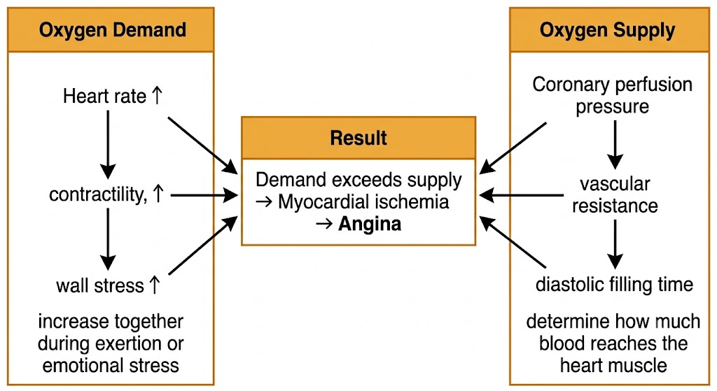
A flow diagram showing oxygen demand factors (heart rate, contractility, wall stress) and oxygen supply factors (coronary perfusion pressure, vascular resistance, diastolic filling time) converging to produce myocardial ischemia when demand exceeds supply.
The myocardial oxygen supply-demand balance underlying angina.

Section 2
Stable Exertional Angina
A fixed supply limitation that becomes symptomatic only when demand rises.

Stable exertional angina is the most common form of angina and the form most antianginal drugs are designed to treat. It results from a fixed atherosclerotic plaque that narrows a coronary artery enough to limit how much additional blood flow the heart can recruit when demand increases.

Mechanism and Clinical Pattern

At rest, the narrowed artery still delivers enough blood to meet the heart muscle's modest oxygen requirement. During exertion or emotional stress, heart rate, contractility, and wall stress all rise together, increasing oxygen demand beyond what the fixed stenosis can supply. The mismatch produces ischemia and the characteristic chest discomfort of angina.

Because the limiting factor is a fixed anatomic narrowing, stable angina follows a reproducible pattern: a given patient reaches their symptom threshold at roughly the same level of exertion each time, and symptoms resolve predictably within two to five minutes of rest or within one to three minutes of sublingual nitroglycerin.

Canadian Cardiovascular Society Classification
Class Clinical Description
Class IAngina only with strenuous or prolonged exertion; ordinary activity unrestricted
Class IISlight limitation; angina with brisk walking, climbing stairs, or after meals
Class IIIMarked limitation; angina after walking only one or two blocks on level ground
Class IVInability to perform any physical activity without symptoms; angina may occur at rest

This classification is a clinical tool for describing symptom severity and tracking response to therapy. A higher class generally indicates a need for more intensive antianginal treatment.


Section 3
Vasospastic Angina
Also called Prinzmetal angina or variant angina — a pure failure of supply, not a rise in demand.

Vasospastic angina results from transient, focal spasm of an epicardial coronary artery. Unlike stable angina, myocardial oxygen demand is normal; the problem is an abrupt drop in supply caused by the artery itself constricting.

Mechanism

Coronary smooth muscle becomes hyperreactive to vasoconstrictor mediators, and reduced availability of nitric oxide from the endothelium removes a normal protective brake on vascular tone. Spasm can occur in coronary arteries that appear angiographically normal or at sites of only mild, non-obstructive plaque.

Recognized triggers include cold exposure, cocaine use, and emotional stress, all of which increase sympathetic tone and can provoke spasm in a susceptible artery.

Clinical Pattern

Episodes occur predominantly at rest, classically in the early morning hours when sympathetic tone is naturally higher. The electrocardiogram during an episode characteristically shows transient ST-segment elevation rather than the ST depression typical of demand-driven ischemia, because the spasm produces transmural rather than subendocardial ischemia. Symptoms relieve rapidly with sublingual nitroglycerin.

High-Yield Contraindication

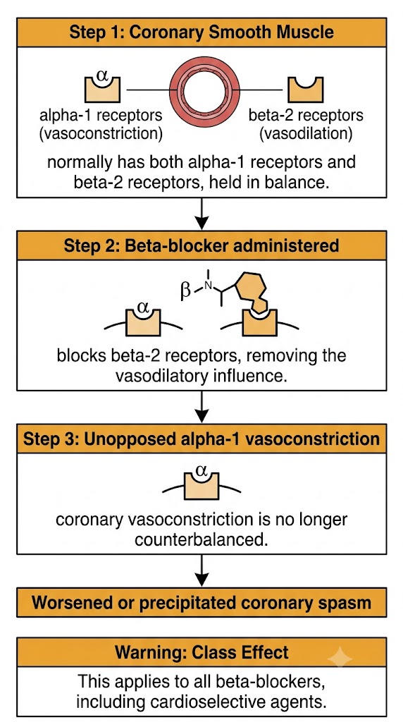
Beta-blockers are contraindicated in vasospastic angina. Blocking beta-2 receptors removes a normal vasodilatory influence on coronary smooth muscle, leaving alpha-1-mediated vasoconstriction unopposed and potentially worsening spasm. This is a class effect that applies to all beta-blockers, including those described as cardioselective.

A flow diagram showing how beta-blockade removes beta-2-mediated coronary vasodilation, leaving alpha-1-mediated vasoconstriction unopposed and worsening coronary spasm.
Why beta-blockade can worsen coronary vasospasm.

Calcium channel blockers are the first-line treatment because they directly counteract the calcium-mediated smooth muscle hyperreactivity that causes the spasm. Long-acting nitrates are used as an adjunct.


Section 4
Microvascular Angina
Sometimes called cardiac syndrome X — ischemia from small vessels that cannot dilate adequately.

Microvascular angina results from dysfunction of the small coronary resistance vessels rather than the larger epicardial arteries. These vessels are normally responsible for adjusting coronary blood flow to match demand, and in microvascular angina they fail to dilate adequately, producing ischemia despite normal-appearing major coronary arteries.

Clinical Recognition

Patients have typical anginal symptoms and a positive stress test, but coronary angiography shows no significant obstruction in the major epicardial arteries. Specialized functional testing can confirm reduced coronary flow reserve when the diagnosis is uncertain. Microvascular angina is more common in postmenopausal women and in patients with hypertension or diabetes.

Sublingual nitroglycerin, which works primarily on larger vessels and on the venous system, may be less effective in microvascular angina than in stable exertional angina. Management typically requires a combination of agents, including beta-blockers and calcium channel blockers, with response that varies between patients.


Section 5
Hemodynamic Targets — The Four Pharmacological Levers
Every antianginal drug class works by manipulating one or more of four hemodynamic variables.

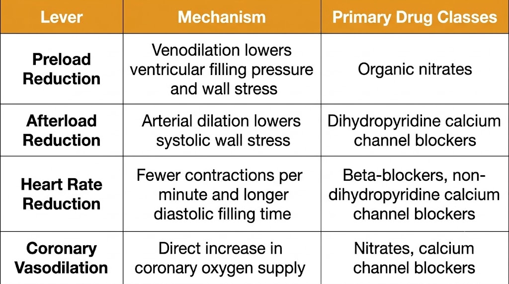
Because myocardial ischemia results from an imbalance between oxygen demand and oxygen supply, every antianginal drug class works by shifting one or both sides of that balance. Organizing the major drug classes by which hemodynamic lever they pull is the single most useful framework for understanding how this entire chapter fits together.

The Rate-Pressure Product as a Bedside Concept

The rate-pressure product, calculated as heart rate multiplied by systolic blood pressure, is a simple bedside estimate of myocardial oxygen demand. A patient with stable angina tends to reach their symptom threshold at a reproducible rate-pressure product. Effective antianginal therapy works by lowering this product during everyday activity, so that ordinary exertion no longer reaches the ischemic threshold.

Four Levers, Four Mechanisms
Lever 1
Preload Reduction
  • Venodilation lowers ventricular filling pressure
  • Reduces wall stress and oxygen demand
  • Primary mechanism: organic nitrates
Lever 2
Afterload Reduction
  • Arterial dilation lowers systolic wall stress
  • Reduces oxygen demand
  • Primary agents: dihydropyridine calcium channel blockers
Lever 3
Heart Rate Reduction
  • Fewer contractions per minute lowers demand
  • Longer diastole improves coronary filling time
  • Primary agents: beta-blockers, non-dihydropyridine calcium channel blockers
Lever 4
Coronary Vasodilation
  • Directly increases oxygen supply
  • Primary agents: nitrates, calcium channel blockers
A reference table listing the four hemodynamic levers of antianginal therapy: preload reduction, afterload reduction, heart rate reduction, and coronary vasodilation, with their mechanisms and primary drug classes.
The four hemodynamic levers of antianginal pharmacotherapy.
Why Combination Therapy Is Common

No single drug class addresses all four levers at once, and every class has dose-limiting adverse effects that prevent unlimited dose escalation. Combining drug classes that act on different levers allows greater total reduction in oxygen demand using lower, better-tolerated doses of each individual agent.

A frequently used combination pairs a beta-blocker with a dihydropyridine calcium channel blocker: the beta-blocker contributes heart rate reduction and also blunts the reflex tachycardia that the calcium channel blocker's vasodilation would otherwise trigger, while the calcium channel blocker contributes afterload reduction and coronary vasodilation. A long-acting nitrate can be added for additional preload reduction. This four-lever framework returns throughout the rest of this chapter as each drug class is covered individually.


Suggested References
Author / Organization Title Source
Libby P, Bonow RO, Mann DL, et al.Braunwald's Heart Disease: A Textbook of Cardiovascular Medicine, 12th ed., Chapter 38: Stable Ischemic Heart DiseaseElsevier, 2022
Knuuti J, Wijns W, Saraste A, et al.2019 ESC Guidelines for the diagnosis and management of chronic coronary syndromesEuropean Heart Journal, 2020
Fihn SD, Gardin JM, Abrams J, et al.2012 ACCF/AHA Guideline for the diagnosis and management of patients with stable ischemic heart diseaseJournal of the American College of Cardiology, 2012
Parker JD, Parker JO.Nitrate therapy for stable angina pectorisNew England Journal of Medicine, 1998
Deedwania PC, Carbajal EV.Role of myocardial oxygen demand in the pathophysiology of silent myocardial ischemiaAmerican Journal of Cardiology, 1992
Cohn PF.Silent myocardial ischemiaAnnals of Internal Medicine, 1988
Beltrame JF, Crea F, Kaski JC, et al.International standardization of diagnostic criteria for vasospastic anginaEuropean Heart Journal, 2017
Yasue H, Nakagawa H, Itoh T, et al.Coronary artery spasm: clinical features, diagnosis, pathogenesis, and treatmentJournal of Cardiology, 2008
Crea F, Bairey Merz CN, Beltrame JF, et al.The parallel tales of microvascular angina and heart failure with preserved ejection fractionEuropean Heart Journal, 2017
Amsterdam EA, Wenger NK, Brindis RG, et al.2014 AHA/ACC Guideline for the management of patients with non-ST-elevation acute coronary syndromesJournal of the American College of Cardiology, 2014
Campeau L.The Canadian Cardiovascular Society grading of angina pectoris revisited 30 years laterCanadian Journal of Cardiology, 2002
Opie LH.Heart Physiology: From Cell to Circulation, 4th ed., Chapter 11: Oxygen Consumption and the Supply-Demand RatioLippincott Williams and Wilkins, 2004