Introduction to Medical Pharmacology
Module 5 — Class IV Agents and Other Nodal Modulators
AARR · Module 5 of 9Section 1
Verapamil and diltiazem — nodal calcium channel blockade for rate control and supraventricular tachycardia
Class IV antiarrhythmic agents block L-type calcium channels in cardiac tissue. Because nodal cells (sinoatrial and atrioventricular) depend on calcium current for their upstroke and pacemaker function, these drugs slow the sinoatrial rate and atrioventricular nodal conduction selectively. The two agents in clinical use are verapamil and diltiazem — both non-dihydropyridine calcium channel blockers with similar indications but meaningfully different hemodynamic profiles.
An important distinction: dihydropyridine calcium channel blockers (amlodipine, nifedipine) act predominantly on vascular smooth muscle and have negligible cardiac electrophysiologic effects. They are not antiarrhythmic agents and must never be substituted for verapamil or diltiazem in arrhythmia management.
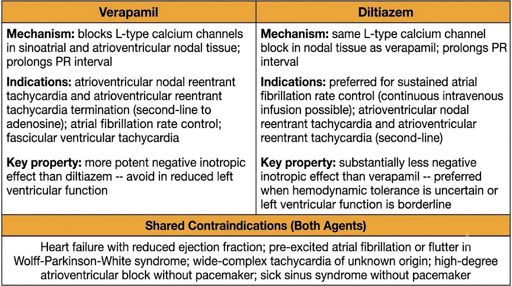
Both verapamil and diltiazem block L-type calcium channels in nodal tissue, slowing atrioventricular nodal conduction and prolonging atrioventricular nodal refractoriness. The electrocardiogram effect is PR interval prolongation. Both also slow the sinoatrial node rate by reducing calcium-dependent automaticity. At therapeutic concentrations, their effect on ventricular myocardium (fast-response tissue) is minimal.
Verapamil produces potent atrioventricular nodal blockade and is effective for acute termination of atrioventricular node-dependent re-entrant tachycardias, including atrioventricular nodal reentrant tachycardia and atrioventricular reentrant tachycardia. Success rates approach 90 percent when given intravenously. It is second-line to adenosine for acute termination but preferred when adenosine is contraindicated (severe asthma).
Verapamil also terminates a specific ventricular tachycardia subtype called fascicular ventricular tachycardia (also known as verapamil-sensitive ventricular tachycardia or Belhassen ventricular tachycardia), which arises from the left posterior fascicle and is driven by a calcium-dependent mechanism. This is one of the very few situations where a calcium channel blocker is appropriate for ventricular tachycardia.
Verapamil has significant negative inotropic effects and moderate peripheral vasodilation. Its use in patients with reduced left ventricular function carries risk of hemodynamic deterioration.
Diltiazem shares verapamil's atrioventricular nodal blocking mechanism and indications but has substantially less negative inotropic effect and comparable peripheral vasodilation. This more favorable hemodynamic profile makes diltiazem the preferred non-dihydropyridine calcium channel blocker for rate control in atrial fibrillation, particularly when hemodynamic tolerance is uncertain or when the patient has borderline left ventricular function. Intravenous diltiazem can be run as a continuous infusion for sustained rate control in atrial fibrillation or flutter, an option not practical with intravenous verapamil.
Critical Contraindication — Both Agents
Never give intravenous verapamil or diltiazem for wide-complex tachycardia of unknown origin. If the rhythm is ventricular tachycardia (not supraventricular tachycardia with aberrant conduction), atrioventricular nodal blockade eliminates the rate-slowing benefit while negative inotropy and vasodilation precipitate hemodynamic collapse. When in doubt, treat wide-complex tachycardia as ventricular tachycardia.
Both agents are also contraindicated in: heart failure with reduced ejection fraction (ejection fraction below 40%), pre-excited atrial fibrillation or flutter in Wolff-Parkinson-White syndrome, sick sinus syndrome without pacemaker, and high-degree atrioventricular block without pacemaker.
Section 2
Ultrashort atrioventricular nodal block — first-line for supraventricular tachycardia termination and diagnosis
Adenosine is a naturally occurring purine nucleoside with an elimination half-life of less than ten seconds in the bloodstream. This extraordinary brevity makes it uniquely suited for acute termination and diagnosis of supraventricular tachycardias — any adverse effects are transient, and the drug is gone almost as quickly as it acts.
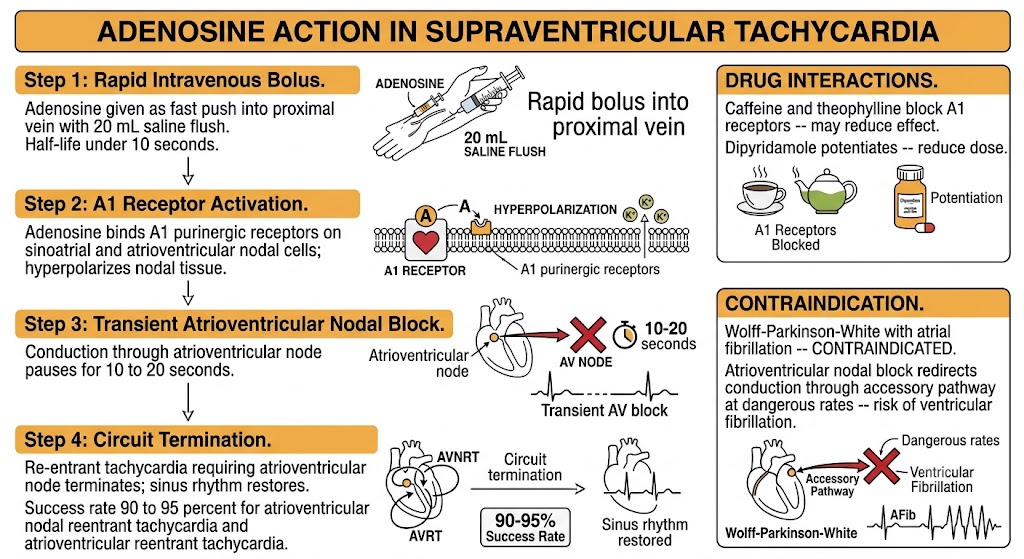
Adenosine activates A1 purinergic receptors on sinoatrial and atrioventricular nodal cells, producing profound transient hyperpolarization that slows or completely blocks atrioventricular nodal conduction for approximately 10 to 20 seconds. This brief window of atrioventricular block is sufficient to terminate re-entrant tachycardias that require the atrioventricular node as a limb of the circuit.
Adenosine must be given as a rapid intravenous bolus followed immediately by a rapid saline flush of at least 20 milliliters, using the largest available proximal peripheral vein (antecubital preferred). Slow administration or distal vein delivery results in drug degradation before reaching the heart. The initial dose is 6 milligrams; if no response, 12 milligrams can be repeated up to twice.
Two drug interactions alter the dose. Methylxanthines — caffeine and theophylline — are competitive antagonists at adenosine receptors and can block adenosine's effect entirely; higher doses may be needed in patients who have recently consumed caffeine. Dipyridamole and carbamazepine block adenosine uptake or breakdown, potentiating its effect; reduce the initial dose to 3 milligrams in patients on these agents. In cardiac transplant recipients with denervated hearts, adenosine sensitivity is dramatically increased and even 3 milligrams can produce prolonged asystole.
Adenosine is first-line for acute termination of atrioventricular nodal reentrant tachycardia and atrioventricular reentrant tachycardia, with success rates of 90 to 95 percent. It is also used as a diagnostic tool in wide-complex tachycardia: if the rhythm is supraventricular tachycardia with aberrant conduction, adenosine terminates it or reveals the underlying atrial rhythm; if ventricular tachycardia, adenosine is ineffective but causes no harm because of its ultrashort half-life.
Flushing, chest tightness, dyspnea, and a sensation of impending doom occur in the majority of patients and resolve within 30 to 60 seconds. Patients should be warned beforehand. Transient atrioventricular block and a brief pause of several seconds are expected and usually self-limiting. Continuous cardiac monitoring and resuscitation equipment must be available.
Bronchospasm is a risk in patients with asthma — adenosine constricts bronchial smooth muscle through its receptor effects. It is relatively contraindicated in symptomatic asthma; use verapamil instead.
Adenosine is absolutely contraindicated in pre-excited atrial fibrillation or flutter in Wolff-Parkinson-White syndrome. Blocking atrioventricular nodal conduction in this setting redirects all conduction through the accessory pathway, which can conduct atrial fibrillation at extremely rapid ventricular rates and degenerate into ventricular fibrillation.
Section 3
Sodium-potassium pump inhibition — rate control adjunct with a narrow therapeutic index
Digoxin is a cardiac glycoside with over two centuries of clinical use. Its antiarrhythmic action is vagotonic rather than direct electrophysiologic, which creates a fundamental limitation: it loses effectiveness during sympathetic activation, making it poorly suited as a sole rate control agent in active patients.
Digoxin inhibits the sodium-potassium pump (sodium-potassium adenosine triphosphatase) on cardiac myocyte membranes. Pump inhibition raises intracellular sodium, which indirectly raises intracellular calcium by reducing the calcium-extruding activity of the sodium-calcium exchanger. The resulting calcium accumulation increases contractile force — the basis of digoxin's positive inotropic effect.
Its atrioventricular nodal slowing effect is primarily vagotonic: digoxin sensitizes cardiac baroreceptors and stimulates vagal outflow, increasing parasympathetic tone to the atrioventricular node. This slows atrioventricular nodal conduction and is effective at rest. During exercise or other sympathetic activation, catecholamines overwhelm the vagotonic effect and heart rate control is lost — explaining why digoxin is less effective than beta-blockers or calcium channel blockers for rate control during physical activity.
For ventricular rate control in atrial fibrillation, digoxin is used as an adjunct to beta-blockers or calcium channel blockers, particularly in sedentary or bedridden patients where exercise rate control is not a priority. Current guidelines recommend it as add-on therapy rather than monotherapy.
In heart failure with reduced ejection fraction, digoxin reduces hospitalizations but does not improve survival. Its inotropic benefit is modest, and toxicity risk limits its use. It is generally reserved for patients who remain symptomatic despite optimal neurohormonal therapy.
Digoxin is renally eliminated with a half-life of 36 to 48 hours in normal renal function, which extends to days or weeks in renal impairment. Dose reduction is required as kidney function declines. The therapeutic range for rate control is narrow: levels above 2 nanograms per milliliter are associated with toxicity, and levels should be drawn at least six hours after a dose.
Toxicity can occur even at levels within the traditional therapeutic range, particularly when predisposing factors are present. Hypokalemia is the most important precipitant — low intracellular potassium enhances digoxin binding to the sodium-potassium pump and amplifies toxicity. Other predisposing conditions include hypomagnesemia, hypercalcemia, hypothyroidism, renal impairment, and advanced age.
Non-cardiac manifestations appear early: nausea, vomiting, and anorexia are the most common initial symptoms. Visual disturbances — yellow-green xanthopsia and halos — are classic but not universal. Confusion and delirium occur in elderly patients.
Cardiac manifestations include virtually any arrhythmia. Bradyarrhythmias and atrioventricular block are most common. Paroxysmal atrial tachycardia with atrioventricular block is a classically described pattern. Bidirectional ventricular tachycardia — alternating axis on each beat — is a sign of severe toxicity.
Management begins with stopping digoxin, correcting hypokalemia (target potassium 4.0 to 5.0 mEq/L), and correcting hypomagnesemia. Calcium must be avoided as it worsens toxicity by raising intracellular calcium further. For life-threatening arrhythmias, digoxin-specific antibody fragments (Digibind or DigiFab) bind and inactivate digoxin rapidly; they are also indicated when serum potassium exceeds 5.5 mEq/L (a sign of severe toxicity) or after massive overdose.
Multiple drugs raise digoxin levels through inhibition of the transport protein that clears digoxin from the body. Amiodarone roughly doubles digoxin levels — halve the digoxin dose when amiodarone is added. Verapamil raises digoxin levels by approximately 70 percent — reduce and monitor. Quinidine approximately doubles digoxin levels — halve the dose. These interactions require active management, not just awareness.
Section 4
Membrane stabilization — first-line for torsades de pointes regardless of serum level
Intravenous magnesium sulfate is a membrane-stabilizing agent with a specific and high-yield antiarrhythmic role: it is the first-line treatment for torsades de pointes, effective even when the patient's serum magnesium level is normal.
Magnesium suppresses early afterdepolarization formation by blocking the calcium current that sustains oscillatory membrane depolarizations during phase 3. This calcium-blocking activity terminates the triggered mechanism driving torsades de pointes, regardless of whether hypomagnesemia was the precipitating cause. The drug is given as a rapid intravenous bolus for acute torsades de pointes episodes.
As an adjunct in digoxin toxicity, magnesium may partially restore sodium-potassium pump activity and reduce intracellular calcium overload. It is given more slowly in this context to avoid worsening atrioventricular block, which can accompany digoxin toxicity.
Magnesium is also administered empirically during cardiopulmonary resuscitation for refractory ventricular fibrillation when torsades de pointes or hypomagnesemia is suspected.
Rapid intravenous administration of magnesium produces flushing and nausea. Loss of deep tendon reflexes is the first clinical sign of magnesium toxicity and signals that plasma levels are approaching a dangerous range. Respiratory paralysis occurs at higher levels. Deep tendon reflexes must be checked during infusion. Calcium gluconate reverses magnesium toxicity and should be immediately available.
Section 5
Choosing among adenosine, calcium channel blockers, beta-blockers, digoxin, and magnesium
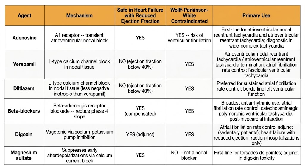
The nodal-modulating agents covered in this module and in Module 3 (beta-blockers) form the toolkit for atrioventricular nodal rate control and supraventricular tachycardia management. Each has a distinct mechanism, onset profile, and clinical niche.
Acute Supraventricular Tachycardia Termination
First- and Second-Line Agents
Atrial Fibrillation Rate Control
Agent Selection by Clinical Context
The Wolff-Parkinson-White Contraindication — Applies to All Nodal-Blocking Agents
Adenosine, verapamil, diltiazem, digoxin, and beta-blockers are all contraindicated in pre-excited atrial fibrillation or flutter in Wolff-Parkinson-White syndrome. Blocking the atrioventricular node in this setting redirects conduction exclusively through the accessory pathway, which can conduct atrial fibrillation at extremely rapid ventricular rates (200 to 300 beats per minute) and degenerate into ventricular fibrillation. The treatment for pre-excited atrial fibrillation in Wolff-Parkinson-White syndrome is intravenous procainamide or electrical cardioversion — not nodal-blocking agents.
| Author / Organization | Title | Source |
|---|---|---|
| Abernethy DR, Schwartz JB | Calcium-antagonist drugs | N Engl J Med. 1999;341(19):1447–1457 |
| Yeh YH, Wakili R, Qi XY, et al | Calcium-handling abnormalities underlying atrial and ventricular arrhythmias in congestive heart failure | Circ Arrhythm Electrophysiol. 2008;1(3):193–202 |
| Delacretaz E | Clinical practice: supraventricular tachycardia | N Engl J Med. 2006;354(10):1039–1051 |
| Lerman BB, Belardinelli L | Cardiac electrophysiology of adenosine: basic and clinical concepts | Circulation. 1991;83(5):1499–1509 |
| Digitalis Investigation Group | The effect of digoxin on mortality and morbidity in patients with heart failure (DIG trial) | N Engl J Med. 1997;336(8):525–533 |
| Hauptman PJ, Kelly RA | Digitalis | Circulation. 1999;99(9):1265–1270 |
| Tzivoni D, Banai S, Schuger C, et al | Treatment of torsade de pointes with magnesium sulfate | Circulation. 1988;77(2):392–397 |
| Brugada J, Katritsis DG, Arbelo E, et al | 2019 ESC Guidelines for the management of patients with supraventricular tachycardia | Eur Heart J. 2020;41(5):655–720 |
| Blomstrom-Lundqvist C, Scheinman MM, Aliot EM, et al | ACC/AHA/ESC guidelines for the management of patients with supraventricular arrhythmias | J Am Coll Cardiol. 2003;42(8):1493–1531 |
| Murgatroyd FD, Camm AJ | Atrial arrhythmias | Lancet. 1993;341(8856):1317–1322 |
| Dorian P, Cass D, Schwartz B, Cooper R, Gelaznikas R, Barr A | Amiodarone as compared with lidocaine for shock-resistant ventricular fibrillation (ALIVE trial) | N Engl J Med. 2002;346(12):884–890 |