CHAPTER 5 · ADRENERGIC PHARMACOLOGY

Section 1

Adrenergic Receptor Classification

G-protein coupling classes, receptor families, and the pharmacological logic of the alpha/beta division

Adrenergic receptors are the molecular targets through which epinephrine and norepinephrine produce their effects throughout the body. Every adrenergic drug works by activating or blocking one or more of these receptors, so predicting a drug's cardiovascular, pulmonary, and metabolic effects requires knowing which receptor subtypes it engages and what those receptors do. The classification of adrenergic receptors into alpha and beta families — and the further subdivision into alpha-1, alpha-2, beta-1, beta-2, and beta-3 — is the foundation of the entire chapter.

The Alpha / Beta Division

All adrenergic receptors belong to the G-protein-coupled receptor superfamily. The defining feature of each subtype is which G-protein it engages, because the identity of that G-protein determines the intracellular second messenger that follows and therefore the functional response in that tissue.

The original division of adrenergic receptors into alpha and beta classes was based on the observation that different tissues respond differently to the same catecholamines. Norepinephrine is the primary agonist at alpha receptors and has modest beta-1 activity. Epinephrine activates both alpha and beta receptors. Isoproterenol, a synthetic catecholamine, is a pure non-selective beta agonist with negligible alpha activity. This differential sensitivity remains clinically relevant today — it explains why norepinephrine is a vasoconstrictor while isoproterenol is a bronchodilator and cardiac stimulant.

G-Protein Coupling by Receptor Subtype

Three G-protein coupling classes cover all adrenergic receptor subtypes. Understanding which class each subtype uses is the key to predicting downstream effects in any organ.

Alpha-1

Gq → Phospholipase C

  • Activates phospholipase C-beta
  • Generates IP3 and diacylglycerol
  • IP3 releases calcium from intracellular stores
  • Net effect: smooth muscle contraction
  • Key locations: vascular smooth muscle, iris dilator, urethral sphincter, prostate

Alpha-2

Gi → Adenylyl Cyclase Inhibition

  • Inhibits adenylyl cyclase
  • Decreases cyclic adenosine monophosphate
  • Key location: presynaptic nerve terminals (autoreceptor)
  • Activation reduces norepinephrine release
  • Also: platelets, pancreatic beta cells, central nervous system

Beta-1 and Beta-2

Gs → Adenylyl Cyclase Activation

  • Activates adenylyl cyclase
  • Increases cyclic adenosine monophosphate
  • Activates protein kinase A
  • Beta-1: heart and kidney
  • Beta-2: bronchi, uterus, skeletal muscle vasculature, liver

Beta-3

Gs → Atypical Distribution

  • Also Gs-coupled, increases cyclic adenosine monophosphate
  • Restricted to adipose tissue and bladder detrusor
  • Adipose: lipolysis and thermogenesis
  • Bladder: detrusor relaxation (target of mirabegron)
  • Not a significant target of standard adrenergic drugs
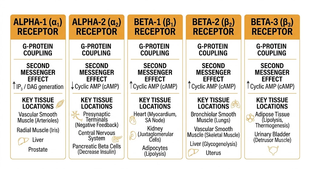
Diagram comparing the five adrenergic receptor subtypes by G-protein coupling class, second messenger pathway, and key tissue locations.
Adrenergic receptor subtypes: G-protein coupling, second messenger pathways, and key tissue locations. Source: Gemini AI, generated for educational use.

Section 2

Alpha Receptor Subtypes

Alpha-1 Gq/calcium cascade and functional consequences; alpha-2 presynaptic autoreceptor function and clinical pharmacology

The two alpha receptor subtypes use opposite signaling strategies and serve distinct physiological roles. Alpha-1 receptors drive smooth muscle contraction through a calcium-dependent pathway. Alpha-2 receptors suppress neurotransmitter release through cyclic adenosine monophosphate inhibition. Drugs targeting each subtype produce characteristically different clinical effects, and distinguishing between them is essential for understanding the pharmacology of phenylephrine, clonidine, and the alpha-blocker drug classes.

Alpha-1 Receptors — Signaling and Effects

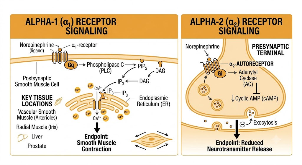
Alpha-1 receptors couple to Gq proteins, which activate phospholipase C-beta at the inner membrane. Phospholipase C-beta cleaves a membrane phospholipid into two second messengers: inositol trisphosphate and diacylglycerol. Inositol trisphosphate travels to the endoplasmic reticulum and triggers release of stored calcium into the cytoplasm. This rise in intracellular calcium is the signal that drives smooth muscle contraction throughout the body.

In vascular smooth muscle, the result is vasoconstriction — the dominant effect of norepinephrine and the primary mechanism of drugs like phenylephrine. Alpha-1 receptors are also responsible for mydriasis (contraction of the iris dilator muscle), maintenance of urinary continence (contraction of the internal urethral sphincter), and prostate smooth muscle tone — the pharmacological basis of alpha-1 blockers in treating benign prostatic hyperplasia.

Side-by-side diagram of alpha-1 receptor Gq/IP3/calcium signaling leading to smooth muscle contraction, and alpha-2 receptor Gi/cyclic AMP inhibition at a presynaptic terminal leading to reduced norepinephrine release.
Alpha-1 receptor signaling (Gq/IP3/calcium cascade) and alpha-2 receptor signaling (Gi/cyclic AMP inhibition) at the presynaptic autoreceptor. Source: Gemini AI, generated for educational use.
Alpha-2 Receptors — Presynaptic Autoreceptor Function

Alpha-2 receptors couple to Gi proteins, which inhibit adenylyl cyclase and reduce intracellular cyclic adenosine monophosphate. The most pharmacologically important location of alpha-2 receptors is on the presynaptic terminals of adrenergic neurons themselves, where they function as autoreceptors.

When norepinephrine accumulates in the synapse, it activates these presynaptic alpha-2 receptors, suppressing further norepinephrine release. This is a negative feedback loop — a braking mechanism that prevents excessive sympathetic outflow. Drugs that activate alpha-2 autoreceptors (clonidine, dexmedetomidine) exploit this mechanism to reduce sympathetic tone centrally, producing antihypertensive and sedative effects. Yohimbine blocks alpha-2 receptors, removes the brake, and increases norepinephrine release.

Clonidine Withdrawal — Mechanism and Clinical Risk

Chronic clonidine therapy activates alpha-2 autoreceptors, reducing sympathetic outflow and lowering blood pressure. With prolonged use, the adrenergic system adapts by upregulating postsynaptic receptors. Abrupt discontinuation removes the alpha-2 brake suddenly, allowing a surge of norepinephrine release onto these supersensitive receptors. The result is a rebound hypertensive crisis — potentially severe, especially in patients with underlying cardiovascular disease. This is the mechanistic basis for the requirement to taper clonidine gradually before discontinuation rather than stopping it abruptly.

Alpha-2 Receptor Locations Beyond the Presynaptic Terminal

Alpha-2 receptors are also found postsynaptically in several tissues. In platelets, alpha-2 activation promotes aggregation by inhibiting the cyclic adenosine monophosphate signaling that would otherwise keep platelets quiescent. In pancreatic beta cells, alpha-2 activation suppresses insulin secretion — one mechanism by which epinephrine-mediated stress responses cause hyperglycemia. In the central nervous system, alpha-2 receptors in the locus coeruleus are the primary target of dexmedetomidine, explaining its sedative and anxiolytic effects without respiratory depression.


Section 3

Beta Receptor Subtypes

Beta-1 cardiac and renal effects; beta-2 bronchodilation, uterine relaxation, and metabolic effects; beta-3 adipose and bladder pharmacology

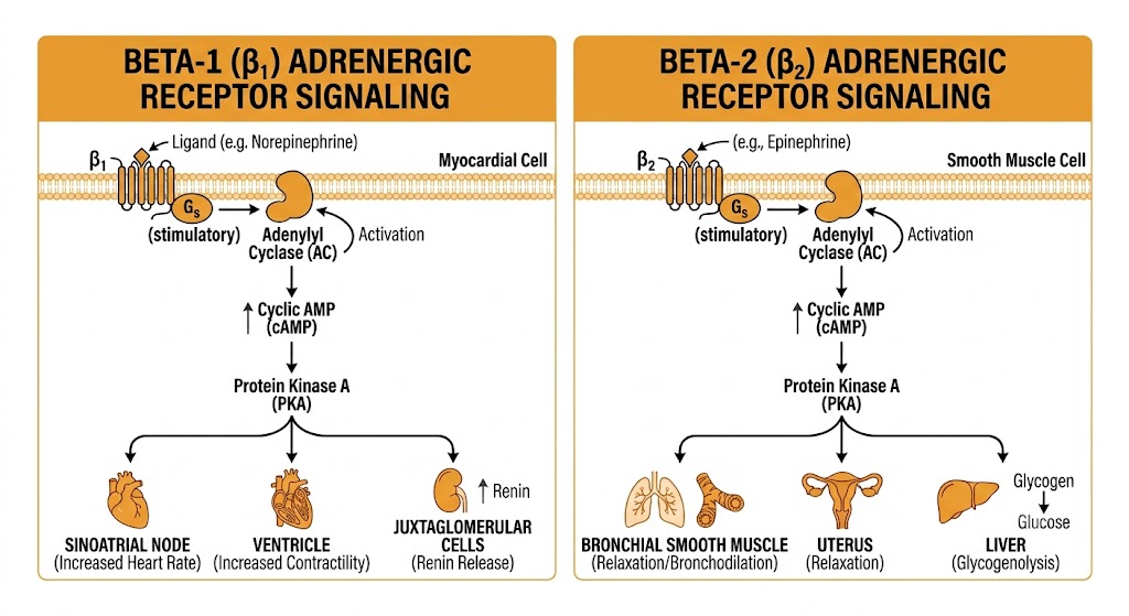
All three beta receptor subtypes couple to Gs proteins and raise intracellular cyclic adenosine monophosphate, but their tissue distributions differ markedly. Beta-1 receptors dominate in the heart and kidney. Beta-2 receptors predominate in bronchial smooth muscle, uterine smooth muscle, and skeletal muscle vasculature. Beta-3 receptors are restricted to adipose tissue and the bladder. These distributions explain why selective versus non-selective beta agonists and antagonists produce different organ-level effects.

Diagram showing beta-1 and beta-2 receptor Gs/cyclic AMP/protein kinase A signaling with tissue-specific effects: cardiac stimulation and renin release for beta-1, and bronchodilation, uterine relaxation, and glycogenolysis for beta-2.
Beta-1 and beta-2 receptor signaling: shared Gs/cyclic AMP/protein kinase A cascade with organ-specific downstream effects. Source: Gemini AI, generated for educational use.
Beta-1 Receptors — Heart and Kidney

Beta-1 receptors are the predominant adrenergic receptor in the myocardium. When activated by catecholamines, they raise cyclic adenosine monophosphate and activate protein kinase A, which phosphorylates several cardiac targets simultaneously. In the sinoatrial node, this speeds spontaneous depolarization and increases heart rate (positive chronotropy). In ventricular muscle, it increases contractile force (positive inotropy) and speeds relaxation (positive lusitropy). In the atrioventricular node, it increases conduction velocity (positive dromotropy), shortening the pulse rate conduction interval on an electrocardiogram.

Beyond the heart, beta-1 receptors in juxtaglomerular cells of the kidney stimulate renin release. This connects sympathetic activation to the renin-angiotensin-aldosterone system, amplifying the blood pressure response to adrenergic stimulation. Beta-1 blockade reduces renin secretion, which is one mechanism contributing to the antihypertensive effect of beta-blockers in addition to their cardiac effects.

Beta-2 Receptors — Bronchi, Uterus, and Metabolism

Beta-2 receptors are the dominant adrenergic receptor in bronchial smooth muscle. Their activation causes bronchodilation, which is the pharmacological rationale for selective beta-2 agonists (albuterol, salmeterol) in asthma and chronic obstructive pulmonary disease. The same mechanism operates in uterine smooth muscle, where beta-2 activation causes relaxation — the basis for terbutaline's use as a tocolytic agent to inhibit preterm labor.

Beta-2 receptors also mediate important metabolic effects. In the liver, beta-2 activation promotes glycogenolysis and gluconeogenesis, raising blood glucose during the sympathetic stress response. In skeletal muscle, beta-2 activation stimulates the sodium-potassium-adenosine triphosphatase pump, shifting potassium into cells and lowering serum potassium. This mechanism explains the hypokalemia seen in severe asthma exacerbations treated with high-dose beta-2 agonists and during states of catecholamine excess such as pheochromocytoma crisis.

Beta-2 Receptor Distribution in the Heart — Clinical Relevance

Beta-2 receptors are present in the heart at lower density than beta-1 receptors, normally accounting for roughly 20 to 25 percent of cardiac beta receptor activity. In chronic heart failure, beta-1 receptors are selectively downregulated due to sustained catecholamine exposure, and the proportion of cardiac beta-2 receptors increases substantially. This shift has implications for drug therapy: cardioselective beta-1 blockers may not fully block the adrenergic drive in a failing heart with upregulated beta-2 receptors, which is one rationale for using non-selective agents such as carvedilol in heart failure management.

Beta-3 Receptors — Adipose Tissue and Bladder

Beta-3 receptors are expressed primarily in adipose tissue, where their activation drives lipolysis (releasing free fatty acids from stored triglycerides) and thermogenesis in brown adipose tissue. They are also expressed in the bladder detrusor muscle, where activation causes smooth muscle relaxation, increasing the bladder's storage capacity. This peripheral location is the therapeutic target of mirabegron, a selective beta-3 agonist approved for overactive bladder. Because beta-3 receptors are relatively resistant to desensitization compared to beta-2 receptors, mirabegron maintains efficacy during long-term use — a practical advantage over muscarinic antagonists, which also treat overactive bladder.


Section 4

Dopamine Receptors — Peripheral Pharmacology

D1 renal and mesenteric vasodilation, D2 presynaptic inhibition, and the dose-dependent receptor engagement of intravenous dopamine

Dopamine acts on distinct receptor families in the periphery, producing cardiovascular and renal effects that are entirely separate from its central nervous system functions. The peripheral pharmacology of intravenous dopamine is dose-dependent: different receptor populations are engaged as the infusion rate increases, producing a clinical profile that shifts from renal vasodilation at low doses to cardiac stimulation at moderate doses to vasoconstriction at high doses. Understanding this receptor hierarchy is essential for interpreting dopamine's use in shock management.

D1 Receptors — Renal and Splanchnic Vasodilation

D1 receptors couple to Gs proteins, raising cyclic adenosine monophosphate similarly to beta receptors. They are expressed in the renal afferent arteriole and mesenteric vasculature, where their activation causes vasodilation. In the kidney, this vasodilation increases renal blood flow. D1 receptors in the proximal tubule also reduce sodium reabsorption, producing a natriuretic effect. These two actions — renal vasodilation and natriuresis — formed the rationale for the now-abandoned practice of low-dose dopamine infusion in patients at risk for acute kidney injury.

Multiple randomized controlled trials, including a landmark trial by the Australian and New Zealand Intensive Care Society, demonstrated definitively that low-dose dopamine does not protect the kidneys from injury and should not be used for renal protection. This is a well-established negative finding in critical care medicine.

D2 Receptors — Presynaptic Inhibition and Nausea

D2 receptors couple to Gi proteins and inhibit adenylyl cyclase, analogous to alpha-2 receptors. In the periphery, D2 receptors on sympathetic nerve terminals inhibit norepinephrine release. D2 receptors in the gut inhibit gastrointestinal motility — the target of metoclopramide's prokinetic effect, which works partly by blocking these peripheral D2 receptors. In the chemoreceptor trigger zone of the area postrema — a region at the brainstem surface that lacks a functional blood-brain barrier — D2 receptor activation stimulates nausea and vomiting. This explains why dopamine infusions and dopamine agonist drugs commonly cause nausea, and why D2 antagonists such as metoclopramide and prochlorperazine are effective antiemetics.

Dose-Dependent Receptor Engagement of Intravenous Dopamine

Low dose (approximately 1–3 micrograms per kilogram per minute): Primarily D1 receptor activation. Renal and mesenteric vasodilation, natriuresis. Note: does not protect against acute kidney injury despite this mechanism.

Moderate dose (approximately 3–10 micrograms per kilogram per minute): Beta-1 receptor activation dominates. Increased cardiac contractility and heart rate, increased cardiac output. Norepinephrine release from sympathetic terminals also contributes.

High dose (above 10 micrograms per kilogram per minute): Alpha-1 receptor activation. Systemic vasoconstriction, increased systemic vascular resistance. Clinical profile resembles norepinephrine. Arrhythmia risk increases substantially at these doses.

These dose ranges are approximations. Significant patient-to-patient variability means the transitions between receptor-dominant phases are not sharp in clinical practice. For septic shock, norepinephrine is preferred over dopamine based on superior outcome data and a lower arrhythmia rate.


Section 5

Organ-Level Tissue Distribution

Receptor subtype predominance in heart, vasculature, lung, kidney, eye, and genitourinary tract — and the net functional response in each organ

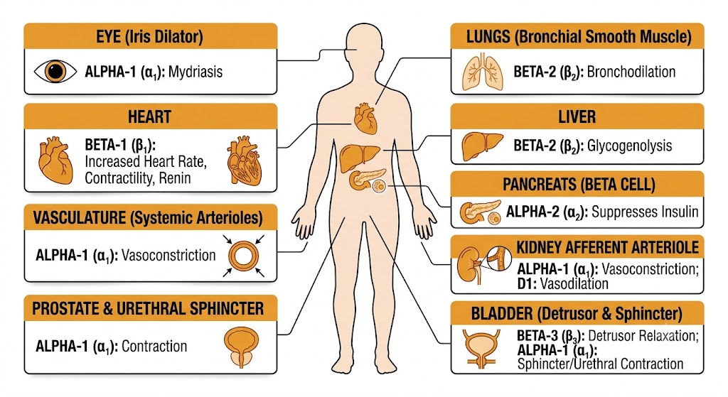
Knowing the signaling pathway of each receptor subtype is necessary but not sufficient for predicting drug effects. You also need to know which subtype predominates in each organ, because the same drug can have opposite effects in different tissues depending on which receptors are present. This section integrates receptor subtype knowledge with organ distribution to support clinical prediction.

Anatomical diagram showing dominant adrenergic receptor subtypes and their functional effects in the heart, vasculature, bronchi, kidney, prostate, bladder, iris, liver, and pancreas.
Organ-level distribution of adrenergic receptor subtypes and net functional effect of activation at each site. Source: Gemini AI, generated for educational use.
Organ / Tissue Dominant Receptor Effect of Activation Clinical Relevance
Heart (sinoatrial node, ventricle) Beta-1 Increased heart rate, contractility, conduction velocity Target of beta-blockers in hypertension, heart failure, arrhythmia
Systemic arterioles Alpha-1 Vasoconstriction, increased blood pressure Target of alpha-1 blockers in hypertension and benign prostatic hyperplasia
Skeletal muscle vasculature Beta-2 Vasodilation Contributes to epinephrine-mediated fall in total peripheral resistance at low doses
Bronchial smooth muscle Beta-2 Bronchodilation Target of albuterol and salmeterol in asthma and chronic obstructive pulmonary disease
Kidney afferent arteriole Alpha-1 (constricts); D1 (dilates) Net vasoconstriction during sympathetic activation Reduced glomerular filtration rate in shock; dopamine-mediated vasodilation at low doses
Juxtaglomerular cells Beta-1 Renin release Connects sympathetic activation to renin-angiotensin-aldosterone system; blocked by beta-blockers
Uterine smooth muscle Beta-2 Relaxation Terbutaline used as tocolytic in preterm labor
Prostate, internal urethral sphincter Alpha-1 Contraction — maintains urinary continence; contributes to outflow obstruction Alpha-1 blockers (prazosin, tamsulosin) relax prostate and sphincter to relieve benign prostatic hyperplasia symptoms
Bladder detrusor Beta-3 Relaxation — increases storage capacity Target of mirabegron in overactive bladder
Iris dilator muscle Alpha-1 Contraction — mydriasis (pupil dilation) Phenylephrine eye drops for mydriasis during ophthalmologic examination
Liver Beta-2, Alpha-1 Glycogenolysis; gluconeogenesis (beta-2) Catecholamine-mediated hyperglycemia in stress, pheochromocytoma, severe asthma
Pancreatic beta cells Alpha-2 (dominant over beta-2) Suppression of insulin secretion Epinephrine-mediated hyperglycemia in the stress response

Section 6

Receptor Regulation

Desensitization with chronic agonist exposure, upregulation with chronic antagonist exposure, and heart failure receptor remodeling

Adrenergic receptor responsiveness is not fixed. Prolonged exposure to an agonist reduces receptor density and sensitivity — a process called downregulation that underlies clinical tachyphylaxis. Conversely, prolonged blockade increases receptor density, which explains why abrupt withdrawal of a beta-blocker can be dangerous. Three clinical scenarios illustrate these principles at the level expected for a second-year student.

Tachyphylaxis with Beta-2 Agonist Overuse

Regular use of short-acting beta-2 agonists such as albuterol exposes bronchial smooth muscle receptors to sustained agonist stimulation. Over days to weeks, this drives receptor internalization and reduced gene expression, decreasing the density of beta-2 receptors on the cell surface. The clinical result is tachyphylaxis — shorter duration of bronchodilation, reduced peak effect, and progressively less relief from the same inhaler dose. Patients who use rescue inhalers more than twice a week are exhibiting a pattern that leads to this downregulation, which is one reason why inhaled corticosteroids — not increasing the dose of short-acting agonists — are the appropriate escalation in persistent asthma.

Beta-Blocker Withdrawal Syndrome

The reciprocal process occurs with chronic beta-blocker therapy. When beta-1 receptors are chronically blocked, the adrenergic system compensates by upregulating receptor density — increasing the number of receptors on cardiac myocytes. If the beta-blocker is then stopped abruptly, normal circulating catecholamine levels suddenly encounter a supersensitive, upregulated receptor population. The result is rebound tachycardia, hypertension, angina, and in patients with coronary artery disease, a real risk of myocardial infarction.

This is the pharmacological basis for the clinical rule that beta-blockers must always be tapered gradually over one to two weeks before discontinuation — never stopped abruptly, especially in patients with known coronary artery disease or those in the perioperative period.

Receptor Remodeling in Heart Failure

Chronic heart failure is characterized by persistent sympathetic activation and chronically elevated norepinephrine levels in the cardiac interstitium. This sustained catecholamine exposure drives progressive downregulation of cardiac beta-1 receptors, which can fall to 50 percent or less of normal density in advanced heart failure. The result is impaired contractile reserve — the heart cannot mount an adequate adrenergic response to increased demand.

Paradoxically, the treatment for this receptor depletion is not more sympathetic stimulation but less: carvedilol and metoprolol succinate, by blocking the chronically elevated adrenergic drive, allow gradual recovery of beta-1 receptor density over months of therapy. This receptor resensitization is one mechanism contributing to the well-established mortality benefit of beta-blockers in heart failure with reduced ejection fraction.

Three Regulatory Scenarios — Summary

Beta-2 agonist tachyphylaxis: Chronic short-acting agonist overuse drives beta-2 receptor downregulation in airway smooth muscle. Prevent by using inhaled corticosteroids as the primary controller with short-acting agonists reserved for rescue only.

Beta-blocker withdrawal: Chronic beta-1 blockade upregulates cardiac beta-1 receptors. Abrupt discontinuation exposes supersensitive receptors to normal catecholamine levels, precipitating rebound tachycardia, angina, and potential myocardial infarction. Always taper gradually.

Heart failure remodeling: Chronic norepinephrine excess downregulates cardiac beta-1 receptors, impairing contractile reserve. Beta-blockers (carvedilol, metoprolol succinate) reverse this over months of therapy by reducing the chronic adrenergic stimulus.


Suggested References
Author / Organization Title Source
Lefkowitz RJ Seven transmembrane receptors: something old, something new Acta Physiol (Oxf). 2007;190(1):9–19
Ahlquist RP A study of the adrenotropic receptors Am J Physiol. 1948;153(3):586–600
Bylund DB, Eikenberg DC, Hieble JP, et al. International Union of Pharmacology nomenclature of adrenoceptors Pharmacol Rev. 1994;46(2):121–136
Brodde OE, Michel MC Adrenergic and muscarinic receptors in the human heart Pharmacol Rev. 1999;51(4):651–690
Brede M, Philipp M, Knaus A, Muthig V, Hein L Alpha2-adrenergic receptor subtypes — novel functions uncovered in gene-targeted mouse models Biol Cell. 2004;96(5):343–348
Bristow MR Beta-adrenergic receptor blockade in chronic heart failure Circulation. 2000;101(5):558–569
Johnson M Molecular mechanisms of beta2-adrenergic receptor function, response, and regulation J Allergy Clin Immunol. 2006;117(1):18–24
Missale C, Nash SR, Robinson SW, Jaber M, Caron MG Dopamine receptors: from structure to function Physiol Rev. 1998;78(1):189–225
Bellomo R, Chapman M, Finfer S, Hickling K, Myburgh J; ANZICS Clinical Trials Group Low-dose dopamine in patients with early renal dysfunction: a placebo-controlled randomised trial Lancet. 2000;356(9248):2139–2143
Tonini M, Cipollina L, Poluzzi E, Crema F, Corazza GR, De Ponti F Review article: clinical implications of enteric and central D2 receptor blockade by antidopaminergic gastrointestinal prokinetics Aliment Pharmacol Ther. 2004;19(4):379–390
De Backer D, Biston P, Devriendt J, et al. Comparison of dopamine and norepinephrine in the treatment of shock N Engl J Med. 2010;362(9):779–789
Krupnick JG, Benovic JL The role of receptor kinases and arrestins in G protein-coupled receptor regulation Annu Rev Pharmacol Toxicol. 1998;38:289–319
Bangalore S, Makani H, Radford M, et al. Clinical outcomes with beta-blockers for myocardial infarction: a meta-analysis of randomized trials Am J Med. 2014;127(10):939–953