Introduction to Medical Pharmacology
Epinephrine, norepinephrine, dopamine, and dobutamine — receptor profiles, clinical uses, and drug interactions
Chapter 5 · Module 2 of 6Section 1
Biosynthetic pathway from tyrosine to epinephrine, rate-limiting step, enzymatic inactivation by monoamine oxidase and catechol-O-methyltransferase, and pharmacokinetic consequences of the catechol structure
Epinephrine, norepinephrine, and dopamine share a common biosynthetic pathway and are inactivated by the same two enzymatic systems. This shared biochemistry explains why drugs that block catecholamine synthesis, storage, reuptake, or degradation have predictable and often dramatic effects on sympathetic tone — and why certain drug combinations produce life-threatening interactions.
Catecholamine synthesis begins with the dietary amino acid tyrosine. The rate-limiting step is the conversion of tyrosine to L-DOPA (L-3,4-dihydroxyphenylalanine) by the enzyme tyrosine hydroxylase. Because this step is rate-limiting, it is the primary point of autoregulation — catecholamines themselves inhibit tyrosine hydroxylase through end-product inhibition, preventing runaway synthesis during sympathetic activation.
L-DOPA is then decarboxylated to dopamine by DOPA decarboxylase. In dopaminergic neurons, synthesis stops here. In noradrenergic neurons, dopamine is transported into vesicles and converted to norepinephrine by dopamine beta-hydroxylase. In the adrenal medulla, norepinephrine undergoes one final methylation step to become epinephrine, catalyzed by phenylethanolamine N-methyltransferase. This enzyme is induced by glucocorticoids from the adjacent adrenal cortex — explaining why the adrenal medulla predominantly produces epinephrine rather than norepinephrine, and why adrenal cortex destruction impairs epinephrine synthesis.
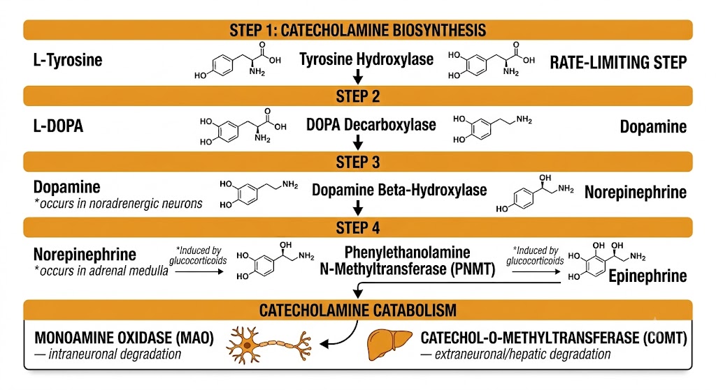
Catecholamines are inactivated by two enzymes working in complementary locations. Monoamine oxidase is located intraneuronally — within the presynaptic nerve terminal — where it oxidizes catecholamines that are not stored in vesicles. Two isoforms exist with different substrate preferences: monoamine oxidase type A preferentially inactivates norepinephrine and epinephrine; monoamine oxidase type B preferentially inactivates dopamine. Drugs that inhibit these enzymes (monoamine oxidase inhibitors) prevent the normal breakdown of catecholamines, dramatically amplifying their effects — the basis of one of the most dangerous drug interactions in pharmacology.
Catechol-O-methyltransferase operates primarily in the liver, kidney, and gut wall rather than at the synapse. It is the primary route of inactivation for circulating catecholamines — those that escape reuptake and reach the systemic circulation. Together, monoamine oxidase and catechol-O-methyltransferase account for the extremely short plasma half-life of administered catecholamines (typically one to three minutes for intravenous epinephrine and norepinephrine).
All clinically used catecholamines are administered intravenously or by injection. Oral bioavailability is negligible because monoamine oxidase and catechol-O-methyltransferase in the gut wall and liver destroy essentially all orally ingested catecholamine before it reaches the systemic circulation. The catechol structure is also susceptible to oxidation at gastric pH. Because of their extremely short half-lives after intravenous administration, all catecholamines are given as continuous infusions when sustained effects are required, allowing precise titration of dose to hemodynamic effect.
Biosynthesis Pathway — Key Enzymes at a Glance
Tyrosine → L-DOPA: Tyrosine hydroxylase — rate-limiting step; subject to end-product inhibition by catecholamines
L-DOPA → Dopamine: DOPA decarboxylase — requires vitamin B6
Dopamine → Norepinephrine: Dopamine beta-hydroxylase — requires vitamin C and copper; occurs in noradrenergic neurons and adrenal medulla
Norepinephrine → Epinephrine: Phenylethanolamine N-methyltransferase — requires S-adenosylmethionine; induced by glucocorticoids; occurs only in adrenal medulla chromaffin cells
Catabolism: Monoamine oxidase (intraneuronal) + catechol-O-methyltransferase (extraneuronal, hepatic) working in concert produce the one-to-three minute plasma half-life
Section 2
Full alpha and beta agonism, dose-dependent cardiovascular effects, anaphylaxis, cardiac arrest, and local anesthetic adjuvancy
Epinephrine is the endogenous hormone of the acute stress response and the most pharmacologically versatile of the catecholamines. It activates all five adrenergic receptor subtypes, and its clinical applications span anaphylaxis, cardiac arrest, and local anesthesia — three entirely different clinical contexts, each exploiting a different facet of its receptor profile.
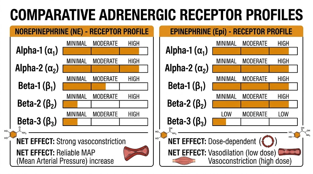
Epinephrine is a full agonist at alpha-1, alpha-2, beta-1, beta-2, and beta-3 receptors. The cardiovascular effect at any given dose reflects which receptor populations predominate in the target tissue at that plasma concentration. At low infusion rates, beta-2-mediated vasodilation in skeletal muscle vasculature is significant enough to reduce total peripheral resistance, so mean arterial pressure may remain stable or fall slightly despite increased cardiac output. At higher doses or during bolus injection, alpha-1 activation becomes dominant in most vascular beds, producing net vasoconstriction, rising blood pressure, and widening pulse pressure.
Beta-1 stimulation at all doses increases heart rate, contractility, and atrioventricular node conduction velocity. Epinephrine also lowers the threshold for ventricular arrhythmias — an effect that is amplified by hypoxia, acidosis, and certain anesthetics, and that limits its use to genuine emergencies in patients with coronary artery disease.
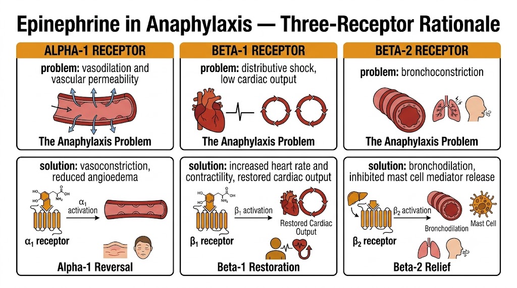
Epinephrine is the only first-line treatment for anaphylaxis, with no absolute contraindications in that setting. Its value lies precisely in the breadth of its receptor activity — it simultaneously addresses all three pathophysiological components of anaphylaxis. Alpha-1 activation reverses the vasodilation and vascular permeability that produce hypotension and angioedema. Beta-1 activation supports cardiac output in the setting of distributive shock. Beta-2 activation reverses bronchoconstriction and, importantly, inhibits further release of histamine and leukotrienes from mast cells and basophils, blunting the ongoing allergic response.
The correct route is intramuscular injection into the mid-anterolateral thigh (vastus lateralis muscle) — not the deltoid, not subcutaneous. The thigh produces higher peak plasma concentrations more rapidly. The dose for adults is 0.3 to 0.5 mg of the 1:1,000 solution (1 mg per milliliter), repeated every 5 to 15 minutes if there is no response. Intravenous epinephrine in anaphylaxis is reserved for patients already in cardiac arrest or in refractory shock with continuous hemodynamic monitoring — an intravenous bolus in an awake patient can precipitate severe hypertension or ventricular arrhythmias.
During cardiopulmonary resuscitation, epinephrine 1 mg is administered intravenously every 3 to 5 minutes. The primary mechanism during cardiac arrest is alpha-1-mediated vasoconstriction, which raises aortic diastolic pressure and thereby increases coronary perfusion pressure during the compression phase — improving the likelihood of restoring spontaneous circulation. The beta-1 cardiac stimulation becomes relevant after return of spontaneous circulation rather than during the arrest itself.
Evidence from the PARAMEDIC2 trial (2018) showed that epinephrine increased return of spontaneous circulation and survival to hospital discharge but did not improve neurologically favorable survival at 30 days — a nuance that has shifted the framing of epinephrine in cardiac arrest from an unqualified benefit to a drug that improves short-term survival with uncertain neurological implications.
Epinephrine is added to local anesthetic solutions at concentrations of 1:100,000 to 1:200,000 to produce local alpha-1-mediated vasoconstriction at the injection site. This slows systemic absorption of the local anesthetic, prolonging its duration of action and reducing peak plasma concentration — which in turn reduces the risk of local anesthetic systemic toxicity. It also reduces bleeding in the surgical field.
Epinephrine is contraindicated as a local anesthetic additive in anatomical locations supplied by end arteries — the digits, the penis, the tip of the nose, and the ear lobes. In these locations, alpha-1-mediated vasoconstriction can cause complete arterial occlusion and ischemic necrosis, since there are no collateral vessels to maintain perfusion.
Section 3
Alpha-1/alpha-2/beta-1 receptor profile, hemodynamic effects, first-line use in septic shock, extravasation management
Norepinephrine is the primary vasopressor for distributive shock and the standard of care for septic shock. Its receptor profile differs from epinephrine in one clinically critical way: norepinephrine has minimal beta-2 activity. This means it produces reliable vasoconstriction without the beta-2-mediated vasodilation that can complicate epinephrine's hemodynamic profile at lower doses.
Norepinephrine is a potent agonist at alpha-1, alpha-2, and beta-1 receptors, with comparatively weak beta-2 activity. The dominant clinical effect is alpha-1-mediated arteriolar and venous vasoconstriction throughout the systemic circulation, producing reliable increases in systemic vascular resistance, mean arterial pressure, and both systolic and diastolic blood pressure. Because there is no meaningful beta-2-mediated vasodilation to counterbalance this effect, mean arterial pressure rises predictably — unlike epinephrine, where the net vascular effect depends on dose.
The increase in afterload from alpha-1 vasoconstriction tends to reduce cardiac output modestly unless beta-1-mediated inotropy compensates. Reflex bradycardia through baroreceptor activation frequently accompanies the pressor response. The rise in diastolic blood pressure increases coronary perfusion pressure, which is beneficial in patients with coronary artery disease who need vasopressor support.
Norepinephrine is the vasopressor of first choice for septic shock, supported by surviving sepsis campaign guidelines and multiple randomized controlled trials. The Sepsis Occurrence in Acutely Ill Patients II trial demonstrated that norepinephrine was associated with significantly fewer arrhythmias than dopamine (12 percent versus 24 percent arrhythmia rate) and improved outcomes in the cardiogenic shock subgroup. Current practice initiates norepinephrine at 0.01 to 0.05 micrograms per kilogram per minute and titrates upward to maintain mean arterial pressure above 65 millimeters of mercury.
Norepinephrine Extravasation — Immediate Management
Extravasation of norepinephrine from a peripheral intravenous line causes intense local alpha-1-mediated vasoconstriction, producing tissue ischemia and necrosis if untreated. Immediate management: stop the infusion, then infiltrate the affected area with phentolamine 5 to 10 mg diluted in 10 to 15 milliliters of normal saline using a fine-gauge needle. Phentolamine competitively blocks alpha-1 receptors locally, reversing vasoconstriction. Treatment is most effective within 12 hours of extravasation. Norepinephrine should be administered through central venous access whenever possible to prevent this complication.
Section 4
Dose-dependent receptor engagement, the renal protection myth, current clinical positioning, and adverse effects
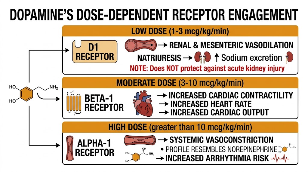
Dopamine is unique among the catecholamine vasopressors in that its receptor profile changes qualitatively with dose — producing renal vasodilation at low doses, cardiac stimulation at moderate doses, and vasoconstriction at high doses. This complexity, combined with a higher arrhythmia rate than norepinephrine, has progressively narrowed its clinical role in shock management.
At low infusion rates (approximately 1 to 3 micrograms per kilogram per minute), dopamine activates D1 receptors in the renal and mesenteric vasculature, producing regional vasodilation and natriuresis. At moderate rates (approximately 3 to 10 micrograms per kilogram per minute), beta-1 receptor activation dominates, increasing cardiac contractility and heart rate and raising cardiac output. At high rates (above 10 micrograms per kilogram per minute), alpha-1 activation produces systemic vasoconstriction, and the hemodynamic profile resembles norepinephrine.
These dose ranges are approximations, not pharmacological guarantees. Individual variability in receptor density, volume status, and baseline sympathetic tone means the transitions between these phases are not sharp in clinical practice.
For decades, low-dose dopamine (1 to 3 micrograms per kilogram per minute) was used clinically to protect the kidneys from acute injury, based on the physiological rationale that D1 receptor activation produces renal vasodilation and natriuresis. Multiple randomized controlled trials have definitively disproven this rationale. The landmark Australian and New Zealand Intensive Care Society trial found no difference in peak creatinine, need for renal replacement therapy, or intensive care unit length of stay between low-dose dopamine and placebo in patients with early renal dysfunction. Low-dose dopamine does not prevent acute kidney injury in any clinical context and should not be used for that purpose.
The Sepsis Occurrence in Acutely Ill Patients II trial established norepinephrine as the preferred vasopressor over dopamine for septic shock, driven by dopamine's significantly higher arrhythmia rate (24 percent versus 12 percent) and worse outcomes in the cardiogenic shock subgroup. Dopamine retains a limited role in bradycardia with hypotension when atropine has failed and a chronotropic effect is specifically desired.
Beyond arrhythmias, dopamine causes nausea and vomiting through D2 receptor activation in the chemoreceptor trigger zone, causes tissue necrosis on extravasation (managed with phentolamine, same as norepinephrine), and suppresses pituitary hormone secretion during prolonged infusion. Central venous access is required for administration.
Dopamine — Current Clinical Positioning
Not recommended as first-line vasopressor for septic shock — norepinephrine is preferred (Sepsis Occurrence in Acutely Ill Patients II trial).
Not recommended for renal protection in any clinical context — multiple randomized controlled trials show no benefit.
Retained indication: bradycardia with hypotension when atropine has failed and a chronotropic effect is needed.
Use with caution in patients prone to arrhythmias. Central venous access required. Avoid in pheochromocytoma.
Section 5
Selective beta-1 inotrope, mild afterload reduction, cardiogenic shock use combined with norepinephrine, pharmacological stress testing, and tachyphylaxis
Dobutamine is a synthetic catecholamine designed to provide positive inotropy with minimal vasopressor activity. It fills the clinical gap between the non-selective catecholamines and the need for pure cardiac output augmentation — particularly in patients with low cardiac output whose blood pressure is preserved or only modestly reduced.
Dobutamine acts predominantly at beta-1 receptors, producing positive inotropy and modest positive chronotropy. It also has mild beta-2 activity, which causes a degree of peripheral vasodilation and afterload reduction — augmenting forward flow by reducing the resistance the heart pumps against. The combined effect is increased cardiac output and reduced pulmonary capillary wedge pressure (filling pressure), with little or no increase in mean arterial pressure. This last point is critical: dobutamine does not reliably raise blood pressure, making it unsuitable as a sole agent in hypotensive patients.
Alpha-1 effects are minimal — the result of a racemic mixture in which the alpha effects of the two enantiomers cancel. Dobutamine has no dopaminergic receptor activity and does not release norepinephrine from nerve terminals; its inotropic effect is entirely direct.
Dobutamine is the inotrope of choice for cardiogenic shock or acute decompensated heart failure with reduced cardiac output, particularly when systolic blood pressure is above approximately 90 millimeters of mercury. In frank cardiogenic shock with significant hypotension, dobutamine is combined with norepinephrine: norepinephrine provides vasopressor support to maintain perfusion pressure while dobutamine augments cardiac output and reduces filling pressures. This combination — independently titratable agents for two different hemodynamic problems — is now preferred over dopamine alone, which provides mixed vasopressor and inotropic effects that cannot be independently adjusted.
Tachyphylaxis develops with continuous dobutamine infusions lasting beyond approximately 72 hours, due to beta-1 receptor downregulation. Dose escalation may be required over time. Continuous dobutamine infusions have not been shown to improve survival in chronic heart failure and may increase mortality by increasing arrhythmia risk in an already vulnerable myocardium.
Dobutamine stress echocardiography is a widely used non-invasive test for coronary artery disease in patients who cannot exercise adequately (due to orthopedic limitations, peripheral vascular disease, or poor exercise tolerance). Dobutamine is infused at increasing doses to raise heart rate and myocardial oxygen demand, simulating the physiological stress of exercise. Regions of myocardium supplied by significantly stenotic coronary arteries develop wall motion abnormalities as demand outstrips supply — detectable by echocardiography. A biphasic response (improved contractility at low dose, worsening at high dose) identifies viable but hibernating myocardium that may recover function after revascularization.
Dobutamine vs. Dopamine in Cardiogenic Shock
Dobutamine: Primarily inotropic. Reduces afterload (beta-2 vasodilation). Does not reliably raise mean arterial pressure. Use when blood pressure is preserved and the primary problem is low cardiac output.
Dopamine (moderate dose): Mixed inotropic and vasopressor. Use when both low output and hypotension coexist and norepinephrine alone is insufficient — though this role is now largely replaced by norepinephrine plus dobutamine as separately titratable agents.
Preferred strategy for cardiogenic shock: Norepinephrine (vasopressor) + dobutamine (inotrope) as independently dosed infusions.
Section 6
Monoamine oxidase inhibitors, tricyclic antidepressants, beta-blockers in anaphylaxis, and cocaine — mechanisms and clinical management
The catecholamines interact with several drug classes through mechanisms that can produce life-threatening consequences. These interactions arise in emergency medicine, anesthesia, and psychiatry practice and must be anticipated before catecholamines are administered to any patient on interacting medications.
Monoamine oxidase inhibitors irreversibly inhibit the enzymes responsible for catecholamine degradation at the nerve terminal and in peripheral tissues. When catecholamines — or drugs that cause catecholamine release (indirect sympathomimetics such as ephedrine, amphetamines, or dietary tyramine) — are given to a patient taking a monoamine oxidase inhibitor, the inability to catabolize the excess catecholamine leads to prolonged, severely exaggerated sympathomimetic effects. The result can be a hypertensive crisis with severe headache, hypertensive encephalopathy, intracerebral hemorrhage, pulmonary edema, and death.
Because monoamine oxidase inhibitor-mediated enzyme inhibition is irreversible, new enzyme must be synthesized before normal catecholamine metabolism is restored. This takes approximately two weeks. Catecholamines and indirect sympathomimetics must be avoided or used with extreme caution for the full two-week washout period after monoamine oxidase inhibitor discontinuation. When a vasopressor is unavoidable in a monoamine oxidase inhibitor-exposed patient, phenylephrine — a pure direct alpha-1 agonist with no indirect effects — is the safest choice, used at the lowest effective dose with continuous monitoring.
Tricyclic antidepressants block the norepinephrine transporter, preventing reuptake of catecholamines from the synapse back into the presynaptic terminal. This reuptake blockade prolongs and amplifies the effect of any direct-acting catecholamine (epinephrine, norepinephrine, phenylephrine) because the drug remains in the synapse longer at higher effective concentration. Standard doses can produce exaggerated and prolonged cardiovascular responses; dose reduction and careful monitoring are required when direct-acting catecholamines must be used in tricyclic antidepressant-treated patients.
The reciprocal effect applies to indirect-acting sympathomimetics (ephedrine, amphetamines): tricyclic antidepressant-mediated transporter blockade reduces catecholamine uptake into nerve terminals, depleting the releasable pool and making indirect agents less effective.
Patients taking non-selective beta-blockers (propranolol, nadolol) present a management challenge during anaphylaxis. Beta-blockade prevents epinephrine from activating beta-2 receptors (blocking bronchodilation) while leaving its alpha-1-mediated vasoconstrictive effects unopposed. The result is paradoxical severe hypertension with persistent bronchospasm — the opposite of what is needed.
The pharmacological rescue is glucagon, 1 to 2 milligrams given intravenously. Glucagon activates its own receptor, which couples to Gs and increases cardiac cyclic adenosine monophosphate independently of beta-adrenergic receptors. This provides positive inotropy and bronchodilation through a pathway that completely bypasses the blocked beta receptors. Higher and repeated doses of epinephrine are also required, and nebulized ipratropium provides additional bronchodilation through a cholinergic-independent mechanism.
Cocaine blocks the norepinephrine transporter and dopamine transporter, potentiating catecholamine effects by the same reuptake inhibition mechanism as tricyclic antidepressants. In a cocaine-intoxicated patient already in a state of catecholamine excess (tachycardia, hypertension, hyperthermia), administering additional catecholamines risks precipitating hypertensive crisis or ventricular arrhythmias. Beta-blockers are contraindicated in cocaine-associated cardiovascular toxicity because blocking beta receptors in a state of catecholamine excess leaves alpha-1 vasoconstriction unopposed, worsening hypertension and coronary vasospasm. Benzodiazepines (to reduce central sympathetic outflow), phentolamine (non-selective alpha blocker for hypertension), and nitrates are the preferred agents.
High-Risk Catecholamine Interactions — Summary
Monoamine oxidase inhibitors + catecholamines: Hypertensive crisis risk. Avoid for two weeks after monoamine oxidase inhibitor discontinuation. If vasopressor needed, use phenylephrine at lowest effective dose.
Tricyclic antidepressants + direct catecholamines: Enhanced and prolonged effects from reuptake inhibition. Reduce doses and monitor carefully. Indirect sympathomimetics are less effective.
Non-selective beta-blockers + epinephrine in anaphylaxis: Paradoxical hypertension and persistent bronchospasm. Use glucagon 1 to 2 milligrams intravenously to bypass blocked receptors. Add ipratropium for bronchodilation.
Cocaine + sympathomimetics: Avoid — risk of hypertensive crisis and arrhythmias. Beta-blockers contraindicated (unopposed alpha-1). Use benzodiazepines and phentolamine.
| Author / Organization | Title | Source |
|---|---|---|
| Goldstein DS | Catecholamines 101 | Clin Auton Res. 2010;20(6):331–352 |
| Eisenhofer G, Kopin IJ, Goldstein DS | Catecholamine metabolism: a contemporary view with implications for physiology and medicine | Pharmacol Rev. 2004;56(3):331–349 |
| Lenders JW, Duh QY, Eisenhofer G, et al. | Pheochromocytoma and paraganglioma: an Endocrine Society clinical practice guideline | J Clin Endocrinol Metab. 2014;99(6):1915–1942 |
| Westfall TC, Westfall DP | Adrenergic agonists and antagonists. In: Brunton LL, et al., eds. Goodman & Gilman’s: The Pharmacological Basis of Therapeutics, 13th ed. | McGraw-Hill; 2018:191–224 |
| Simons FE, Ardusso LR, Bil MB, et al. | World Allergy Organization guidelines for the assessment and management of anaphylaxis | World Allergy Organ J. 2011;4(2):13–37 |
| Perkins GD, Ji C, Deakin CD, et al. | A randomized trial of epinephrine in out-of-hospital cardiac arrest (PARAMEDIC2) | N Engl J Med. 2018;379(8):711–721 |
| Levy B | Bench-to-bedside review: is there a place for epinephrine in septic shock? | Crit Care. 2005;9(6):561–565 |
| De Backer D, Biston P, Devriendt J, et al. | Comparison of dopamine and norepinephrine in the treatment of shock (SOAP II) | N Engl J Med. 2010;362(9):779–789 |
| Missale C, Nash SR, Robinson SW, Jaber M, Caron MG | Dopamine receptors: from structure to function | Physiol Rev. 1998;78(1):189–225 |
| Bellomo R, Chapman M, Finfer S, Hickling K, Myburgh J; ANZICS Clinical Trials Group | Low-dose dopamine in patients with early renal dysfunction: a placebo-controlled randomised trial | Lancet. 2000;356(9248):2139–2143 |
| Ruffolo RR Jr | The pharmacology of dobutamine | Am J Med Sci. 1987;294(4):244–248 |
| O’Connor CM, Gattis WA, Uretsky BF, et al. | Continuous intravenous dobutamine is associated with an increased risk of death in patients with advanced heart failure (FIRST trial) | Am Heart J. 1999;138(1):78–86 |
| Gillman PK | Monoamine oxidase inhibitors, opioid analgesics and serotonin toxicity | Br J Anaesth. 2005;95(4):434–441 |
| Hondeghem LM, Katzung BG | Antiarrhythmic agents: the modulated receptor mechanism of action of sodium and calcium channel-blocking drugs | Annu Rev Pharmacol Toxicol. 1984;24:387–423 |