Introduction to Medical Pharmacology
Module 1 — ANS Organization and Functional Anatomy
ANSI · Module 1 of 4Section 1
The three-division framework and the two-neuron efferent arc
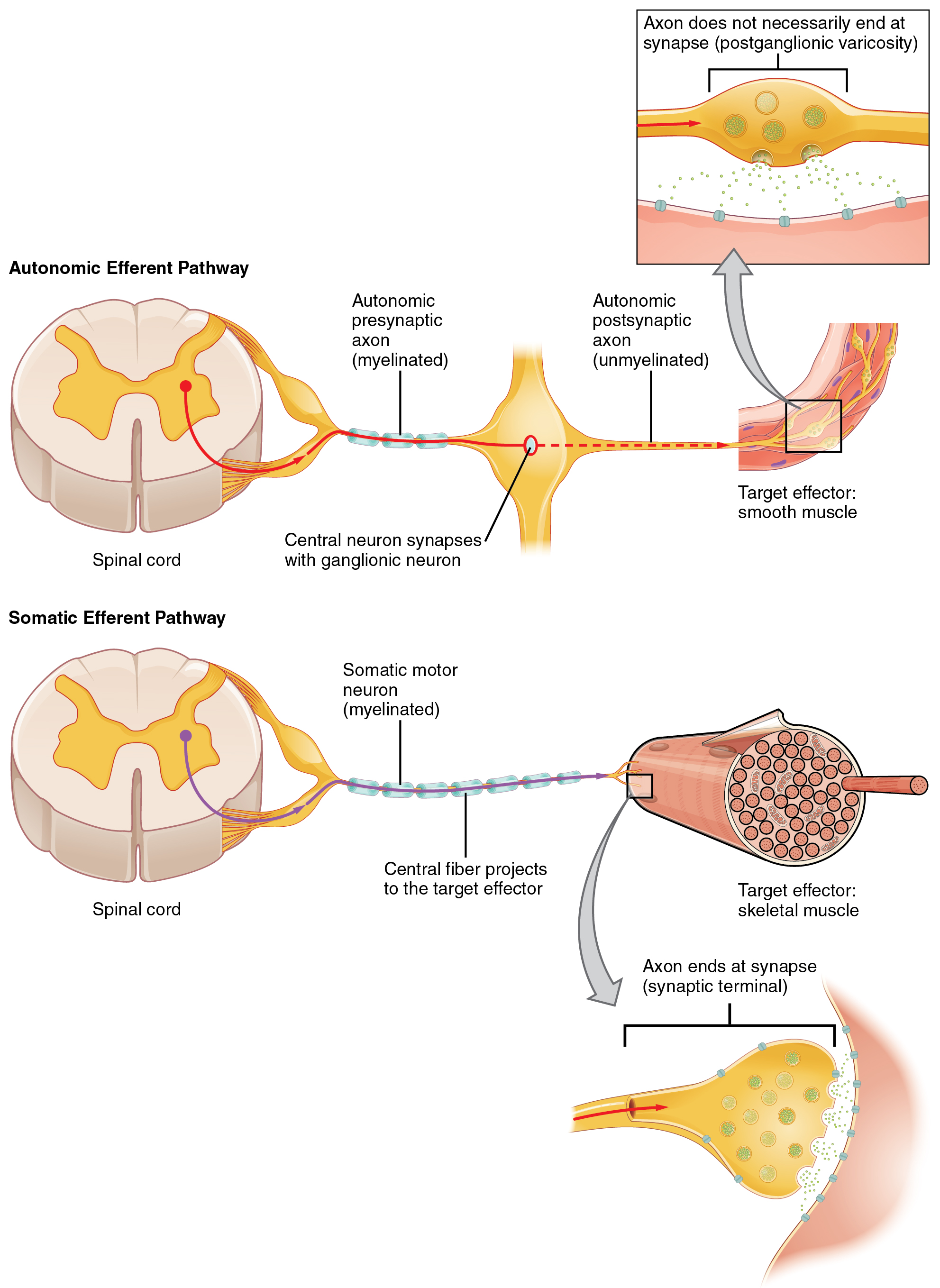
The autonomic nervous system is the principal pathway through which the central nervous system regulates visceral organs, glands, smooth muscle, and cardiac muscle. Unlike the somatic motor system, which drives skeletal muscle through a single motor neuron projecting directly from the spinal cord to the target, the autonomic nervous system inserts a ganglion between the central nervous system and the target organ. This creates the two-neuron efferent arc that defines all autonomic outflow and establishes one of pharmacology's most clinically exploited anatomical features.
The two-neuron arc consists of a preganglionic neuron, whose cell body lies within the central nervous system (in the brainstem or spinal cord), and a postganglionic neuron, whose cell body resides in a peripheral ganglion and whose axon reaches the target tissue. The synapse between these two neurons — the ganglionic synapse — uses acetylcholine as the neurotransmitter in both divisions, acting on nicotinic acetylcholine receptors of the ganglionic subtype (designated N-N). This shared cholinergic chemistry at the ganglionic level has a direct pharmacological consequence: drugs that block ganglionic nicotinic receptors disrupt both sympathetic and parasympathetic outflow simultaneously.
The autonomic nervous system is organized into three components. The sympathetic division arises from the thoracic and lumbar spinal cord and coordinates responses to stress and exercise. The parasympathetic division arises from the brainstem and sacral spinal cord and governs restorative, vegetative functions. The enteric nervous system is a semi-autonomous neuronal network embedded in the wall of the gastrointestinal tract, capable of regulating intestinal motility and secretion independently of the other two divisions. Each of these is examined in detail in subsequent sections.
The Ganglionic Synapse — Why It Matters for Pharmacology
All preganglionic-to-postganglionic communication in both the sympathetic and parasympathetic divisions uses acetylcholine acting on nicotinic N-N receptors. Ganglionic blocking drugs such as trimethaphan and mecamylamine block these receptors non-selectively, abolishing output from both divisions simultaneously. The clinical result is orthostatic hypotension (loss of sympathetic vascular tone), paralytic ileus (loss of parasympathetic gut tone), urinary retention, and anhidrosis. This indiscriminate profile limits ganglionic blockers to a narrow range of clinical indications — primarily hypertensive emergencies and deliberate controlled hypotension during surgery. Understanding this bilateral-blockade consequence is fundamental to predicting the effects of any drug acting at or upstream of the ganglionic synapse.
Section 2
Preganglionic origin, paravertebral ganglia, and the adrenal medulla as a modified ganglion
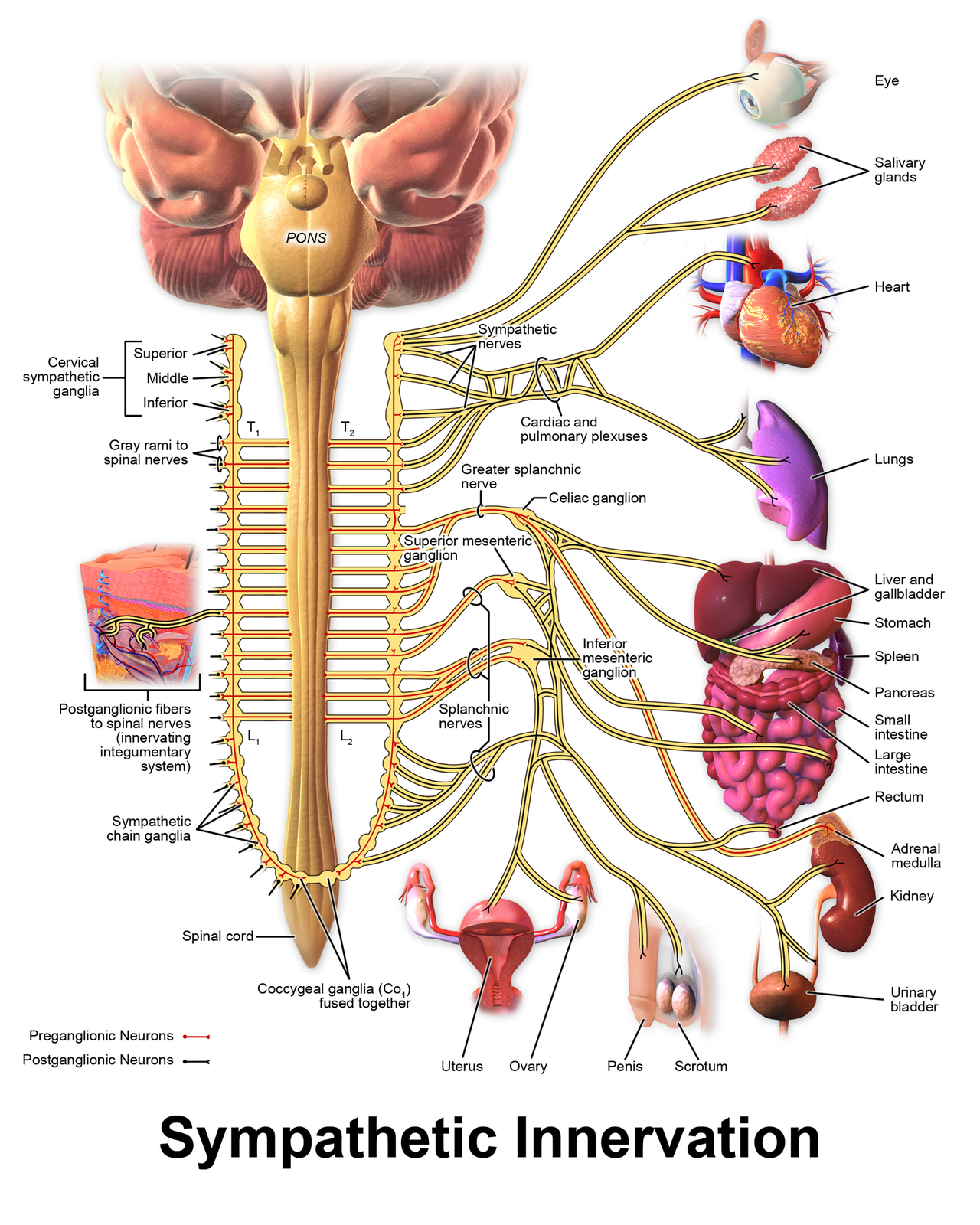
The sympathetic division originates from preganglionic neurons located in the lateral horn of the spinal cord from approximately the first thoracic to the second lumbar segment — the thoracolumbar outflow. These preganglionic neurons send relatively short myelinated axons to ganglia located close to the vertebral column, either in the paired paravertebral sympathetic chain (a longitudinal string of ganglia running on either side of the spine) or in prevertebral ganglia located in the abdomen near the major blood vessels.
The pattern of short preganglionic neurons synapsing on ganglia near the spinal column means that postganglionic neurons project long, unmyelinated axons from these ganglia to distant target organs throughout the body. A single preganglionic fiber can synapse on many postganglionic neurons, which in turn project to multiple organs — an anatomical arrangement that enables the broad, coordinated sympathetic activation that characterizes the fight-or-flight response. Heart rate increases, blood vessels in skeletal muscle dilate, skin and gut vessels constrict, the pupils dilate, and bronchioles dilate — all simultaneously.
Postganglionic sympathetic neurons release norepinephrine as their primary neurotransmitter, which acts on adrenergic receptors in target tissues. The adrenal medulla is a functionally unique structure: it is a modified sympathetic ganglion in which the postganglionic cells have differentiated into chromaffin cells. Rather than extending long axons to target organs, these cells release their catecholamines — approximately 80 percent epinephrine and 20 percent norepinephrine — directly into the bloodstream, effectively creating a hormonal amplification of sympathetic activation.
Architecture
Sympathetic Division
Adrenal Medulla
Modified Sympathetic Ganglion
Section 3
Long preganglionic neurons, terminal ganglia, and acetylcholine at the target
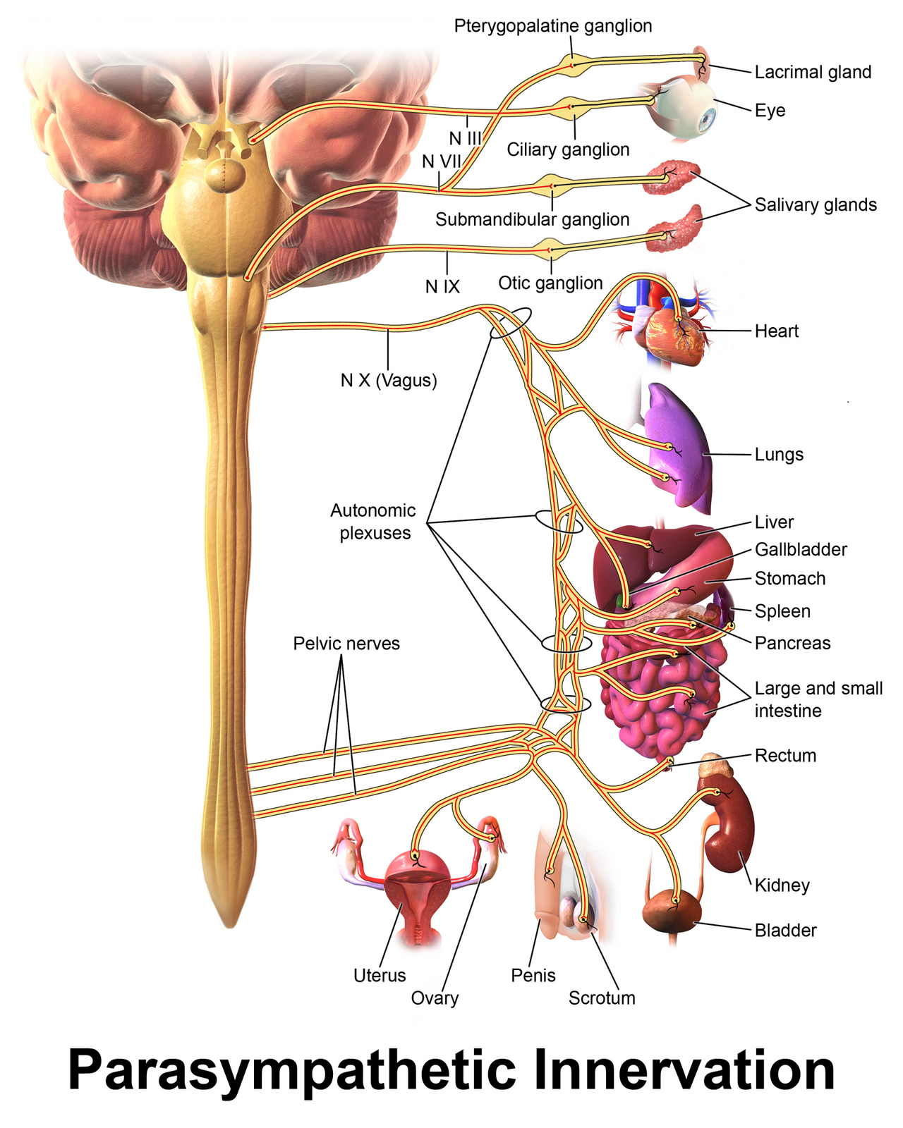
The parasympathetic division arises from two anatomically separate regions — the cranial outflow and the sacral outflow — giving it the designation craniosacral. Cranial parasympathetic preganglionic neurons originate in nuclei associated with four cranial nerves: the oculomotor nerve (cranial nerve III, controlling pupil constriction and lens accommodation), the facial nerve (cranial nerve VII, governing lacrimal and salivary gland secretion), the glossopharyngeal nerve (cranial nerve IX, innervating the parotid gland), and the vagus nerve (cranial nerve X, supplying the heart, lungs, and most abdominal viscera). Sacral parasympathetic outflow arises from spinal cord segments S2 through S4 and reaches the pelvic viscera — including the bladder, distal colon, and reproductive organs — via the pelvic nerves.
In contrast to the sympathetic division, parasympathetic preganglionic neurons project long axons all the way to ganglia located in or immediately adjacent to the target organ — the terminal ganglia. From these terminal ganglia, postganglionic neurons project only short distances to the effector cells. This organizational difference has pharmacological significance: because parasympathetic ganglia are embedded within target organs, they are largely inaccessible to drugs applied systemically at ganglionic doses without also blocking the sympathetic ganglia in the paravertebral chain.
The postganglionic neurotransmitter of the parasympathetic division is acetylcholine, acting on muscarinic receptors (as distinct from the nicotinic receptors at the ganglionic synapse). The clinical effects of parasympathetic activation follow the mnemonic SLUDD: salivation, lacrimation, urination, defecation, and digestion — along with bradycardia, bronchoconstriction, and pupillary constriction. The vagus nerve carries the vast majority of cranial parasympathetic outflow and accounts for resting heart rate regulation, bronchoconstriction, and gastrointestinal motility.
Comparing the Two Efferent Divisions
Sympathetic: thoracolumbar origin, short preganglionic fibers, ganglia near the spine, long postganglionic fibers, norepinephrine at the target (epinephrine from adrenal medulla). Widespread divergence enables coordinated mass activation.
Parasympathetic: craniosacral origin, long preganglionic fibers, terminal ganglia at or near the target organ, short postganglionic fibers, acetylcholine at the target (muscarinic receptors). Discrete, organ-specific activation is the functional result.
Section 4
A semi-autonomous neural network governing gastrointestinal function
The enteric nervous system consists of an estimated 200 to 500 million neurons embedded within the wall of the gastrointestinal tract, from the esophagus to the anus. Its neuronal population exceeds that of the spinal cord, and it can maintain coordinated intestinal motility and secretion even when all extrinsic connections to the central nervous system are severed — hence the designation "second brain." Two major networks make up the enteric nervous system: the myenteric plexus (Auerbach's plexus), located between the circular and longitudinal muscle layers and governing motility, and the submucosal plexus (Meissner's plexus), located closer to the intestinal lumen and regulating secretion and local blood flow.
Serotonin (5-hydroxytryptamine) is the dominant signaling molecule coordinating enteric nervous system activity, with approximately 95 percent of the body's total serotonin stored in enterochromaffin cells of the gut epithelium. Serotonin activates enteric neurons to initiate the peristaltic reflex. This pharmacological role of serotonin in the gut is the basis for prokinetic drugs and explains why selective serotonin reuptake inhibitors can produce gastrointestinal side effects. Acetylcholine and nitric oxide are the primary excitatory and inhibitory neurotransmitters, respectively, within the enteric neural circuits that coordinate propulsion.
The enteric nervous system is modulated by — but not wholly dependent on — the sympathetic and parasympathetic divisions. Sympathetic input generally inhibits gut motility and secretion; parasympathetic input generally promotes them. Opioid receptors are densely expressed throughout the enteric nervous system, and activation of these receptors reduces propulsive motility and increases sphincter tone, producing the constipation that accompanies opioid therapy. Peripherally restricted opioid antagonists such as methylnaltrexone and naloxegol are designed to reverse this effect at the enteric nervous system without reversing central analgesia.
Clinical Relevance: Opioids and the Enteric Nervous System
Opioid-induced bowel dysfunction occurs because mu-opioid receptors in the enteric nervous system reduce gut propulsion and increase sphincter tone — distinct from the central analgesic mechanism. Peripherally restricted opioid antagonists (methylnaltrexone, naloxegol, naldemedine) block enteric mu receptors without crossing the blood-brain barrier, reversing constipation without antagonizing analgesia. This drug class exemplifies using anatomical compartmentalization — central versus peripheral receptor access — as a pharmacological design principle.
Section 5
Competing inputs and the dominant division at each organ
Most visceral organs receive innervation from both the sympathetic and parasympathetic divisions — a principle called dual innervation. In organs with dual innervation, the two divisions generally produce opposing effects: the sympathetic division increases heart rate and blood pressure while the parasympathetic decreases them; the sympathetic dilates the pupil while the parasympathetic constricts it; the sympathetic relaxes bronchial smooth muscle while the parasympathetic contracts it. This reciprocal arrangement allows fine-tuned moment-to-moment regulation of organ function.
Under resting conditions, most organs are not at a neutral set point — they operate under the sustained influence of one division more than the other, a condition called tonic autonomic tone. The heart is the most clinically relevant example: resting heart rate is held below the intrinsic rate of the sinoatrial node by dominant resting parasympathetic (vagal) tone. This is why blocking muscarinic receptors with atropine accelerates the heart, and why enhanced vagal tone in well-trained athletes produces resting bradycardia.
Blood vessels, in contrast, lack functional parasympathetic innervation and are maintained under continuous resting sympathetic vasoconstrictor tone. Blockade of alpha-1 adrenergic receptors with an alpha blocker removes this tonic vasoconstriction, producing vasodilation and a fall in blood pressure. Understanding which division holds tonic dominance at each organ is essential for predicting the clinical effect of autonomic drugs.
Parasympathetic Dominance
Resting Tone
Sympathetic Dominance
Resting Tone
Section 6
Linking anatomical site to drug class and selectivity
The anatomical organization of the autonomic nervous system maps directly onto the pharmacological classification of drugs acting on it. Every autonomic drug acts at one of three anatomical levels: the ganglionic synapse, the neuroeffector junction (where the postganglionic neuron meets its target), or the central nervous system above the ganglia. Correctly identifying the anatomical target of a drug immediately predicts whether it will affect one or both autonomic divisions and which organ systems will be involved.
Drugs acting at the ganglionic synapse block nicotinic N-N receptors and disrupt both sympathetic and parasympathetic outflow simultaneously. Their effects are bilateral and difficult to control, making them poorly tolerated for most clinical applications. Drugs acting at the neuroeffector junction are division-specific: muscarinic receptor agonists and antagonists affect tissues innervated by parasympathetic postganglionic fibers; adrenergic receptor agonists and antagonists affect tissues innervated by sympathetic postganglionic fibers and tissues responding to adrenal medullary catecholamines.
The location of ganglia further predicts accessibility. Sympathetic ganglia in the paravertebral chain are anatomically accessible to regional anesthetics and surgical interventions (sympathetic nerve blocks). Parasympathetic ganglia embedded within target organs are not accessible at normal drug concentrations without systemic effects. This anatomical distinction is why selective peripheral parasympathetic modulation is achieved pharmacologically through muscarinic receptor targeting rather than ganglionic targeting.
Drug Target Anatomy — A Predictive Framework
Ganglionic blockers (trimethaphan, mecamylamine): block nicotinic N-N receptors, disrupt both divisions simultaneously. Indiscriminate profile limits clinical use.
Muscarinic drugs (atropine, pilocarpine): act at parasympathetic neuroeffector junctions. Predict effects by knowing which organs have parasympathetic postganglionic fibers releasing acetylcholine onto muscarinic receptors.
Adrenergic drugs (epinephrine, propranolol): act at sympathetic neuroeffector junctions and on adrenal-medullary hormone targets. Predict effects by knowing the receptor subtype distribution (alpha-1, alpha-2, beta-1, beta-2) at each organ.
| Author / Organization | Title | Source |
|---|---|---|
| Janig W, McLachlan EM | Characteristics of function-specific pathways in the sympathetic nervous system | Trends in Neurosciences. 1992;15(12):475-481. |
| Katzung BG, ed | Basic and Clinical Pharmacology. 15th ed. Chapters 6-9 (Autonomic Nervous System Pharmacology) | McGraw-Hill; 2021. |
| Breit S, Kupferberg A, Rogler G, Hasler G | Vagus nerve as modulator of the brain-gut axis in psychiatric and inflammatory disorders | Frontiers in Psychiatry. 2018;9:44. |
| Jansen ASP, Van Nguyen X, Karpitskiy V, Mettenleiter TC, Loewy AD | Central command neurons of the sympathetic nervous system: basis of the fight-or-flight response | Science. 1995;270(5236):644-646. |
| Goldstein DS, Eisenhofer G, Kopin IJ | Sources and significance of plasma levels of catechols and their metabolites in humans | Journal of Pharmacology and Experimental Therapeutics. 2003;305(3):800-811. |
| Wehrwein EA, Orer HS, Barman SM | Overview of the anatomy, physiology, and pharmacology of the autonomic nervous system | Comprehensive Physiology. 2016;6(3):1239-1278. |
| Furness JB | The enteric nervous system and neurogastroenterology | Nature Reviews Gastroenterology and Hepatology. 2012;9(5):286-294. |
| Bornstein JC, Costa M, Grider JR | Enteric motor and interneuronal circuits controlling motility | Neurogastroenterology and Motility. 2004;16(Suppl 1):34-38. |
| Becker G, Blum HE | Novel opioid antagonists for opioid-induced bowel dysfunction and postoperative ileus | Lancet. 2009;373(9670):1198-1206. |
| Saper CB | The central autonomic nervous system: conscious visceral perception and autonomic pattern generation | Annual Review of Neuroscience. 2002;25:433-469. |
| Michel MC, Vrydag W | Alpha1-, alpha2- and beta-adrenoceptors in the urinary bladder, urethra and prostate | British Journal of Pharmacology. 2006;147(Suppl 2):S88-S119. |
| Cazzola M, Page CP, Calzetta L, Matera MG | Pharmacology and therapeutics of bronchodilators | Pharmacological Reviews. 2012;64(3):450-504. |