CHAPTER 4 · ANS INTRODUCTION

Section 1

Cholinergic Transmission — Synthesis, Storage, and Termination

Acetylcholine lifecycle and the drugs that interrupt each step

Acetylcholine is the neurotransmitter at all autonomic ganglia, all parasympathetic postganglionic synapses, the neuromuscular junction, and some central nervous system pathways. Its synthesis, storage, release, and inactivation each represent discrete pharmacological targets that are exploited by drugs in clinical use.

Synthesis occurs in the presynaptic terminal when choline acetyltransferase catalyzes the combination of choline (taken up from the extracellular fluid by a sodium-dependent choline transporter) with acetyl-coenzyme A to form acetylcholine. The newly synthesized acetylcholine is then packaged into synaptic vesicles by the vesicular acetylcholine transporter. Hemicholinium-3 is an experimental drug that blocks the choline transporter, preventing choline uptake and depleting acetylcholine stores — it is not used clinically but illustrates the vulnerability of this synthesis step.

Release is triggered when an action potential depolarizes the nerve terminal, opening voltage-gated calcium channels. The influx of calcium drives fusion of acetylcholine-containing vesicles with the presynaptic membrane, releasing acetylcholine into the synaptic cleft. Botulinum toxin — produced by Clostridium botulinum — is a protease that cleaves proteins required for vesicular fusion (the SNARE complex components), blocking acetylcholine release at all cholinergic synapses. Clinically, botulinum toxin is used in purified form for focal muscle spasticity, hyperhidrosis, cosmetic applications, and certain movement disorders.

Inactivation of acetylcholine is achieved almost entirely by enzymatic hydrolysis, not by reuptake. Acetylcholinesterase, located on the postsynaptic membrane and within the synaptic cleft, rapidly hydrolyzes acetylcholine into choline and acetate, terminating its action within milliseconds. Choline is then transported back into the presynaptic terminal for resynthesis. Drugs that inhibit acetylcholinesterase — the anticholinesterases — prolong and amplify cholinergic transmission at all acetylcholine-releasing synapses.

Acetylcholinesterase Inhibitors

Clinical and Toxic

  • Reversible (carbamate): neostigmine, pyridostigmine — myasthenia gravis, reversal of neuromuscular blockade
  • Reversible (tertiary): physostigmine, donepezil — glaucoma, Alzheimer disease
  • Irreversible (organophosphate): sarin, VX (nerve agents), malathion, parathion (insecticides) — bind covalently; atropine + pralidoxime treatment

Presynaptic Targets

Blocking Release or Synthesis

  • Botulinum toxin: cleaves SNARE proteins, blocks vesicle fusion and acetylcholine release
  • Clinical uses: focal dystonia, spasticity, hyperhidrosis, cosmetic
  • Hemicholinium-3: blocks choline uptake (experimental only)
  • Vesamicol: blocks vesicular acetylcholine transporter (experimental)

Section 2

Catecholamine Biosynthesis — The Enzymatic Pathway

From tyrosine to epinephrine: four steps, four pharmacological targets

The catecholamines — dopamine, norepinephrine, and epinephrine — are synthesized through a linear enzymatic pathway beginning with the amino acid tyrosine. Each step is catalyzed by a distinct enzyme, and each enzyme is a potential drug target. Understanding the pathway allows prediction of which drugs deplete catecholamine stores, which block specific steps, and which alter the ratio of products.

Step 1: Tyrosine is converted to L-dihydroxyphenylalanine (L-DOPA) by tyrosine hydroxylase. This is the rate-limiting step of the entire pathway and is the primary point of feedback regulation — catecholamines inhibit tyrosine hydroxylase at high concentrations, providing end-product inhibition. The drug metyrosine (alpha-methyltyrosine) inhibits tyrosine hydroxylase and is used in the management of pheochromocytoma to reduce catecholamine synthesis preoperatively.

Step 2: L-DOPA is converted to dopamine by aromatic L-amino acid decarboxylase (also called DOPA decarboxylase), which requires pyridoxal phosphate (vitamin B6) as a cofactor. This reaction occurs in all catecholamine-producing neurons and in peripheral tissues. In Parkinson disease therapy, levodopa (L-DOPA) is co-administered with carbidopa, a peripheral DOPA decarboxylase inhibitor, to prevent peripheral conversion of levodopa to dopamine before it can cross the blood-brain barrier.

Step 3: Dopamine is converted to norepinephrine by dopamine beta-hydroxylase, located within synaptic vesicles. This reaction requires ascorbic acid and copper. Dopamine beta-hydroxylase deficiency is a rare congenital disorder causing severe orthostatic hypotension and inability to produce norepinephrine or epinephrine.

Step 4: Norepinephrine is converted to epinephrine by phenylethanolamine-N-methyltransferase, an enzyme found almost exclusively in the chromaffin cells of the adrenal medulla. This enzyme requires S-adenosylmethionine as a methyl donor and is induced by high local glucocorticoid concentrations from the adrenal cortex — which is why adrenal medullary epinephrine production depends on an intact adrenal cortex.

Clinical Drug Connections to the Biosynthetic Pathway

Metyrosine inhibits tyrosine hydroxylase (step 1) — used to deplete catecholamines in pheochromocytoma.

Carbidopa inhibits peripheral DOPA decarboxylase (step 2) — given with levodopa to increase brain dopamine delivery in Parkinson disease.

Reserpine blocks the vesicular monoamine transporter, preventing storage of dopamine, norepinephrine, and epinephrine in vesicles — depletes all catecholamine stores over time. Historical antihypertensive; causes depression at higher doses.
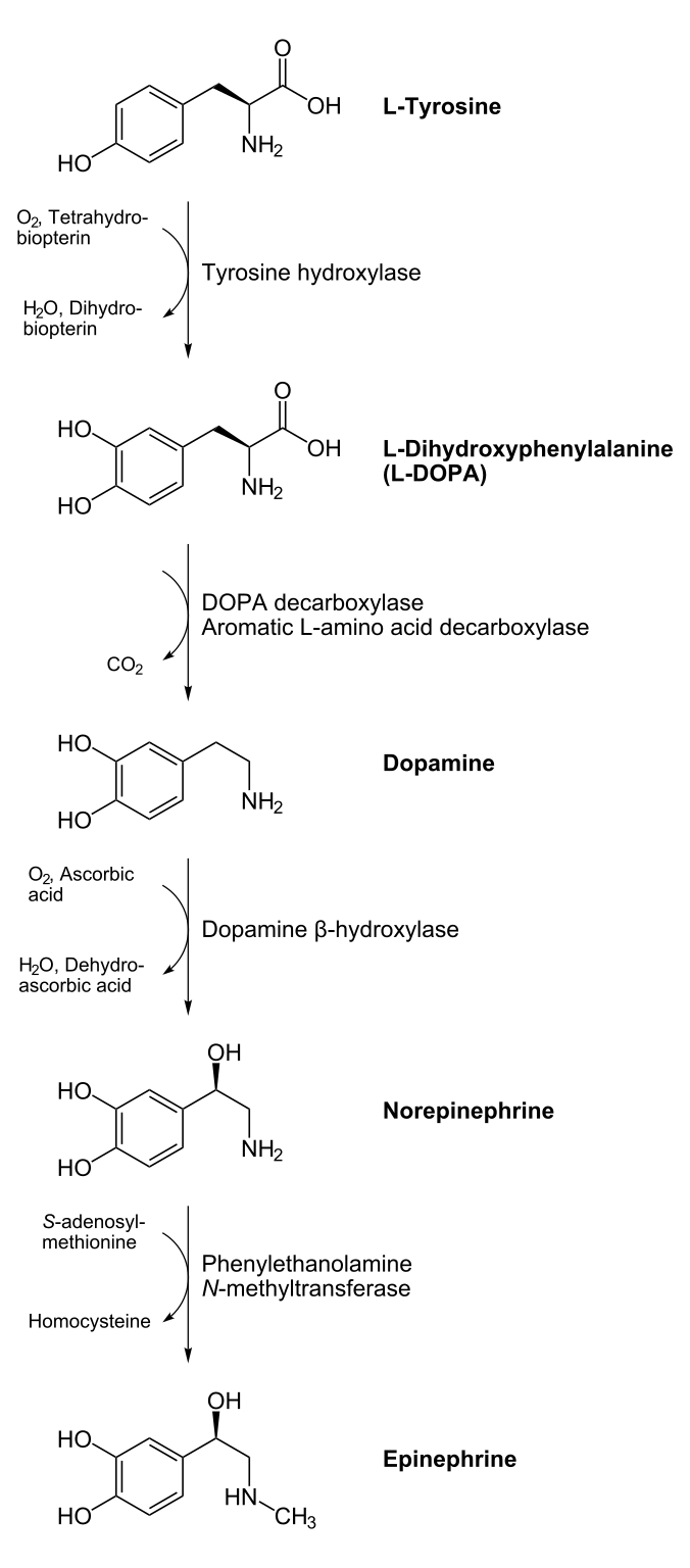
Diagram of the catecholamine biosynthetic pathway showing the sequential enzymatic conversion of tyrosine to L-DOPA to dopamine to norepinephrine to epinephrine, with each catalyzing enzyme labeled at each step.
NEUROtiker. Catecholamine Biosynthesis Pathway. Source: Wikimedia Commons. Public domain.

Section 3

Vesicular Storage, Exocytotic Release, and Autoreceptor Modulation

How catecholamines are stored, released, and regulated at the presynaptic terminal

After synthesis, dopamine and norepinephrine are actively transported into dense-core vesicles by the vesicular monoamine transporter-2 (VMAT-2), a membrane protein powered by a proton gradient. Vesicular storage serves two purposes: it concentrates the neurotransmitter to achieve high local release concentrations during exocytosis, and it protects the monoamines from degradation by monoamine oxidase in the cytoplasm. Reserpine irreversibly blocks VMAT-2, causing catecholamines to leak out of vesicles into the cytoplasm where they are degraded by monoamine oxidase. The result is depletion of norepinephrine stores over days, producing antihypertensive effects and, at higher doses, depression. Tetrabenazine (VMAT-2 inhibitor) is used to reduce abnormal involuntary movements in Huntington disease.

Release is triggered by action potentials that depolarize the presynaptic terminal and open voltage-gated calcium channels. Calcium influx drives vesicle-membrane fusion and exocytosis of catecholamine into the synaptic cleft. Indirectly acting sympathomimetics such as amphetamine work by entering the presynaptic terminal — they are substrates for the norepinephrine transporter — and then reversing the direction of the transporter and VMAT-2, causing non-exocytotic release of catecholamine stores into the cleft. This mechanism is independent of calcium and accounts for the ability of amphetamine to release catecholamines even in the absence of action potentials.

Release is finely regulated by presynaptic autoreceptors — alpha-2 adrenergic receptors on the sympathetic nerve terminal that detect norepinephrine in the cleft and, when activated, reduce further release through inhibition of adenylyl cyclase and decreased calcium channel opening. This negative-feedback mechanism limits the magnitude of adrenergic activation. The drug clonidine — an alpha-2 agonist — exploits these presynaptic autoreceptors to reduce sympathetic outflow, producing its antihypertensive effect.


Section 4

Termination of Adrenergic Signaling — Reuptake and Metabolism

The norepinephrine transporter, monoamine oxidase, and catechol-O-methyltransferase

The primary mechanism terminating norepinephrine signaling at sympathetic synapses is reuptake: norepinephrine is transported back into the presynaptic terminal by the norepinephrine transporter (NET), a sodium-dependent carrier protein in the presynaptic membrane. This reuptake mechanism accounts for approximately 70 to 80 percent of norepinephrine clearance from the synaptic cleft. Once inside the terminal, norepinephrine can be repackaged into vesicles (via VMAT-2) for reuse, or degraded by monoamine oxidase.

Monoamine oxidase is a mitochondrial enzyme present in the presynaptic terminal (and in the liver, intestinal wall, and other tissues) that oxidatively deaminates catecholamines and serotonin, producing inactive metabolites. There are two isoforms: monoamine oxidase-A preferentially metabolizes norepinephrine and serotonin; monoamine oxidase-B preferentially metabolizes dopamine and phenylethylamine. Monoamine oxidase inhibitors — including phenelzine and tranylcypromine (non-selective, irreversible) and selegiline (monoamine oxidase-B selective) — are used as antidepressants or in Parkinson disease. Their clinical limitations arise from the accumulation of dietary tyramine (normally metabolized in the gut and liver by monoamine oxidase-A), which can trigger hypertensive crises.

Catechol-O-methyltransferase is a cytoplasmic enzyme present in postsynaptic cells, liver, and kidney that methylates the catechol ring of norepinephrine, epinephrine, and dopamine using S-adenosylmethionine. It is a secondary pathway of catecholamine inactivation but becomes clinically important in Parkinson disease therapy: catechol-O-methyltransferase inhibitors (entacapone, tolcapone) block peripheral conversion of levodopa and dopamine, extending the duration of effect when used adjunctively with levodopa-carbidopa.

Reuptake Inhibitors (NET)

Clinical Drugs Blocking Norepinephrine Transporter

  • Cocaine: blocks NET and dopamine transporter — vasoconstriction, tachycardia, euphoria
  • Tricyclic antidepressants (amitriptyline, imipramine): block NET and serotonin transporter
  • Atomoxetine: selective NET inhibitor — attention-deficit/hyperactivity disorder
  • Effect: accumulate norepinephrine and/or dopamine in synaptic cleft, prolonging and amplifying signaling

Metabolic Enzyme Targets

Monoamine Oxidase and Catechol-O-methyltransferase

  • Monoamine oxidase-A inhibitors (phenelzine): antidepressant; tyramine interaction risk
  • Monoamine oxidase-B inhibitors (selegiline, rasagiline): Parkinson disease adjuncts
  • Catechol-O-methyltransferase inhibitors (entacapone, tolcapone): extend levodopa effect in Parkinson disease

Section 5

Clinical Drug Target Map — Neurotransmission Step by Step

Linking each step in the neurotransmitter lifecycle to a drug class

The neurotransmitter lifecycle — synthesis, storage, release, receptor activation, and inactivation — provides a systematic map of drug targets. Every major class of autonomic drug acts at one of these steps, and knowing which step a drug targets immediately predicts its mechanism of action and its likely adverse effects.

For the cholinergic system: acetylcholinesterase inhibitors block inactivation (the most clinically important target class), botulinum toxin blocks release, and direct muscarinic or nicotinic agonists bypass the entire presynaptic apparatus to activate receptors directly. For the adrenergic system: the most important targets are the postsynaptic adrenergic receptors themselves (alpha and beta agonists and antagonists), followed by the presynaptic reuptake transporter (norepinephrine transporter inhibitors such as cocaine and tricyclic antidepressants), storage depletion (reserpine, tetrabenazine via VMAT-2), and biosynthesis inhibition (metyrosine, carbidopa).

One principle worth noting: drugs acting at postsynaptic receptors produce immediate, predictable effects whose direction depends on the receptor subtype and organ. Drugs acting presynaptically (depleting stores, blocking synthesis, blocking reuptake) have effects that evolve over hours to days as stores change and often produce paradoxical early responses. Reserpine, for example, may briefly release catecholamines as vesicles are disrupted before depletion takes hold. Understanding which side of the synapse a drug acts on is as important as knowing its receptor target.

Quick Reference — Adrenergic Drug Target by Mechanism

Biosynthesis inhibition: metyrosine (tyrosine hydroxylase), carbidopa (DOPA decarboxylase peripherally)

Storage depletion: reserpine, tetrabenazine (VMAT-2 blockers)

Release promotion (indirect sympathomimetics): amphetamine, ephedrine

Release inhibition (presynaptic alpha-2 agonists): clonidine, alpha-methyldopa (converts to alpha-methylnorepinephrine)

Reuptake inhibition: cocaine, tricyclic antidepressants, atomoxetine (NET blockers)

Metabolism inhibition: monoamine oxidase inhibitors (phenelzine, selegiline), catechol-O-methyltransferase inhibitors (entacapone)

Postsynaptic receptor agonists/antagonists: phenylephrine, propranolol, prazosin — the largest and most clinically used category


Suggested References
Author / OrganizationTitleSource
Wess JMolecular biology of muscarinic acetylcholine receptorsCritical Reviews in Neurobiology. 1996;10(1):69-99.
Schiavo G, Benfenati F, Poulain B, et alTetanus and botulinum-B neurotoxins block neurotransmitter release by proteolytic cleavage of synaptobrevinNature. 1992;359(6398):832-835.
Dolly JO, Aoki KRThe structure and mode of action of different botulinum toxinsEuropean Journal of Neurology. 2006;13(Suppl 4):1-9.
Taylor PAnticholinesterase agents. In: Brunton LL, Hilal-Dandan R, Knollmann BC, eds. Goodman and Gilman's The Pharmacological Basis of Therapeutics. 13th ed.McGraw-Hill; 2018:163-186.
Eddleston M, Buckley NA, Eyer P, Dawson AHManagement of acute organophosphorus pesticide poisoningLancet. 2008;371(9612):597-607.
Nagatsu T, Levitt M, Udenfriend STyrosine hydroxylase: the initial step in norepinephrine biosynthesisJournal of Biological Chemistry. 1964;239:2910-2917.
Pacak K, Linehan WM, Eisenhofer G, Walther MM, Goldstein DSRecent advances in genetics, diagnosis, localization, and treatment of pheochromocytomaAnnals of Internal Medicine. 2001;134(4):315-329.
Robertson D, Haile V, Perry SE, Robertson RM, Phillips JA 3rd, Biaggioni IDopamine beta-hydroxylase deficiency: a genetic disorder of cardiovascular regulationHypertension. 1991;18(1):1-8.
Schuldiner S, Shirvan A, Linial MVesicular neurotransmitter transporters: from bacteria to humansPhysiological Reviews. 1995;75(2):369-392.
Bhidayasiri R, Jitkritsadakul O, Friedman JH, Fahn SUpdating the recommendations for treatment of tardive syndromes: a systematic review of new evidence and practical treatment algorithmJournal of the Neurological Sciences. 2018;389:67-75.
Eisenhofer G, Kopin IJ, Goldstein DSCatecholamine metabolism: a contemporary view with implications for physiology and medicinePharmacological Reviews. 2004;56(3):331-349.
Goldstein DSThe Autonomic Nervous System in Health and DiseaseMarcel Dekker; 2001.
Lundberg JMPharmacology of cotransmission in the autonomic nervous systemPharmacological Reviews. 1996;48(1):113-178.
Burnstock GCotransmissionCurrent Opinion in Pharmacology. 2004;4(1):47-52.
Goldstein DS, Eisenhofer G, Kopin IJSources and significance of plasma levels of catechols and their metabolites in humansJournal of Pharmacology and Experimental Therapeutics. 2003;305(3):800-811.