Introduction to Medical Pharmacology
Adverse Effects and Systematic Clinical Management
Chapter 16 · Module 5 of 6Section 1
A consolidated reference: classification, time course, mechanisms, and management of all four extrapyramidal syndromes
Extrapyramidal side effects are among the most clinically significant adverse effects of antipsychotic therapy and the primary reason patients discontinue treatment without medical advice. This section provides a consolidated reference covering all four syndromes — three reversible and one potentially permanent — organized by time course, mechanism, and management.
The four extrapyramidal syndromes differ fundamentally in their onset timing, and this time course is one of the most testable features at the second-year medical student level. Acute dystonia appears within hours to days. Akathisia appears within days to weeks. Drug-induced parkinsonism appears within weeks. Tardive dyskinesia appears after months to years of exposure and is addressed in detail in Section 2.
Hours to Days
Acute Dystonia
Days to Weeks
Akathisia
Weeks
Drug-Induced Parkinsonism
High-potency first-generation antipsychotics carry the highest extrapyramidal symptom risk. Second-generation antipsychotics with high serotonin 5-HT2A to D2 ratios — quetiapine, clozapine — carry essentially no extrapyramidal symptom risk at standard doses. Partial agonists carry low extrapyramidal symptom risk but meaningful akathisia risk. Risk is dose-dependent for all agents: staying within the recommended dose range and avoiding unnecessary dose escalation is the most effective prevention strategy. When extrapyramidal symptoms develop, the preferred management sequence is dose reduction first, then agent switch, then pharmacological management — not adding anticholinergics as the default first step.
Section 2
Pathophysiology, risk stratification, and vesicular monoamine transporter 2 inhibitor therapy
Tardive dyskinesia is the most feared long-term complication of antipsychotic therapy because it may be irreversible. It arises from a compensatory change in dopamine receptor density that develops in response to chronic D2 blockade, and it can persist indefinitely even after the causative drug is stopped. Prevention through minimizing unnecessary exposure is the most effective strategy; when tardive dyskinesia develops, the first approved pharmacological treatments are now available.
Chronic D2 receptor blockade triggers a compensatory response in the striatum: postsynaptic D2 receptors upregulate in number and sensitivity. As long as the antipsychotic maintains sufficient blockade, this supersensitivity is concealed. When blockade falls — between doses, after dose reduction, or upon discontinuation — the supersensitive receptors are activated by endogenous dopamine, producing hyperkinetic involuntary movements.
This mechanism explains one of the most clinically deceptive features of tardive dyskinesia: withdrawal-emergent dyskinesia. When the antipsychotic dose is reduced or stopped, the movements may first appear or dramatically worsen — because reducing the blockade unmasks the underlying supersensitivity. This can lead to the error of concluding that dose reduction caused the dyskinesia and resuming or increasing the antipsychotic, which temporarily suppresses the movements while worsening the underlying supersensitivity further.
The movements of tardive dyskinesia are repetitive, involuntary, and purposeless. The orofacial region is most commonly affected: lip smacking, tongue protrusion, chewing movements, and facial grimacing are characteristic. Choreiform movements of the limbs and trunk rocking also occur. Risk factors include older age, female sex, longer cumulative duration of antipsychotic exposure, higher doses, use of high-potency first-generation antipsychotics, early appearance of extrapyramidal symptoms during treatment, and a history of mood disorder as the indication for antipsychotic use. The annual incidence with first-generation antipsychotics is approximately 5 percent per year for the first several years, with cumulative risk approaching 20 to 30 percent after 5 years. Second-generation antipsychotics carry substantially lower but nonzero risk — approximately 0.5 to 1 percent per year.
Vesicular monoamine transporter 2 is the transporter responsible for packaging dopamine into synaptic vesicles before release. Inhibiting this transporter depletes presynaptic dopamine stores, reducing the amount of dopamine available to activate supersensitive striatal D2 receptors. Two vesicular monoamine transporter 2 inhibitors — valbenazine and deutetrabenazine — are approved specifically for tardive dyskinesia by the Food and Drug Administration and represent the first pharmacological treatments with regulatory approval for this condition.
Valbenazine is administered once daily and has demonstrated significant reduction in involuntary movement scores in randomized trials. Deutetrabenazine is a deuterium-substituted derivative of tetrabenazine with twice-daily dosing and a more favorable adverse effect profile than tetrabenazine. Both carry a class adverse effect of somnolence and a risk of worsening depression or suicidality — requiring monitoring for depressive symptoms, and both are contraindicated in patients with untreated depression or active suicidality. Crucially, both agents treat tardive dyskinesia without requiring discontinuation of the antipsychotic, resolving the clinical dilemma of treating the movement disorder while maintaining psychiatric stability.
Once established, tardive dyskinesia may be permanent even after stopping the drug. Complete resolution is uncommon. Vesicular monoamine transporter 2 inhibitors suppress rather than cure the movements. The only truly effective strategy is prevention: use the lowest effective antipsychotic dose, reassess the need for continued therapy at regular intervals, and switch to a lower-risk agent whenever clinically feasible.
Clozapine has the lowest tardive dyskinesia liability of any antipsychotic due to its low, fast-dissociating D2 occupancy and may actually suppress existing tardive dyskinesia in patients switched from other agents.
Section 3
Diagnosis, emergency management, and distinction from serotonin syndrome
Neuroleptic malignant syndrome is rare but potentially fatal, and its recognition requires pattern identification under pressure in an acutely ill patient. The clinical challenge is twofold: recognizing the syndrome promptly and distinguishing it from other hyperthermia syndromes — particularly serotonin syndrome — whose management differs substantially.
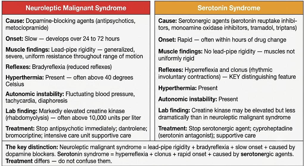
Neuroleptic malignant syndrome is characterized by a tetrad of four findings developing together in a patient on antipsychotic therapy: hyperthermia (often above 40 degrees Celsius), severe generalized lead-pipe muscle rigidity, autonomic instability (fluctuating blood pressure, tachycardia, diaphoresis, tachypnea), and altered consciousness ranging from confusion to coma. It develops over 24 to 72 hours — a relatively slow onset compared with serotonin syndrome. Any dopamine-blocking antipsychotic can cause it, though high-potency first-generation agents are most frequently implicated. It occurs most commonly in the first weeks of therapy or after rapid dose escalation.
The laboratory hallmark is markedly elevated creatine kinase from rhabdomyolysis — levels above 10,000 units per liter are common in severe cases, and levels above 100,000 have been reported. Leukocytosis, elevated liver transaminases, metabolic acidosis, and myoglobinuria threatening acute kidney injury are additional findings. Elevated creatine kinase in a febrile, rigid patient on an antipsychotic should immediately raise suspicion for neuroleptic malignant syndrome.
The distinction between neuroleptic malignant syndrome and serotonin syndrome is one of the highest-yield clinical differentials in pharmacology. Both present with hyperthermia, autonomic instability, and altered consciousness in patients on psychotropic medications. The key differentiating features are the nature of the neuromuscular findings and the onset timing.
Neuroleptic malignant syndrome produces lead-pipe rigidity — generalized, severe, uniform resistance throughout the range of motion — with bradyreflexia. It develops slowly over 24 to 72 hours. It is caused by dopamine-blocking agents (antipsychotics, metoclopramide, prochlorperazine). Serotonin syndrome produces hyperreflexia and clonus — rhythmic involuntary muscle contractions — rather than rigidity. It develops rapidly, often within hours of a serotonergic drug change. It is caused by agents that increase serotonin activity (serotonin reuptake inhibitors, monoamine oxidase inhibitors, tramadol, linezolid, triptans).
Management of neuroleptic malignant syndrome is primarily supportive and must begin immediately upon recognition. Stop all antipsychotics and other dopamine-blocking agents immediately. Initiate aggressive cooling — cooling blankets, ice packs to major vessels, cold intravenous fluids. Provide vigorous intravenous fluid resuscitation to protect renal function from myoglobin precipitation. Monitor creatine kinase and renal function at minimum every 6 hours during the acute phase. Intensive care unit admission is required for most cases.
Specific pharmacological interventions include dantrolene — a direct-acting skeletal muscle relaxant that inhibits calcium release from the sarcoplasmic reticulum, reducing rigidity and thereby heat production. Bromocriptine and amantadine restore central dopaminergic tone and may accelerate recovery. Benzodiazepines address agitation and reduce peripheral sympathetic activity. When antipsychotic therapy must be restarted after full recovery, wait at least 2 weeks after complete resolution, use the lowest-potency agent available at the lowest effective dose, and monitor closely for recurrence.
Neuroleptic malignant syndrome: Lead-pipe rigidity + bradyreflexia + slow onset (24–72 hours) + caused by dopamine blockers + markedly elevated creatine kinase
Serotonin syndrome: Hyperreflexia + clonus + rapid onset (hours) + caused by serotonergic agents + creatine kinase less dramatically elevated
Treatment differs: Neuroleptic malignant syndrome — stop antipsychotic, dantrolene, bromocriptine. Serotonin syndrome — stop serotonergic agent, cyproheptadine (serotonin antagonist).
Section 4
Monitoring, prevention, and pharmacological management of antipsychotic-induced weight gain, glucose dysregulation, and dyslipidemia
Antipsychotic-induced metabolic syndrome is the most prevalent long-term adverse effect burden of the class. It encompasses weight gain, glucose dysregulation, dyslipidemia, and hypertension, driven primarily by histamine H1 blockade (appetite stimulation) and serotonin 5-HT2C blockade (impaired satiety signaling). The clinical consequences extend beyond cosmetic concerns: patients with schizophrenia already have substantially elevated rates of cardiovascular disease and diabetes, and antipsychotic-induced metabolic syndrome compounds these risks, contributing to the 15 to 25-year reduction in life expectancy documented in this population.
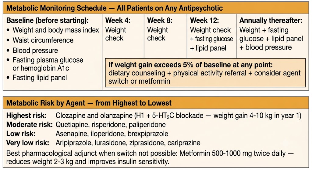
Metabolic risk varies substantially across agents and is predicted largely by histamine H1 and serotonin 5-HT2C receptor affinity. The highest-risk agents are clozapine and olanzapine, both of which produce mean weight gains of 4 to 10 kilograms or more in the first year and carry significantly elevated rates of new-onset type 2 diabetes. Quetiapine and risperidone carry intermediate metabolic risk. Aripiprazole, lurasidone, ziprasidone, and brexpiprazole carry the lowest metabolic risk among second-generation agents. First-generation antipsychotics generally carry lower metabolic risk than clozapine and olanzapine, though chlorpromazine and thioridazine produce meaningful weight gain through histamine H1 blockade.
Systematic metabolic monitoring is the standard of care for all patients on any antipsychotic, regardless of the agent's perceived metabolic risk. Baseline assessment before initiating any antipsychotic should include weight and body mass index, waist circumference, blood pressure, fasting plasma glucose or hemoglobin A1c, and a fasting lipid panel. Follow-up weight is assessed at 4, 8, and 12 weeks after initiation. Fasting glucose and lipids are repeated at 12 weeks and then annually. Any patient who gains more than 5 percent of baseline body weight at any monitoring point warrants a clinical response — dietary counseling, physical activity referral, and consideration of pharmacological intervention or antipsychotic switch.
The most effective strategy for managing antipsychotic-induced metabolic syndrome is switching to a metabolically more favorable agent when the clinical situation permits. Switching from olanzapine or clozapine to aripiprazole, lurasidone, or ziprasidone consistently produces weight loss, improved insulin sensitivity, and favorable lipid changes in randomized trials — though the trade-off between metabolic improvement and potential loss of antipsychotic efficacy must be assessed individually for each patient.
When a switch is not feasible because the current agent provides superior efficacy, metformin has the strongest pharmacological evidence base as an adjunct, producing mean weight reductions of 2 to 3 kilograms and improvements in insulin sensitivity in randomized trials. It is generally well tolerated and is the recommended first-line pharmacological adjunct for antipsychotic-induced weight gain when an agent switch is not possible. Topiramate has shown efficacy in some trials but carries cognitive adverse effects — word-finding difficulties and concentration impairment — that are particularly burdensome in patients with schizophrenia. Aripiprazole augmentation of clozapine or olanzapine regimens produces modest weight attenuation and is used clinically.
Section 5
Three distinct adverse effect domains with different risk distributions, clinical consequences, and management approaches
Beyond motor side effects and metabolic complications, three additional adverse effect domains require systematic understanding: QTc interval prolongation, hyperprolactinemia, and sedation. Each operates through a distinct mechanism, affects a different subset of patients, and requires a different monitoring and management strategy.
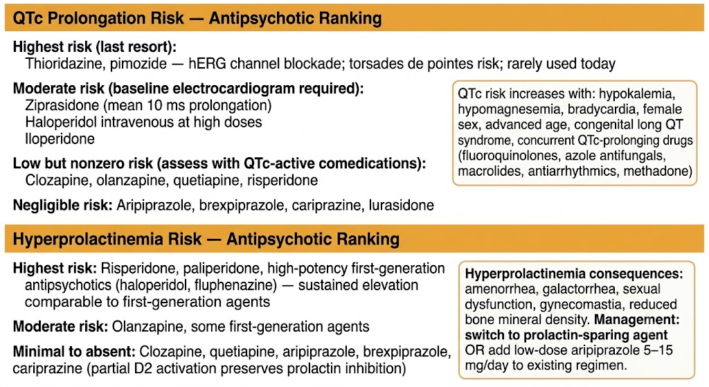
Antipsychotics prolong the cardiac QTc interval by blocking the rapidly activating delayed rectifier potassium current — the current responsible for cardiac repolarization. Reduced repolarization reserve increases the risk of torsades de pointes, a polymorphic ventricular tachycardia that can degenerate into ventricular fibrillation and sudden cardiac death. QTc prolongation liability varies substantially across antipsychotics. Thioridazine and pimozide carry the highest risk and are now rarely used for this reason. Ziprasidone produces mean QTc prolongation of approximately 10 milliseconds. Haloperidol administered intravenously at high doses carries substantial risk. Iloperidone and amisulpride are intermediate. Clozapine, olanzapine, quetiapine, and risperidone carry low but nonzero risk. The partial agonists — aripiprazole, brexpiprazole, and cariprazine — produce negligible QTc prolongation.
The clinical threshold for concern is a QTc exceeding 500 milliseconds or an increase from baseline of more than 60 milliseconds. Risk is compounded by electrolyte abnormalities (hypokalemia and hypomagnesemia independently lower the threshold for torsades de pointes), bradycardia, congenital or acquired long QT syndrome, female sex, advanced age, and co-administration of other QTc-prolonging agents. A baseline electrocardiogram is recommended before initiating any antipsychotic with meaningful QTc liability. Correction of electrolyte abnormalities before initiating or continuing these agents is a mandatory and often overlooked intervention.
Antipsychotic-induced hyperprolactinemia results from D2 blockade in the tuberoinfundibular pathway, removing the dopaminergic inhibition of prolactin secretion from pituitary lactotroph cells. Clinical consequences include amenorrhea and menstrual irregularity, galactorrhea, sexual dysfunction in both sexes (reduced libido, erectile dysfunction, anorgasmia), gynecomastia in men, and with sustained elevation, reduced bone mineral density and increased fracture risk. Hyperprolactinemia is most pronounced and sustained with risperidone, paliperidone, and high-potency first-generation antipsychotics. It is essentially absent with clozapine, quetiapine, and the partial agonists — aripiprazole, brexpiprazole, and cariprazine — which maintain sufficient D2 activation to preserve dopaminergic inhibition of prolactin secretion.
Management begins with serum prolactin measurement to confirm diagnosis. Switching to a prolactin-sparing agent is the most effective intervention. Adding low-dose aripiprazole (5 to 15 milligrams per day) to an existing regimen of a prolactin-elevating antipsychotic — particularly risperidone or paliperidone — reduces prolactin levels substantially without requiring a full agent switch, supported by several randomized trials.
Antipsychotic-induced sedation is mediated primarily by histamine H1 receptor blockade, with contributions from alpha-1 adrenergic and muscarinic M1 blockade. Sedation is most prominent with clozapine, quetiapine, olanzapine, and chlorpromazine — all agents with high histamine H1 affinity. In the acute phase, sedation can be therapeutically useful for managing agitation and improving sleep. In the maintenance phase, persistent sedation impairs cognitive performance, occupational functioning, quality of life, and long-term medication adherence. Management strategies include dose reduction, shifting the full dose to bedtime when once-daily dosing permits, and switching to a less sedating agent such as aripiprazole, ziprasidone, or lurasidone.
Section 6
A systematic review of the unique adverse effect burden requiring specialized monitoring and management
Clozapine's adverse effect profile is qualitatively distinct from all other antipsychotics. Its unique risks — agranulocytosis, myocarditis, dose-dependent seizures, sialorrhea, and pronounced metabolic effects — require a systematic monitoring approach that goes beyond the standard antipsychotic framework. Each of these effects has a specific mechanism, a defined monitoring protocol, and a management strategy that differs from what would be applied with other agents.
Clozapine-induced agranulocytosis — absolute neutrophil count below 500 cells per microliter — occurs in approximately 0.8 to 1 percent of patients with peak risk in the first 3 to 6 months. The mechanism involves toxic clozapine metabolites damaging neutrophil precursors in the bone marrow, with a possible immune-mediated component. The Risk Evaluation and Mitigation Strategy program mandates absolute neutrophil count monitoring before every dispensing: weekly for 6 months, biweekly for months 6 to 12, then monthly thereafter. If the absolute neutrophil count falls below 1000 cells per microliter, clozapine must be interrupted or discontinued depending on severity. Permanent discontinuation is required if the count falls below 500 cells per microliter — rechallenge is not permitted.
Granulocyte colony-stimulating factor can be used adjunctively in severe agranulocytosis. Benign ethnic neutropenia — prevalent in patients of African, Middle Eastern, and Afro-Caribbean ancestry — produces chronically lower baseline absolute neutrophil count values without increased infection risk. Updated Risk Evaluation and Mitigation Strategy guidelines provide adjusted monitoring thresholds for this population to prevent inappropriate clozapine discontinuation.
Clozapine lowers the seizure threshold in a dose-dependent manner: risk is approximately 1 to 2 percent at doses below 300 milligrams per day, rising to approximately 5 percent at doses exceeding 600 milligrams per day. When a patient on clozapine develops a seizure, the first response is to exclude metabolic causes, reduce the clozapine dose if possible, and determine whether an antiseizure drug is needed. Valproate is the preferred antiseizure drug to add — it provides mood stabilization as a co-benefit in schizoaffective disorder and does not significantly affect clozapine plasma levels. Carbamazepine is contraindicated with clozapine because it induces cytochrome P450 enzymes (reducing clozapine levels to subtherapeutic concentrations) and independently suppresses bone marrow (additive agranulocytosis risk). Lamotrigine is a reasonable alternative that is metabolically neutral and does not interact pharmacokinetically with clozapine.
Sialorrhea — excessive salivation — occurs in approximately 30 to 80 percent of clozapine-treated patients and is one of the most common reasons for patient distress. The mechanism is paradoxical: despite clozapine's significant muscarinic M1 antagonism (which would be expected to reduce salivation), excessive salivation occurs because clozapine acts as an agonist at muscarinic M4 receptors in the submandibular glands. Management options include glycopyrrolate (an anticholinergic that does not cross the blood-brain barrier, avoiding central cognitive effects), low-dose clonidine, and sublingual ipratropium. Nocturnal sialorrhea may be managed with scopolamine patches applied at bedtime.
Myocarditis is a rare but potentially fatal complication occurring predominantly in the first 6 to 8 weeks of treatment. Presentation includes fever, chest pain, dyspnea, tachycardia, and elevation of troponin and C-reactive protein. If myocarditis is suspected, clozapine must be discontinued immediately — rechallenge is generally contraindicated. Monitoring troponin and C-reactive protein during the first 4 weeks of treatment is recommended by several international guidelines. Orthostatic hypotension from alpha-1 adrenergic blockade is particularly prominent during initiation and requires slow titration starting at 12.5 milligrams once or twice daily with gradual dose escalation.
Use valproate: Does not affect clozapine levels; provides mood stabilization co-benefit.
Never use carbamazepine: Induces cytochrome P450 1A2 (reduces clozapine to subtherapeutic levels) AND independently suppresses bone marrow (additive agranulocytosis risk). This combination is contraindicated — both mechanisms operate simultaneously.
Lamotrigine is acceptable: Metabolically neutral, no pharmacokinetic interaction with clozapine.
| Author / Organization | Title | Source |
|---|---|---|
| Stahl SM | Stahl's Essential Psychopharmacology: Neuroscientific Basis and Practical Applications. 4th ed. | Cambridge University Press; 2013:129–237 |
| Kane JM, Fleischhacker WW, Hansen L, Perlis R, Pikalov A, Assunção-Talbott S | Akathisia: an updated review focusing on second-generation antipsychotics | J Clin Psychiatry. 2009;70(5):627–643 |
| Correll CU, Leucht S, Kane JM | Lower risk for tardive dyskinesia associated with second-generation antipsychotics: a systematic review of 1-year studies | Am J Psychiatry. 2004;161(3):414–425 |
| Hauser RA, Factor SA, Marder SR, et al | KINECT 3: a phase 3 randomized, double-blind, placebo-controlled trial of valbenazine for tardive dyskinesia | Am J Psychiatry. 2017;174(5):476–484 |
| Anderson KE, Stamler D, Davis MD, et al | Deutetrabenazine for treatment of involuntary movements in patients with tardive dyskinesia (AIM-TD): a double-blind, randomised, placebo-controlled, phase 3 trial | Lancet Psychiatry. 2017;4(8):595–604 |
| Strawn JR, Keck PE Jr, Caroff SN | Neuroleptic malignant syndrome | Am J Psychiatry. 2007;164(6):870–876 |
| De Hert M, Detraux J, van Winkel R, Yu W, Correll CU | Metabolic and cardiovascular adverse effects associated with antipsychotic drugs | Nat Rev Endocrinol. 2011;8(2):114–126 |
| Fiedorowicz JG, Miller DD, Bishop JR, et al | Systematic review and meta-analysis of pharmacological interventions for weight gain from antipsychotics and mood stabilizers | Curr Psychiatry Rev. 2012;8(1):25–36 |
| Glassman AH, Bigger JT Jr | Antipsychotic drugs: prolonged QTc interval, torsade de pointes, and sudden death | Am J Psychiatry. 2001;158(11):1774–1782 |
| Meltzer HY, Massey BW | The role of serotonin receptors in the action of atypical antipsychotic drugs | Curr Opin Pharmacol. 2011;11(1):59–67 |
| Citrome L | Clozapine for schizophrenia: life-threatening or life-saving treatment? | Curr Psychiatry. 2009;8(12):56–63 |