CHAPTER 36  ·  ANTIVIRAL PHARMACOLOGY
1. Herpesvirus Biology · 2. Acyclovir & Valacyclovir · 3. Ganciclovir & Foscarnet · 4. Cidofovir & Brincidofovir · 5. HSV/VZV Special Populations · 6. CMV in Transplant & HIV ↑ Top
Module Contents
Section 1
Herpesvirus Biology: Classification, Latency, and Drug Targets
Subfamily organization, latency mechanisms, and why antivirals suppress but cannot cure

The herpesviruses are a large family of double-stranded DNA (dsDNA) viruses unified by their capacity for lifelong latency in the host. This biological strategy has profound clinical consequences: immunosuppression of any cause can reactivate latent virus and produce disease ranging from self-limited mucocutaneous lesions to life-threatening end-organ damage in vulnerable patients.

The family Herpesviridae is divided into three subfamilies. The alphaherpesviruses — herpes simplex virus type 1 (HSV-1), herpes simplex virus type 2 (HSV-2), and varicella-zoster virus (VZV) — replicate rapidly and establish latency in sensory ganglia. The betaherpesviruses — cytomegalovirus (CMV), human herpesvirus 6 (HHV-6), and human herpesvirus 7 (HHV-7) — replicate more slowly and establish latency in hematopoietic cells. The gammaherpesviruses — Epstein-Barr virus (EBV) and Kaposi sarcoma-associated herpesvirus (KSHV, also designated human herpesvirus 8 [HHV-8]) — are lymphotropic and associated with malignant transformation. Antiviral drug spectra correspond closely to subfamily boundaries: most licensed agents target alphaherpesviruses far more potently than betaherpesviruses or gammaherpesviruses.

Herpesvirus replication follows a defined sequence that antivirals exploit at multiple steps. After cell entry, the viral DNA genome circularizes in the nucleus and transcription proceeds in a regulated cascade: immediate-early genes encode regulatory proteins, early genes encode enzymes required for DNA replication including the viral DNA polymerase, and late genes encode structural proteins. The viral thymidine kinase (TK), encoded by HSV-1, HSV-2, and VZV, is the primary target for acyclovir activation; CMV lacks a viral TK entirely, explaining the intrinsic resistance of CMV to acyclovir. Viral DNA polymerase is the final common target shared by acyclovir, ganciclovir, foscarnet, and cidofovir, though each interacts with the polymerase through distinct mechanisms.

Latency is established when the virus enters a transcriptionally restricted state, persisting as episomal DNA without productive replication. HSV-1 and HSV-2 establish latency in trigeminal and dorsal root ganglia; VZV becomes latent in cranial nerve and dorsal root ganglia following primary varicella, reactivating as herpes zoster decades later when cell-mediated immunity declines. CMV establishes latency in CD34+ hematopoietic progenitor cells and monocytes, with reactivation triggered by inflammatory cytokines. No currently available antiviral eradicates latent virus — therapy is therefore directed at suppressing active replication and preventing end-organ damage.

Antiviral Coverage by Herpesvirus Subfamily

Acyclovir and valacyclovir cover HSV-1, HSV-2, and VZV (alphaherpesviruses) with minimal CMV activity. Ganciclovir and valganciclovir are agents of choice for CMV (betaherpesvirus). Foscarnet and cidofovir provide broader coverage including acyclovir-resistant HSV and ganciclovir-resistant CMV. No licensed agent reliably treats EBV or KSHV disease. CMV lacks viral TK — intrinsically resistant to acyclovir.

Section 2
Acyclovir and Valacyclovir: Mechanism, Spectrum, and Resistance
Viral TK-dependent activation, obligate chain termination, and clinical dosing

Acyclovir remains the prototype antiviral against which all herpesvirus agents are compared. Its selectivity for virus-infected cells rests on a two-step activation mechanism that concentrates the active drug precisely where it is needed, leaving uninfected host cells essentially unexposed to inhibitory concentrations.

Acyclovir is an acyclic nucleoside analogue of guanosine. Its selectivity depends on preferential phosphorylation by virus-encoded thymidine kinase (TK) present in HSV-1, HSV-2, and VZV-infected cells — viral TK converts acyclovir to acyclovir monophosphate far more efficiently than cellular kinases, creating a concentration gradient of roughly 40–100-fold in infected versus uninfected cells. Cellular kinases then complete the conversion to acyclovir triphosphate, which inhibits viral DNA polymerase and causes obligate chain termination after incorporation into the growing viral DNA strand.
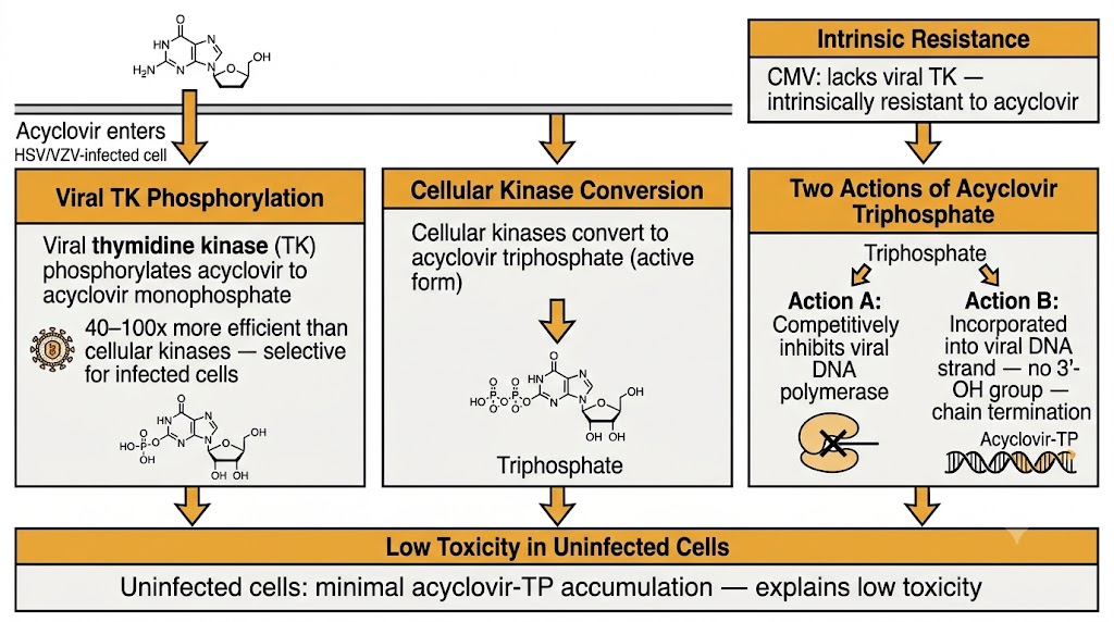
Acyclovir two-step activation mechanism flow diagram

The clinical spectrum of acyclovir encompasses HSV-1, HSV-2, and VZV. HSV-1 and HSV-2 are the most susceptible; VZV is approximately 10-fold less susceptible, requiring higher drug exposures — reflected in the substantially higher acyclovir doses used for varicella and zoster compared with HSV disease. EBV carries a viral TK of low activity and acyclovir has minimal clinically meaningful effect on EBV disease. CMV lacks a viral TK entirely and is intrinsically resistant to acyclovir. Oral bioavailability of acyclovir is poor (15–30%) and saturable at higher doses. Valacyclovir, the L-valyl ester prodrug of acyclovir, is rapidly hydrolyzed after oral administration to acyclovir, achieving plasma concentrations three to five times higher than equivalent oral acyclovir doses and approximating those achieved with intravenous acyclovir at standard doses. Acyclovir distributes widely, achieving therapeutic concentrations in cerebrospinal fluid (CSF) at approximately 50% of plasma levels — critical for treatment of herpes simplex encephalitis (HSE). Renal excretion is the primary elimination route; dose adjustment is required for creatinine clearance (CrCl) below 50 mL/min. Intravenous acyclovir can precipitate in renal tubules if infused rapidly or in hypovolemic patients — adequate hydration during intravenous infusion is essential.

Resistance to acyclovir in clinical practice is caused predominantly by mutations in the viral TK gene that reduce TK activity or alter substrate specificity so that acyclovir is no longer efficiently phosphorylated. TK-null and TK-partial mutants occur almost exclusively in immunocompromised patients receiving prolonged acyclovir therapy. Because TK is required for acyclovir activation but not for foscarnet or cidofovir activity, acyclovir-resistant HSV strains retain full susceptibility to both foscarnet and cidofovir — providing a clear therapeutic escape route. Less commonly, mutations in the viral DNA polymerase reduce acyclovir binding affinity and may confer partial cross-resistance to foscarnet.
Section 3
Ganciclovir, Valganciclovir, and Foscarnet
UL97-dependent activation, myelosuppression, pyrophosphate analogue inhibition, and nephrotoxicity

Cytomegalovirus disease in immunocompromised patients remains one of the most challenging management problems in transplant and HIV medicine. The agents available for CMV treatment and prophylaxis carry significant toxicity profiles that require careful monitoring throughout the course of therapy.

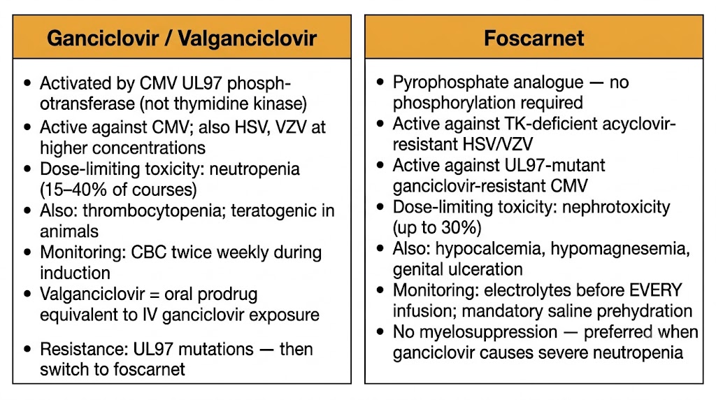
Ganciclovir is an acyclic nucleoside analogue structurally related to acyclovir. CMV encodes a viral phosphotransferase rather than a classical thymidine kinase, and UL97 phosphorylates ganciclovir to its monophosphate form. Cellular kinases complete the conversion to ganciclovir triphosphate, which inhibits CMV DNA polymerase and is incorporated into elongating viral DNA, where it slows and eventually terminates replication. Ganciclovir's selectivity for CMV-infected cells is considerably lower than acyclovir's selectivity for HSV-infected cells — this narrower selectivity underlies ganciclovir's more significant toxicity profile. The dose-limiting toxicity of ganciclovir is myelosuppression, specifically neutropenia, occurring in 15–40% of treatment courses. Granulocyte colony-stimulating factor (G-CSF) is used to support absolute neutrophil count (ANC) during essential ganciclovir therapy. Ganciclovir is teratogenic and potentially carcinogenic in animal studies; use in pregnancy is reserved for life-threatening CMV disease. Valganciclovir, the oral L-valyl ester prodrug, achieves systemic exposure equivalent to intravenous ganciclovir and has replaced oral ganciclovir for prophylaxis and maintenance therapy in transplant recipients and HIV patients with controlled CMV disease.

Foscarnet (phosphonoformic acid) is a pyrophosphate analogue that inhibits viral DNA polymerase by a mechanism entirely distinct from nucleoside analogues. Foscarnet directly inhibits viral DNA polymerase without requiring intracellular phosphorylation and is therefore active against TK-deficient acyclovir-resistant HSV and against UL97-mutant ganciclovir-resistant CMV, provided the polymerase itself remains susceptible. Foscarnet is active against all human herpesviruses including CMV, HSV-1, HSV-2, VZV, and HHV-6. Its major limitation is severe nephrotoxicity, occurring in up to 30% of patients. Vigorous saline prehydration before each infusion is mandatory and substantially reduces nephrotoxicity risk. Electrolyte disturbances are common and potentially severe: hypocalcemia (due to calcium chelation by foscarnet), hypomagnesemia, hypokalemia, and hypophosphatemia all require careful monitoring. Genital ulceration from high concentrations of foscarnet in urine is a distinctive adverse effect. Its lack of myelosuppression makes foscarnet the preferred CMV agent when ganciclovir-related neutropenia is severe.

Ganciclovir and Foscarnet Toxicity — Key Monitoring Points

Ganciclovir/valganciclovir: complete blood count (CBC) with differential twice weekly during induction, weekly during maintenance; serum creatinine twice weekly. Foscarnet: serum electrolytes (calcium, magnesium, potassium, phosphate) before every infusion; serum creatinine twice weekly; 500–1000 mL normal saline prehydration mandatory before each dose; monitor for genital ulceration.

Ganciclovir versus foscarnet mechanism toxicity and resistance coverage
Section 4
Cidofovir and Brincidofovir
Virus-independent phosphorylation, proximal tubular nephrotoxicity, and the lipid conjugate strategy

Cidofovir and brincidofovir occupy a distinct niche in herpesvirus pharmacology, offering broad-spectrum DNA virus coverage extending well beyond the herpesvirus family. Cidofovir's clinical utility is severely limited by nephrotoxicity; brincidofovir uses lipid conjugate chemistry to deliver cidofovir's active metabolite intracellularly while bypassing the renal tubular accumulation that drives toxicity.

Cidofovir is an acyclic nucleoside phosphonate. Its pharmacological uniqueness is that it does not require viral TK or CMV UL97 for initial phosphorylation — cellular enzymes convert cidofovir directly to cidofovir diphosphate, the active moiety, which inhibits viral DNA polymerase causing chain termination after incorporation. Because activation is entirely virus-independent, cidofovir retains full activity against TK-deficient acyclovir-resistant HSV strains and against UL97-mutant ganciclovir-resistant CMV strains, provided the viral DNA polymerase remains susceptible. The antiviral spectrum is broad: CMV, HSV-1, HSV-2, VZV, adenovirus, BK polyomavirus (BKPyV), and orthopoxviruses including smallpox and mpox.

The clinical utility of cidofovir is severely constrained by dose-dependent nephrotoxicity. Cidofovir is concentrated in proximal tubular cells, reaching concentrations that cause mitochondrial dysfunction, tubular apoptosis, and progressive renal failure. The mandatory co-administration of probenecid substantially reduces cidofovir uptake into proximal tubular cells: probenecid 2 g is given orally three hours before each cidofovir dose and 1 g at two and eight hours after infusion. Aggressive intravenous saline pre-loading before each infusion is also required. Despite these precautions, serum creatinine must be checked before every dose, and cidofovir must be withheld if creatinine rises by 0.3 mg/dL or more above baseline or if proteinuria reaches 2+ or greater on dipstick. Uveitis and ocular hypotony are distinctive adverse effects reported primarily in HIV patients receiving systemic cidofovir.

Brincidofovir (CMX001) is a lipid conjugate of cidofovir linked via an ether lipid to improve oral bioavailability and reduce renal tubular uptake. The lipid conjugate enters cells via lipid transport pathways, dramatically reducing proximal tubular exposure and the associated nephrotoxicity. Brincidofovir received Food and Drug Administration (FDA) approval in 2021 for treatment of smallpox (variola virus) and has been used in mpox outbreaks. The primary adverse effects are gastrointestinal: diarrhea, nausea, vomiting, and abdominal pain can be treatment-limiting. Hepatotoxicity requires monitoring. The absence of significant nephrotoxicity compared with cidofovir makes brincidofovir potentially attractive for immunocompromised patients with renal insufficiency.

Cidofovir Mandatory Administration Protocol

Every cidofovir infusion requires: (1) probenecid 2 g orally 3 hours before, then 1 g at 2 and 8 hours post-infusion; (2) 1 L normal saline IV over 1 hour pre-infusion; (3) serum creatinine and urine protein checked within 48 hours of each dose; (4) hold if creatinine rises ≥0.3 mg/dL above baseline or proteinuria ≥2+. Cidofovir is contraindicated if CrCl <55 mL/min or if pre-existing proteinuria ≥100 mg/dL.

Section 5
HSV and VZV in Special Populations
Immunocompromised patients, pregnancy safety, and neonatal HSV management

The pharmacological management of herpesvirus infections is substantially modified by clinical context. Immunocompromised patients, pregnant women, and neonates each present distinct pharmacokinetic, virological, and safety considerations that require departure from standard outpatient treatment paradigms.

In immunocompromised patients, HSV and VZV infections carry substantially greater morbidity than in immunocompetent hosts. Mucocutaneous HSV may progress to extensive necrotic ulceration rather than resolving spontaneously. VZV primary infection and reactivation can disseminate viscerally, causing pneumonitis, hepatitis, and encephalitis. The threshold for intravenous therapy is lower: HSV encephalitis, disseminated HSV, and disseminated zoster in immunocompromised patients are managed with intravenous acyclovir (10–12 mg/kg every 8 hours) rather than oral regimens. Chronic suppressive therapy with valacyclovir or acyclovir is standard practice in solid organ transplant (SOT) recipients, hematopoietic stem cell transplant (HSCT) recipients, and patients with advanced HIV during profound immunosuppression to prevent HSV and VZV reactivation. When acyclovir-resistant HSV or VZV is identified or strongly suspected based on lack of clinical response after 5–7 days of adequate intravenous acyclovir, foscarnet is the agent of choice.

Acyclovir and valacyclovir are classified as safe in pregnancy based on animal safety data and extensive human experience from the Acyclovir in Pregnancy Registry, which found no increase in birth defects among over 1,800 first-trimester exposures. Current guidelines support acyclovir and valacyclovir as safe and effective for primary genital HSV in pregnancy, VZV pneumonia (which carries 40% maternal mortality if untreated), and disseminated herpesvirus infections. Suppressive valacyclovir therapy from 36 weeks gestation reduces the risk of HSV shedding and recurrent lesions at delivery, lowering the rate of cesarean delivery performed for active HSV disease. Ganciclovir and foscarnet are generally avoided in pregnancy given teratogenic potential in animal models, reserving their use for maternal life-threatening CMV disease.

Neonatal HSV is a medical emergency carrying substantial mortality and neurological morbidity even with treatment. Neonates acquire HSV-1 or HSV-2 most commonly through contact with maternal genital secretions at delivery. Disease manifests as three overlapping syndromes: skin, eye, and mouth (SEM) disease; encephalitis; and disseminated disease. All three forms require immediate high-dose intravenous acyclovir at 20 mg/kg every 8 hours, with duration determined by syndrome: 14 days for SEM disease and 21 days for encephalitis or disseminated disease. Following completion of intravenous therapy, oral acyclovir suppression at 300 mg/m² three times daily for six months significantly reduces HSV recurrence and improves neurodevelopmental outcomes in infants with central nervous system (CNS) or disseminated disease — one of the most compelling evidence-based uses of long-term antiviral suppression in any population.

Section 6
CMV Disease in Transplant and HIV
Prophylaxis strategies, end-organ manifestations, resistance management, and letermovir

CMV disease in solid organ transplant (SOT) and hematopoietic stem cell transplant (HSCT) recipients, and in HIV patients with advanced immunodeficiency, requires individualized decisions about prophylaxis strategy, viral load monitoring thresholds, treatment intensity, and duration of secondary prophylaxis based on the patient's evolving immune status.

In solid organ transplantation, the risk of CMV disease is determined primarily by the donor and recipient CMV serostatus. The highest-risk combination is a CMV-seropositive donor organ transplanted into a CMV-seronegative recipient (D+/R-), carrying a 50–80% risk of CMV infection without prophylaxis. Two prophylaxis strategies have been validated: universal prophylaxis (valganciclovir or ganciclovir for a defined period post-transplant, typically 3–6 months) and pre-emptive therapy (monitoring CMV viral load by PCR and initiating therapy when viral load exceeds a predefined threshold before symptoms develop). Universal prophylaxis reduces CMV disease incidence more consistently but is associated with late-onset CMV disease occurring after prophylaxis discontinuation, particularly in D+/R- recipients. Letermovir, a CMV terminase inhibitor, is approved for prophylaxis in CMV-seropositive HSCT recipients from day 0 through day 100 post-transplant. Letermovir has no myelosuppressive activity — a major clinical advantage in HSCT recipients already at risk for prolonged neutropenia following conditioning. It is a moderate inhibitor of cytochrome P450 3A4 (CYP3A4) and increases tacrolimus and cyclosporine concentrations by 40–45% and 15% respectively, requiring dose reduction at initiation with frequent trough level monitoring.

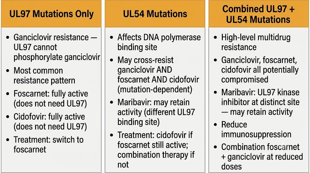
CMV antiviral resistance — UL97, UL54, and combined mutations treatment pivots

CMV end-organ disease encompasses pneumonitis, gastrointestinal disease (esophagitis, colitis), hepatitis, retinitis, and neurological disease. CMV colitis presents with profuse watery or bloody diarrhea in immunocompromised patients; diagnosis requires colonoscopy with biopsy because blood CMV PCR is frequently negative or low-level in isolated gastrointestinal disease — the virus replicates locally without generating detectable systemic viremia in many cases. CMV pneumonitis in HSCT recipients carries the highest mortality, with rates of 30–50% despite antiviral therapy; treatment combines intravenous ganciclovir with intravenous immunoglobulin (IVIG). CMV retinitis in HIV patients with cluster of differentiation 4 (CD4) counts below 50 cells/µL is treated with valganciclovir 900 mg twice daily for induction followed by 900 mg once daily for maintenance; intravitreal ganciclovir or foscarnet injections are used as adjunctive therapy for immediately sight-threatening lesions.

CMV antiviral resistance should be suspected when quantitative CMV viral load fails to decline by at least one log10 copies/mL after two weeks of adequate ganciclovir therapy, or when CMV disease progresses despite treatment. Resistance is confirmed by genotypic testing of the UL97 and UL54 genes. UL97 mutations confer ganciclovir resistance by impairing drug phosphorylation; these strains typically retain foscarnet and cidofovir susceptibility. UL54 mutations may produce cross-resistance between ganciclovir, foscarnet, and cidofovir depending on the specific mutation; combined UL97 and UL54 mutations produce high-level multidrug resistance. Management of UL97-only resistance: switch to foscarnet. UL54 mutations with foscarnet cross-resistance: cidofovir may retain activity. Combined resistance: combination foscarnet plus ganciclovir at reduced doses, reduction of immunosuppression, and consideration of maribavir, which targets the UL97 kinase with a distinct mechanism and retains activity against most UL97 resistance mutations.

Visual Summary  ·  Module 5 of 8
Herpesvirus Pharmacology
Mechanisms, resistance, toxicity, and clinical use across herpesvirus agents
Suggested References
Author(s) Title / Source Focus
Roizman B, Whitley RJ An inquiry into the molecular basis of HSV latency and reactivation. Annu Rev Microbiol. 2013 HSV latency mechanisms
Elion GB Acyclovir: discovery, mechanism of action, and selectivity. J Med Virol. 1993 Acyclovir mechanism — Nobel Prize lecture
Ljungman P et al. Definitions of cytomegalovirus infection and disease in transplant patients for use in clinical trials. Clin Infect Dis. 2017 CMV disease definitions in transplant
Stone KM et al. Pregnancy outcomes following systemic prenatal acyclovir exposure: conclusions from the international acyclovir pregnancy registry, 1984–1999. Birth Defects Res A. 2004 Acyclovir safety in pregnancy
Weller S et al. Pharmacokinetics of the acyclovir pro-drug valaciclovir after escalating single- and multiple-dose administration to normal volunteers. Clin Pharmacol Ther. 1993 Valacyclovir pharmacokinetics
Kimberlin DW et al. Oral acyclovir suppression and neurodevelopment after neonatal herpes. N Engl J Med. 2011 Neonatal HSV suppressive therapy
Kotton CN et al. The Third International Consensus Guidelines on the Management of Cytomegalovirus in Solid-organ Transplantation. Transplantation. 2018 CMV management in SOT
Chou S Approach to drug-resistant cytomegalovirus in transplant recipients. Curr Opin Infect Dis. 2015 CMV resistance testing and management
Lalezari JP et al. Randomized, controlled study of the safety and efficacy of intravenous cidofovir for the treatment of relapsing CMV retinitis in patients with AIDS. J Acquir Immune Defic Syndr. 1998 Cidofovir efficacy in CMV retinitis
Painter W et al. First pharmacokinetic and safety study in humans of the novel lipid antiviral conjugate CMX001, a broad-spectrum oral drug active against double-stranded DNA viruses. Antimicrob Agents Chemother. 2012 Brincidofovir pharmacokinetics and safety
Kenneson A, Cannon MJ Review and meta-analysis of the epidemiology of congenital cytomegalovirus (CMV) infection. Rev Med Virol. 2007 Congenital CMV epidemiology
Back to top