CHAPTER 6 · CHOLINERGIC DRUGS
Section 1

Acetylcholine Synthesis, Storage, and Release

From choline uptake to calcium-triggered exocytosis — each step is a distinct pharmacological target

The cholinergic synapse operates through a tightly regulated cycle: acetylcholine is synthesized, packaged into vesicles, released by calcium-triggered exocytosis, acts on receptors, and is then rapidly hydrolyzed. Understanding where each drug intervenes in this cycle is the foundation of cholinergic pharmacology.

Synthesis and Storage

The rate-limiting step is not synthesis itself but the availability of choline. Choline must be transported into the nerve terminal from the synaptic cleft by a high-affinity choline transporter. When neuronal firing increases, transporter activity rises to meet the higher demand.

Inside the terminal, choline acetyltransferase transfers an acetyl group from acetyl-coenzyme A to choline, producing acetylcholine. This enzyme is the defining marker of cholinergic neurons. In Alzheimer disease, choline acetyltransferase activity is reduced in the cortex and hippocampus, reflecting loss of cholinergic neurons from the basal forebrain — the mechanistic rationale for treating Alzheimer disease with acetylcholinesterase inhibitors.

Newly synthesized acetylcholine is packaged into synaptic vesicles. Storage protects it from cytoplasmic degradation and positions it for rapid calcium-triggered release.
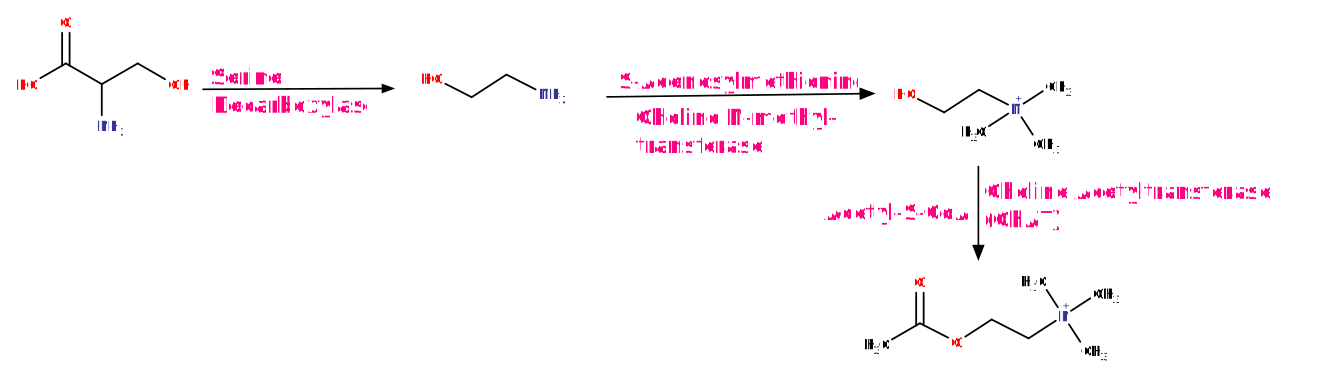
Diagram showing acetylcholine biosynthesis: choline and acetyl-coenzyme A converted to acetylcholine by choline acetyltransferase inside the nerve terminal.
Wikimedia Commons contributor. Acetylcholine biosynthesis. Source: Wikimedia Commons. License: CC BY-SA 4.0.

Release and Termination

When an action potential arrives at the nerve terminal, voltage-gated calcium channels open. The resulting calcium influx triggers fusion of vesicles with the presynaptic membrane and release of acetylcholine into the synaptic cleft.

Acetylcholinesterase in the synaptic cleft then rapidly hydrolyzes acetylcholine into choline and acetate. Choline is recycled back into the terminal for resynthesis.

Because termination depends entirely on enzymatic hydrolysis — not reuptake of intact transmitter — inhibiting acetylcholinesterase prolongs and amplifies every cholinergic signal simultaneously.

Diagram of the cholinergic synapse showing acetylcholine release from vesicles, action at muscarinic and nicotinic receptors, and hydrolysis by acetylcholinesterase in the synaptic cleft.
Wikimedia Commons contributor. Cholinergic synapse. Source: Wikimedia Commons. License: CC BY-SA 4.0.

Two Pharmacological Interventions in This Cycle

Presynaptic Block
Botulinum Toxin
  • Cleaves proteins required for vesicle fusion with the presynaptic membrane
  • Acetylcholine cannot be released
  • Result: flaccid paralysis at the neuromuscular junction (mechanism of botulism)
  • Therapeutic uses: spasticity, dystonia, hyperhidrosis, overactive bladder
Synaptic Cleft
Acetylcholinesterase Inhibitors
  • Block hydrolysis of acetylcholine in the synaptic cleft
  • Prolongs and amplifies all cholinergic signals at all sites
  • Therapeutic: Alzheimer disease, myasthenia gravis, reversal of neuromuscular blockade
  • Toxic: organophosphate poisoning

Section 2

Acetylcholinesterase and Signal Termination

Two cholinesterase enzymes with distinct locations, substrates, and clinical significance

Unlike most neurotransmitters, which are terminated by reuptake of the intact molecule, acetylcholine is terminated entirely by enzymatic hydrolysis. This makes acetylcholinesterase the master regulator of cholinergic signal duration — and makes acetylcholinesterase inhibition one of the most consequential pharmacological interventions in this system.

Acetylcholinesterase — Synaptic Enzyme

Acetylcholinesterase is present at every cholinergic synapse: the neuromuscular junction, autonomic ganglia, parasympathetic neuroeffector junctions, and throughout the central nervous system. Inhibiting it amplifies and prolongs signaling simultaneously at all of these sites — the basis for both its therapeutic applications and its toxicological consequences.

Therapeutic Use
Alzheimer Disease
  • Donepezil, rivastigmine, galantamine
  • Slow acetylcholine breakdown in cortex and hippocampus
  • Partially compensate for loss of basal forebrain cholinergic neurons
Therapeutic Use
Myasthenia Gravis
  • Neostigmine, pyridostigmine
  • Prolong acetylcholine action at the neuromuscular junction
  • Compensate for autoimmune loss of nicotinic receptors at the endplate
Therapeutic Use
Reversal of Blockade
  • Neostigmine given after surgery
  • Accumulated acetylcholine competes with non-depolarizing blocking agent
  • Does not reverse succinylcholine (depolarizing) blockade

Butyrylcholinesterase — Plasma Enzyme

A second cholinesterase, butyrylcholinesterase (also called plasma cholinesterase or pseudocholinesterase), circulates in the plasma and is synthesized by the liver. It has no established role in synaptic neurotransmission. Its primary clinical significance is that it metabolizes succinylcholine, the depolarizing neuromuscular blocking agent used for rapid sequence intubation.

A common genetic variant reduces butyrylcholinesterase activity. Patients with this variant cannot break down succinylcholine at a normal rate, resulting in prolonged neuromuscular blockade lasting hours rather than the expected few minutes — a pharmacogenomic consideration relevant to anesthesia practice.


Section 3

Muscarinic Receptor Subtypes

Five subtypes, two signaling families, and the tissue distribution that determines every drug effect

Muscarinic receptors are G-protein-coupled receptors found at parasympathetic neuroeffector junctions, autonomic ganglia, and throughout the central nervous system. Five subtypes — M1 through M5 — each have a distinct tissue distribution. The clinical effects of every muscarinic drug follow directly from which subtypes it engages and where those subtypes are expressed.

Two Signaling Families

All five subtypes fall into one of two groups. M1, M3, and M5 couple to Gq proteins, activating pathways that raise intracellular calcium and produce excitatory responses. M2 and M4 couple to Gi proteins, which reduce cellular excitability. The odd-even pattern — odd subtypes are Gq, even subtypes are Gi — is a reliable organizing principle for the whole receptor family.

Subtype Profiles and Clinical Relevance

M1 — Gq
Cortex & Ganglia
  • Cerebral cortex, hippocampus, autonomic ganglia
  • Cognitive function; ganglionic modulation
  • M1 blockade causes cognitive impairment and delirium, highest risk in elderly patients
  • Target of benztropine for Parkinson disease tremor
M2 — Gi
Heart
  • Sinoatrial node, atrioventricular node, atria
  • Vagal slowing of heart rate; slows atrioventricular conduction
  • Atropine blocks M2 and removes vagal tone, producing tachycardia
  • Clinical use: atropine for symptomatic bradycardia
M3 — Gq
Smooth Muscle & Glands
  • Gastrointestinal tract: increased motility and secretion
  • Bladder detrusor: contraction — target for overactive bladder drugs
  • Airway smooth muscle: bronchoconstriction
  • Eye: miosis (sphincter pupillae) and accommodation (ciliary muscle)
  • Exocrine glands: salivation, lacrimation, sweating
M4 — Gi
Striatum
  • Striatum (central nervous system)
  • Modulates dopaminergic circuits in movement control
  • Target of muscarinic antagonists for Parkinson disease tremor

How Selectivity Shapes Drug Effect and Adverse Effects

Non-selective muscarinic antagonists such as atropine and scopolamine block all five subtypes, producing the full anticholinergic syndrome: tachycardia, mydriasis, dry mouth, urinary retention, constipation, and — with central nervous system penetration — confusion and delirium.

Selective M3 antagonists limit adverse effects by targeting specific tissues. Darifenacin and solifenacin target M3 in the bladder for overactive bladder, with reduced cardiac and central nervous system effects. Ipratropium and tiotropium are inhaled M3 antagonists producing bronchodilation in chronic obstructive pulmonary disease and asthma with minimal systemic absorption. Benztropine and trihexyphenidyl target M1 and M4 in the striatum for Parkinson disease tremor.


Section 4

Nicotinic Receptor Subtypes

Ligand-gated ion channels at three locations — neuromuscular junction, autonomic ganglia, and central nervous system

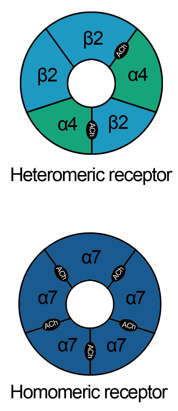
Nicotinic acetylcholine receptors are ligand-gated ion channels, not G-protein-coupled receptors. Acetylcholine binding opens the channel within milliseconds, producing rapid membrane depolarization. This fast, direct mechanism distinguishes nicotinic from muscarinic transmission and makes nicotinic receptors the target for neuromuscular blocking agents, ganglionic blockers, and smoking cessation pharmacotherapy.

Diagram showing homomeric and heteromeric pentameric nicotinic acetylcholine receptor structures with subunit arrangements and central ion channel pore.
Hopur52009. Nicotinic acetylcholine receptor subtypes — homomeric and heteromeric pentameric arrangements. Source: Wikimedia Commons. License: CC BY-SA 3.0.

Muscle-Type Receptors (N-M) at the Neuromuscular Junction

N-M receptors at the neuromuscular junction are the target for neuromuscular blocking agents used in anesthesia. Two fundamentally different mechanisms of block are clinically exploited.

Non-depolarizing block (tubocurarine, rocuronium, vecuronium, pancuronium) is competitive antagonism: acetylcholine cannot open the channel and muscle contraction fails. This block is reversed by acetylcholinesterase inhibitors, which allow acetylcholine to accumulate and compete with the blocking agent.

Depolarizing block (succinylcholine) works differently: the drug activates N-M receptors and holds them in a depolarized state. Initial fasciculations are followed by sustained flaccid paralysis. This block is not reversed by acetylcholinesterase inhibitors.

In myasthenia gravis, autoimmune antibodies destroy N-M receptors, reducing the safety margin for neuromuscular transmission and producing fatigable weakness that worsens with use. Acetylcholinesterase inhibitors are the primary pharmacological treatment.

Neuronal-Type Receptors (N-N) at Autonomic Ganglia

N-N receptors mediate fast transmission through both sympathetic and parasympathetic ganglia. Ganglionic blocking agents (hexamethonium, trimethaphan) interrupt all autonomic transmission simultaneously, producing a predictable syndrome reflecting loss of both divisions:

The inability to selectively block one autonomic division explains why ganglionic blockers were abandoned as antihypertensive agents once more selective drugs became available.

Central Nervous System Nicotinic Receptors and Smoking Cessation

The alpha4-beta2 nicotinic receptor is the primary site of nicotine action in the brain. Nicotine binding to these receptors on dopaminergic neurons in reward pathways enhances dopamine release, producing the reinforcing effects of tobacco use.

Varenicline — Partial Agonism for Smoking Cessation

Varenicline acts as a partial agonist at alpha4-beta2 nicotinic receptors. It provides enough receptor activation to reduce craving and withdrawal symptoms, while competitively blocking the full agonist response to inhaled nicotine.

The result: withdrawal is attenuated and smoking becomes less rewarding simultaneously. This dual action — partial stimulation plus competitive blockade — makes varenicline more effective than nicotine replacement alone.


Section 5

Cholinergic Tone and Autonomic Balance

Dominant parasympathetic targets, two mirror-image clinical syndromes, and three diseases defined by cholinergic imbalance

Knowing which organs are under dominant parasympathetic tone at rest — and what happens when that tone is pharmacologically amplified or removed — predicts the full clinical profile of every drug in this chapter. Cholinergic excess and cholinergic deficit produce recognizable mirror-image syndromes.

Organs Under Dominant Parasympathetic Tone at Rest

Cholinergic Excess — The SLUDGE Toxidrome

Overstimulation from acetylcholinesterase inhibitor toxicity or organophosphate poisoning produces simultaneous overstimulation of muscarinic and nicotinic receptors at all sites in the body.

SLUDGE — Muscarinic Overstimulation

Salivation  —  Lacrimation  —  Urination  —  Defecation  —  Gastrointestinal distress  —  Emesis

Also: bradycardia, bronchospasm, and bronchorrhea (excessive bronchial secretions). At the neuromuscular junction: fasciculations then flaccid paralysis. In the central nervous system: anxiety, seizures, loss of consciousness.

The combination of bronchospasm, bronchorrhea, and diaphragm paralysis is the mechanism of death in severe organophosphate poisoning.

Treatment: atropine (reverses muscarinic effects); pralidoxime (reactivates acetylcholinesterase if given early, before permanent inactivation).

Cholinergic Deficit — The Anticholinergic Syndrome

Anticholinergic Syndrome — Classic Mnemonic

Hot as a hare — hyperthermia from anhidrosis (eccrine sweat glands are cholinergically innervated)

Dry as a bone — dry mouth and dry skin from loss of salivary and sweat gland secretion

Red as a beet — cutaneous flushing from vasodilation

Blind as a bat — mydriasis and cycloplegia from M3 blockade in the eye

Mad as a hatter — confusion, agitation, delirium from central nervous system M1 blockade

Also: tachycardia (M2 blockade), urinary retention, and constipation. Elderly patients and those with existing cognitive impairment are at substantially greater risk of delirium at doses that produce only peripheral effects in younger adults.

Cholinergic Imbalance in Three Disease States

Disease
Alzheimer Disease
  • Selective loss of cholinergic neurons from basal forebrain
  • Deficit in cortex and hippocampus correlates with cognitive decline
  • Treatment: acetylcholinesterase inhibitors
Disease
Parkinson Disease
  • Loss of dopaminergic neurons creates relative cholinergic excess in the striatum
  • Contributes to tremor and rigidity
  • Treatment: muscarinic antagonists (benztropine, trihexyphenidyl) restore balance
Disease
Myasthenia Gravis
  • Autoimmune destruction of N-M nicotinic receptors at the neuromuscular junction
  • Reduced safety margin; fatigable weakness worsens with use
  • Treatment: acetylcholinesterase inhibitors prolong acetylcholine action

Suggested References
Author / Organization Title Source
Ferguson SM, Blakely RDThe choline transporter resurfaces: new roles for synaptic vesicles?Mol Interv. 2004;4(1):22-37
Mesulam MMThe cholinergic lesion of Alzheimer disease: pivotal factor or side show?Learn Mem. 2004;11(1):43-49
Montecucco C, Schiavo GStructure and function of tetanus and botulinum neurotoxinsQ Rev Biophys. 1995;28(4):423-472
Wess J, Eglen RM, Gautam DMuscarinic acetylcholine receptors: mutant mice provide new insights for drug developmentNat Rev Drug Discov. 2007;6(9):721-733
Dvir H, Silman I, Harel M, Rosenberry TL, Sussman JLAcetylcholinesterase: from 3D structure to functionChem Biol Interact. 2010;187(1-3):10-22
Lockridge OReview of human butyrylcholinesterase structure, function, genetic variants, history of use in the clinic, and potential therapeutic usesPharmacol Ther. 2015;148:34-46
Eglen RMMuscarinic receptor subtypes in neuronal and non-neuronal cholinergic functionAuton Autacoid Pharmacol. 2006;26(3):219-233
Taly A, Corringer PJ, Guedin D, Lestage P, Changeux JPNicotinic receptors: allosteric transitions and therapeutic targets in the nervous systemNat Rev Drug Discov. 2009;8(9):733-750
Sine SMEnd-plate acetylcholine receptor: structure, mechanism, pharmacology, and diseasePhysiol Rev. 2012;92(3):1189-1234
Eddleston M, Buckley NA, Eyer P, Dawson AHManagement of acute organophosphorus pesticide poisoningLancet. 2008;371(9612):597-607