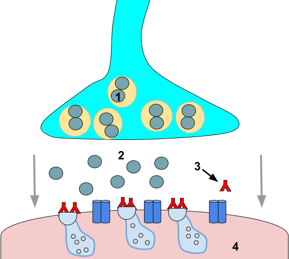
Direct-acting muscarinic agonists bind and activate muscarinic receptors without depending on acetylcholine release or acetylcholinesterase inhibition. As a class, they are structurally related to acetylcholine but modified to resist enzymatic hydrolysis, giving them longer durations of action. Their clinical applications are narrow and defined by the organ-level effects of muscarinic receptor stimulation.
The most clinically useful distinction within this class is the physical chemistry of the drug molecule. Quaternary ammonium compounds carry a permanent positive charge and cannot cross lipid membranes, including the blood-brain barrier. They produce no central nervous system effects at therapeutic doses. Tertiary amines are uncharged at physiologic pH, cross membranes readily, and have the potential for central nervous system activity. This single structural difference determines the adverse effect profile of every drug in this class.
Bethanechol is a quaternary compound with selective muscarinic activity and no meaningful nicotinic effects. It does not cross the blood-brain barrier. Its primary indication is non-obstructive urinary retention, including postoperative and postpartum retention and neurogenic bladder with reduced detrusor contractility. It is given orally or by subcutaneous injection; intravenous use is avoided because it can provoke severe bradycardia and bronchospasm.
Bethanechol is contraindicated in mechanical urinary or bowel obstruction (muscarinic stimulation worsens the obstruction), peptic ulcer disease (increased gastric acid secretion), and asthma or chronic obstructive pulmonary disease (bronchoconstriction through muscarinic subtype 3 receptors in airway smooth muscle).
Pilocarpine is a tertiary amine and the only direct muscarinic agonist in clinical use with meaningful central nervous system penetration. Its actions are mediated primarily through muscarinic subtype 3 receptors: miosis (contraction of the sphincter pupillae), ciliary muscle contraction (shifting the eye toward near-vision accommodation), and stimulation of exocrine gland secretion.
Pilocarpine has three established clinical uses. As topical eye drops, it lowers intraocular pressure in open-angle glaucoma by improving drainage through trabecular meshwork outflow. In acute angle-closure glaucoma, it produces rapid miosis to relieve the pupillary block causing the pressure crisis. Given orally, it treats xerostomia (dry mouth) caused by Sjogren syndrome or radiation therapy to the head and neck, stimulating residual salivary gland secretion. The most common adverse effect is diaphoresis (excessive sweating) from eccrine sweat gland stimulation.
Carbachol is a quaternary compound that activates both muscarinic and nicotinic receptors, distinguishing it from the more selective bethanechol. Its combined receptor activity limits systemic use but makes it useful for intraocular applications. It is instilled directly into the anterior chamber during cataract surgery to produce miosis at the time of lens implantation, and it is used as a second-line topical agent for open-angle glaucoma when pilocarpine is inadequate or not tolerated.
Methacholine is a selective muscarinic agonist with no established therapeutic use. Its only clinical role is as a diagnostic tool: the methacholine bronchoprovocation test. When inhaled in incremental concentrations, methacholine stimulates muscarinic subtype 3 receptors in airway smooth muscle and produces bronchoconstriction in proportion to baseline airway hyperresponsiveness. Patients with asthma show an exaggerated bronchoconstrictor response, while patients with normal airways tolerate much higher concentrations. A negative methacholine challenge effectively excludes active asthma. The test is useful in patients with respiratory symptoms but normal spirometry at rest.
Cevimeline is a tertiary amine with selective activity at muscarinic subtype 1 and muscarinic subtype 3 receptors, developed specifically for xerostomia in Sjogren syndrome. It has a longer duration of action than pilocarpine and a somewhat lower rate of diaphoresis, making it an alternative for patients who cannot tolerate pilocarpine's sweating side effect. Like pilocarpine, it is contraindicated in uncontrolled asthma and narrow-angle glaucoma.
Bethanechol: quaternary; no central nervous system effects; urinary retention and gastrointestinal hypomotility; contraindicated in obstruction, asthma, peptic ulcer disease.
Pilocarpine: tertiary; crosses blood-brain barrier; open-angle and acute angle-closure glaucoma; xerostomia in Sjogren syndrome and radiation injury; adverse effect: diaphoresis.
Carbachol: quaternary; muscarinic and nicotinic activity; intraocular miosis during cataract surgery; second-line glaucoma.
Methacholine: diagnostic only; bronchoprovocation challenge to confirm airway hyperresponsiveness in suspected asthma.
Cevimeline: tertiary; muscarinic subtype 1 and subtype 3 selective; Sjogren xerostomia; longer duration than pilocarpine; less diaphoresis.
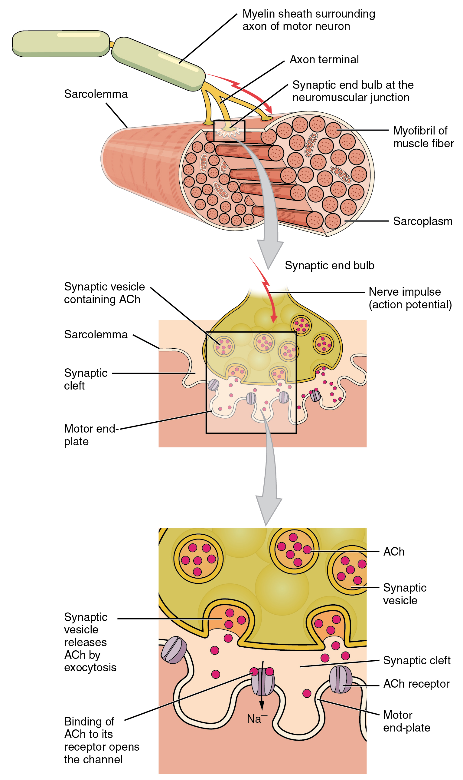
Acetylcholinesterase inhibitors do not activate cholinergic receptors directly. Instead, they block the enzyme that normally hydrolyzes acetylcholine, allowing acetylcholine to accumulate and produce a more sustained, more intense effect at every synapse where it is released. The peripheral agents in this class share a structural feature — they are quaternary ammonium compounds — that prevents them from crossing the blood-brain barrier, confining their effects to the neuromuscular junction, autonomic ganglia, and postganglionic parasympathetic neuroeffector junctions.
Acetylcholinesterase inhibitors are divided by how they interact with the enzyme. Neostigmine and pyridostigmine inhibit the enzyme by carbamylation: the drug transfers a carbamyl group to the active site of acetylcholinesterase, blocking the enzyme for minutes to hours before it can regenerate. This is called reversible inhibition, because the enzyme eventually recovers, but recovery is slow enough to produce a sustained clinical effect. Organophosphates, covered in the next module, inhibit the enzyme by phosphorylation, which is permanent if not treated promptly.
Edrophonium works by a different mechanism: it binds to the active site of acetylcholinesterase through weak, non-covalent electrostatic interactions, without forming a covalent bond at all. The result is an extremely short duration of action — effects begin within seconds and resolve within minutes — which limits its use to diagnostic applications rather than sustained therapy.
Neostigmine is the prototypical peripheral acetylcholinesterase inhibitor. By prolonging acetylcholine action at the neuromuscular junction, it increases the probability that acetylcholine will activate the reduced number of nicotinic receptors remaining in myasthenia gravis, partially compensating for the autoimmune receptor loss. The same mechanism reverses non-depolarizing neuromuscular blockade after surgery: accumulated acetylcholine competes with the blocking agent and gradually restores neuromuscular transmission. Neostigmine does not reverse succinylcholine (depolarizing) blockade and can paradoxically worsen it.
At autonomic postganglionic junctions, neostigmine's accumulation of acetylcholine activates muscarinic receptors, producing bradycardia, increased salivation, bronchospasm, and increased gastrointestinal motility. When neostigmine is used for neuromuscular blockade reversal, it is co-administered with glycopyrrolate to block these muscarinic adverse effects. Glycopyrrolate is preferred over atropine for this purpose because it is also a quaternary compound and therefore has a similar speed of onset to neostigmine and does not produce the tachycardia that can temporarily precede atropine's peak effect. Oral neostigmine is very poorly absorbed from the gastrointestinal tract, so oral doses are substantially higher than parenteral doses to achieve comparable effect.
Beyond myasthenia gravis and neuromuscular blockade reversal, neostigmine is used to treat acute colonic pseudo-obstruction (Ogilvie syndrome), a condition of non-mechanical colonic dilation in severely ill patients where intravenous neostigmine produces rapid decompression by restoring colonic motility.
When neostigmine reverses neuromuscular blockade, acetylcholine accumulates at muscarinic receptors throughout the body — not only at the neuromuscular junction. This produces bradycardia, bronchospasm, and increased secretions.
Glycopyrrolate is given simultaneously to block these muscarinic effects. It is chosen over atropine because it is quaternary (same onset speed as neostigmine, no blood-brain barrier crossing) and does not cause the transient bradycardia that can briefly precede atropine's peak muscarinic blocking effect.
Pyridostigmine is structurally similar to neostigmine and works by the same carbamylation mechanism, but has a longer duration of action. This makes it the preferred oral agent for the long-term management of myasthenia gravis. Patients take it regularly throughout the day to maintain adequate neuromuscular junction acetylcholine levels. A sustained-release formulation is available for overnight dosing, addressing the characteristic weakness patients experience on waking before their first dose takes effect. Muscarinic adverse effects — gastrointestinal cramping, diarrhea, increased secretions — are common dose-limiting problems and are managed by adjusting the dose rather than adding an anticholinergic drug, since blocking muscarinic receptors would not address the patient's underlying therapeutic need at the neuromuscular junction.
Edrophonium's ultrashort duration of action, which prevents its use for ongoing therapy, is precisely what makes it useful diagnostically. When given intravenously, it produces a brief increase in acetylcholine at the neuromuscular junction within seconds, and its effects resolve completely within minutes. This narrow window can be exploited to test whether a patient's weakness is caused by insufficient neuromuscular junction transmission: a transient, measurable improvement in strength after edrophonium suggests the diagnosis of myasthenia gravis. The clinical application of this test is discussed in the next section.
Myasthenia gravis is an autoimmune disease in which antibodies destroy nicotinic receptors at the neuromuscular junction, reducing the safety margin for neuromuscular transmission. The result is fatigable weakness — muscle strength that diminishes with repeated use and recovers with rest. Two pharmacological concepts are central to managing this disease: the use of acetylcholinesterase inhibitors to amplify residual acetylcholine signaling, and the recognition that too much of the same treatment produces a clinical picture that mimics the disease itself.
The edrophonium test exploits the drug's ultrashort duration of action to briefly enhance neuromuscular junction transmission and observe whether a patient's weakness improves. A small dose is given intravenously, and the examiner watches for objectively measurable improvement in a specific sign of weakness: lifting of ptosis (drooping eyelid), improved extraocular muscle movement, increased grip strength, or improved swallowing. A positive result — clear transient improvement that resolves as the drug is eliminated — supports the diagnosis of myasthenia gravis. The test must be performed with atropine and resuscitation capability on hand, because edrophonium's muscarinic effects can produce significant bradycardia, particularly in older patients.
In current clinical practice, the edrophonium test has largely been replaced by serology. Antibodies against the acetylcholine receptor are detected in the large majority of patients with generalized myasthenia gravis and are diagnostic when present. Serological testing is safer, does not require intravenous access and cardiac monitoring, and can be performed in any outpatient setting. The edrophonium test retains a role when rapid bedside confirmation is needed and the clinical situation does not permit waiting for laboratory results.
A hospitalized patient with myasthenia gravis who deteriorates with worsening bulbar and respiratory weakness presents one of the most clinically challenging scenarios in pharmacology: distinguishing between insufficient drug and too much drug. Both myasthenic crisis and cholinergic crisis produce worsening weakness that may require mechanical ventilation. The distinction lies in the accompanying features.
Myasthenic crisis reflects disease progression or under-treatment — there is not enough acetylcholine at the neuromuscular junction to sustain adequate muscle activation. The weakness is not accompanied by signs of muscarinic overstimulation. Cholinergic crisis results from excessive acetylcholinesterase inhibitor dosing: so much acetylcholine accumulates at the neuromuscular junction that the nicotinic receptors become persistently depolarized and unable to respond, producing weakness by a depolarizing block mechanism. At the same time, the same excess acetylcholine overstimulates muscarinic receptors throughout the body, producing the signature features that distinguish cholinergic crisis: copious secretions (bronchorrhea and hypersalivation), bradycardia, miosis, diaphoresis, and gastrointestinal cramping.
Myasthenic crisis: worsening weakness; secretions normal or minimal; heart rate normal or elevated; pupils normal; no gastrointestinal symptoms. Caused by disease progression, infection, surgery, or medications that impair neuromuscular junction transmission. Treatment: respiratory support, plasma exchange or intravenous immunoglobulin, resume acetylcholinesterase inhibitors when stable.
Cholinergic crisis: worsening weakness; copious secretions (bronchorrhea, hypersalivation); bradycardia; miosis; diaphoresis; gastrointestinal cramping and diarrhea. Caused by excessive acetylcholinesterase inhibitor dosing. Treatment: hold all acetylcholinesterase inhibitors; atropine for muscarinic symptoms; respiratory support as needed.
The muscarinic features of cholinergic crisis are the key to the diagnosis. A deteriorating myasthenia gravis patient who is also bradycardic, miotic, and secreting copiously has taken too much pyridostigmine, not too little.
The central nervous system-penetrating acetylcholinesterase inhibitors are tertiary amines that cross the blood-brain barrier and increase acetylcholine levels in the brain. This property is used in two different clinical contexts: physostigmine reverses the central nervous system effects of anticholinergic overdose, while the Alzheimer agents partially compensate for the cholinergic neuron loss that characterizes that disease. All produce peripheral muscarinic adverse effects as a class-wide consequence of their mechanism.
Physostigmine is a naturally occurring carbamate that inhibits acetylcholinesterase by the same carbamylation mechanism as neostigmine, but as a tertiary amine it crosses the blood-brain barrier. This makes it the only acetylcholinesterase inhibitor that reliably reverses the central nervous system manifestations of anticholinergic toxidrome: the confusion, agitation, delirium, and hallucinations produced by overdose of antihistamines, antipsychotics, atropine, scopolamine, and other muscarinic antagonists that penetrate the central nervous system. By raising acetylcholine levels centrally, physostigmine overcomes the muscarinic receptor blockade and clears the delirium. Its duration of action is short, so repeated dosing may be needed.
Physostigmine is contraindicated in anticholinergic toxidrome caused by tricyclic antidepressant overdose. Tricyclic antidepressants block cardiac sodium channels in addition to muscarinic receptors, and physostigmine can provoke seizures and fatal cardiac arrhythmias in this setting. Before administering physostigmine, the electrocardiogram must be reviewed to exclude the prolonged QRS complex that signals tricyclic antidepressant toxicity.
Alzheimer disease is characterized by the selective loss of cholinergic neurons projecting from the basal forebrain to the cerebral cortex and hippocampus. The cholinergic deficit in these projection areas correlates with the severity of cognitive impairment. Acetylcholinesterase inhibitors partially compensate for this loss by slowing the breakdown of acetylcholine released by the surviving neurons, increasing the effective concentration at remaining receptors. This approach produces modest symptomatic improvement in cognition and daily function but does not alter the underlying neurodegenerative process. No currently approved acetylcholinesterase inhibitor slows disease progression.
All three agents produce modest, symptomatic improvement in cognition and daily function. None modifies the underlying disease course. Gastrointestinal adverse effects — nausea, vomiting, diarrhea — are a class-wide consequence of muscarinic stimulation in the gut and are the primary reason patients discontinue treatment. Cholinergic toxicity (bradycardia, excessive secretions, bronchospasm) is possible at high doses or when drug interactions raise plasma levels.
Clinically important drug interactions with muscarinic agonists and acetylcholinesterase inhibitors fall into three categories: enzyme-mediated pharmacokinetic interactions that alter plasma drug levels, additive pharmacodynamic interactions that amplify cholinergic effects at cardiac and other targets, and pharmacodynamic antagonism when anticholinergic drugs are co-prescribed with acetylcholinesterase inhibitors for Alzheimer disease.
Donepezil and galantamine are both metabolized by cytochrome P450 enzymes, specifically the 2D6 and 3A4 isoforms. Potent inhibitors of these enzymes — including fluoxetine and paroxetine for cytochrome P450 2D6, and azole antifungals and clarithromycin for cytochrome P450 3A4 — can substantially raise plasma levels of these drugs when co-administered, increasing the risk of cholinergic adverse effects: nausea, vomiting, diarrhea, and bradycardia. Conversely, cytochrome P450 inducers such as rifampin and carbamazepine accelerate the metabolism of donepezil and galantamine, potentially reducing their therapeutic effect.
Rivastigmine does not undergo cytochrome P450 metabolism and is instead broken down by cholinesterase enzymes directly. This makes rivastigmine the preferred choice for Alzheimer patients who are already taking multiple medications that interact with cytochrome P450 enzymes, since it avoids these pharmacokinetic interactions entirely.
Cardiac glycosides (digoxin) slow the sinoatrial node through a mechanism that includes enhanced vagal tone at muscarinic subtype 2 receptors in the heart. Acetylcholinesterase inhibitors also slow the heart by raising acetylcholine levels at the same muscarinic subtype 2 receptors. When digoxin and an acetylcholinesterase inhibitor are combined, the bradycardic effects are additive, and heart rate monitoring is warranted, particularly when digoxin concentrations are at the high end of the therapeutic range.
Beta-adrenergic blocking agents (beta-blockers) independently slow heart rate by reducing sympathetic input to the sinoatrial node. In combination with an acetylcholinesterase inhibitor, they add another bradycardic influence on top of the cholinergically mediated slowing. The combination requires awareness and heart rate monitoring, particularly in elderly patients with Alzheimer disease who already have a reduced cardiac reserve.
Acetylcholinesterase inhibitors for Alzheimer disease work by raising acetylcholine at central muscarinic receptors. Any drug that blocks muscarinic receptors simultaneously directly opposes this mechanism and negates the therapeutic effect. This pharmacodynamic antagonism is common in clinical practice because many widely used drug classes carry muscarinic blocking activity: tricyclic antidepressants, first-generation antihistamines (diphenhydramine), bladder antimuscarinics (oxybutynin, tolterodine), certain antipsychotics, and antiparkinsonian anticholinergics all have significant muscarinic antagonist properties.
When an acetylcholinesterase inhibitor is initiated for Alzheimer disease, the medication list should be reviewed for drugs with anticholinergic activity. Continuing these agents at the same time as an acetylcholinesterase inhibitor is pharmacologically contradictory and exposes the patient to the adverse effects of both drug classes without obtaining the benefits of either.
Cytochrome P450 pharmacokinetics: Donepezil and galantamine levels rise with cytochrome P450 2D6/3A4 inhibitors (fluoxetine, azole antifungals, clarithromycin) — increased cholinergic toxicity risk. Rivastigmine avoids this category entirely.
Additive bradycardia: Digoxin plus acetylcholinesterase inhibitor — additive muscarinic subtype 2 slowing of sinoatrial node; monitor heart rate. Beta-blockers plus acetylcholinesterase inhibitor — additive bradycardia.
Pharmacodynamic antagonism: Anticholinergic drugs (diphenhydramine, oxybutynin, tricyclic antidepressants) directly oppose acetylcholinesterase inhibitors in Alzheimer disease and should be identified and discontinued when acetylcholinesterase inhibitor therapy begins.
| Author / Organization | Title | Source |
|---|---|---|
| Abrams P, Cardozo L, Fall M, et al. | The standardisation of terminology of lower urinary tract function: report from the Standardisation Sub-committee of the International Continence Society | Neurourol Urodyn. 2002;21(2):167-178 |
| Vivino FB, Al-Hashimi I, Khan Z, et al. | Pilocarpine tablets for the treatment of dry mouth and dry eye symptoms in patients with Sjogren syndrome: a randomized, placebo-controlled, fixed-dose, multicenter trial | Arch Intern Med. 1999;159(2):174-181 |
| Crapo RO, Casaburi R, Coates AL, et al. | Guidelines for methacholine and exercise challenge testing — 1999 | Am J Respir Crit Care Med. 2000;161(1):309-329 |
| Gilhus NE | Myasthenia gravis | N Engl J Med. 2016;375(26):2570-2581 |
| Engel AG | Current status of the congenital myasthenic syndromes | Neuromuscul Disord. 2012;22(2):99-111 |
| Benatar M | A systematic review of diagnostic studies in myasthenia gravis | Neuromuscul Disord. 2006;16(7):459-467 |
| Sanders DB, Wolfe GI, Benatar M, et al. | International consensus guidance for management of myasthenia gravis: executive summary | Neurology. 2016;87(4):419-425 |
| Watkins JW, Schwarz ES, Arroyo-Plasencia AM, Mullins ME | The use of physostigmine by toxicologists in anticholinergic toxicity | J Med Toxicol. 2015;11(2):179-184 |
| Birks JS | Cholinesterase inhibitors for Alzheimer's disease | Cochrane Database Syst Rev. 2006;(1):CD005593 |
| Winblad B, Grossberg G, Frolich L, et al. | IDEAL: a 6-month, double-blind, placebo-controlled study of the first skin patch for Alzheimer disease | Neurology. 2007;69(4 Suppl 1):S14-S22 |
| Noetzli M, Eap CB | Pharmacodynamic, pharmacokinetic and pharmacogenetic aspects of drugs used in the treatment of Alzheimer's disease | Clin Pharmacokinet. 2013;52(4):225-241 |
| Salahudeen MS, Nishtala PS | An overview of pharmacodynamic modelling, ligand-binding approach and its application in clinical practice | Saudi Pharm J. 2017;25(2):165-175 |