CHAPTER 6 · CHOLINERGIC DRUGS
Section 1

Irreversible Acetylcholinesterase Inhibition — Phosphorylation and the Aging Phenomenon

How organophosphates form a covalent bond with the catalytic serine of acetylcholinesterase, why this differs from reversible inhibition, and how the time-dependent aging reaction determines whether pralidoxime can work

Organophosphate compounds are a class of toxic chemicals that includes both agricultural pesticides and chemical warfare nerve agents. All members of this class share a common mechanism: they attack acetylcholinesterase, the enzyme that breaks down acetylcholine at cholinergic synapses, and form a covalent bond with the enzyme that is so stable it is considered functionally irreversible. Understanding why this bond forms, and why it becomes even more irreversible over time through a process called aging, is the foundation for understanding both the clinical severity of organophosphate poisoning and the pharmacology of the antidotes used to treat it.

Mechanism of Inhibition: Phosphorylation of the Active Site Serine

Acetylcholinesterase functions by using a serine residue in its active site to hydrolyze acetylcholine. Under normal conditions, the enzyme forms a transient acetyl-enzyme intermediate that is rapidly cleaved by water, regenerating the free enzyme within milliseconds. Organophosphate compounds exploit this same serine, but with a dramatically different outcome: the phosphorus atom of the organophosphate attacks the serine hydroxyl group and forms a phosphoryl-enzyme bond that is hydrolyzed millions of times more slowly than the normal reaction.

The enzyme is effectively trapped in an inhibited state — spontaneous recovery would take hours to days rather than milliseconds. The result is that acetylcholine accumulates at all cholinergic synapses, muscarinic and nicotinic, central and peripheral, producing continuous uncontrolled receptor stimulation.
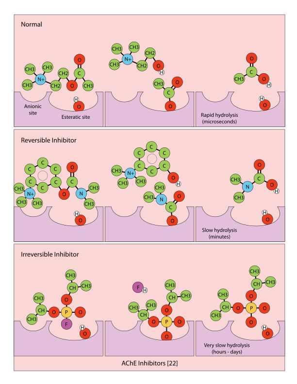
Diagram comparing the mechanisms of reversible and irreversible acetylcholinesterase inhibition, showing carbamate and organophosphate binding to the active site serine
Mechanisms of acetylcholinesterase inhibition: carbamate inhibitors (reversible) vs. organophosphate inhibitors (irreversible). The phosphoryl-enzyme bond formed by organophosphates is hydrolyzed millions of times more slowly than the normal acetyl-enzyme intermediate. Pysicist, CC BY-SA 4.0, via Wikimedia Commons.
Nerve Agents and Pesticide Organophosphates

Organophosphate compounds fall into two clinically important categories. Chemical warfare nerve agents include the G-series compounds (sarin, soman, tabun, and cyclosarin) and the V-series compounds such as VX. G-series agents are volatile and primarily absorbed through inhalation; V-series agents are dense, oily substances with exceptional skin penetration, making dermal contact the dominant exposure route.

Agricultural organophosphate pesticides include parathion, malathion, chlorpyrifos, and diazinon, among many others. Parathion and chlorpyrifos are associated with the majority of severe agricultural poisoning fatalities worldwide. The clinical importance of the category distinction lies in the pharmacokinetics of exposure rather than the mechanism of toxicity, which is the same for all members of the class.

The Aging Phenomenon and Its Clinical Consequences

After the initial phosphorylation of acetylcholinesterase, the phosphoryl-enzyme adduct undergoes a spontaneous secondary reaction called aging, in which one of the alkyl groups attached to the phosphorus is eliminated. This converts the adduct into a form resistant to nucleophilic attack by oxime reactivators such as pralidoxime. Once aging is complete, no antidote can restore the enzyme — it is permanently inactivated and must be replaced by synthesis of new protein, a process that takes days to weeks.

The clinical importance of aging lies entirely in its rate. Different organophosphate compounds age at vastly different speeds, and this determines the window of opportunity for pralidoxime to be effective.

Soman ages within minutes, making pralidoxime of no practical value unless administered in the first few minutes of exposure. Sarin ages over approximately three to five hours, providing a meaningful treatment window if antidotes are available promptly. VX ages over thirty-six to forty-eight hours, giving a wide window for oxime therapy. Most agricultural pesticide organophosphates age over twelve to thirty-six hours or more, which is why pralidoxime can be effective even when treatment is delayed by several hours. The practical implication is that pralidoxime should always be given as early as possible and continued for at least twenty-four to forty-eight hours in pesticide poisoning cases, even when the initial clinical response appears modest.

Aging Half-Lives — The Window for Pralidoxime

Soman (GD): aging within 2 to 6 minutes. Pralidoxime is effectively useless for soman poisoning after the first few minutes. Pyridostigmine pretreatment is the prophylactic strategy for soman exposure. Sarin (GB): aging half-life approximately 3 to 5 hours. Pralidoxime is effective when given early. VX: aging over 36 to 48 hours. Wide treatment window. Agricultural pesticide organophosphates (parathion, chlorpyrifos): aging over 12 to 36 hours or more. Pralidoxime should be given promptly and continued for 24 to 48 hours.

Section 2

Organophosphate Pharmacokinetics — Absorption, Bioactivation, and Central Nervous System Penetration

Routes of entry, why no route of exposure is trivial, the thiono-organophosphate bioactivation step that delays toxicity after pesticide ingestion, and how lipid solubility drives central nervous system toxicity

The clinical presentation of organophosphate poisoning is shaped not only by the mechanism of toxicity but also by how the compound enters the body and how it is metabolized. Organophosphates as a class are highly lipid-soluble molecules capable of crossing virtually every biological membrane, including skin, mucous membranes, lung alveoli, and the blood-brain barrier. This broad penetrability means that decontamination must address all possible routes of exposure and that central nervous system symptoms are expected in serious poisoning, not exceptional findings.

Routes of Absorption

Inhalation is the fastest and most dangerous route of exposure for volatile organophosphate compounds. Nerve agent vapors such as sarin are absorbed across the alveolar-capillary membrane almost instantaneously, producing systemic effects within seconds to minutes of inhalation. Because of this speed, inhalation exposures to nerve agents can be lethal before the victim can take protective action.

Dermal absorption is the dominant route for agents with low volatility, including VX and many agricultural organophosphate pesticides encountered during field spraying. The rate of skin absorption depends on the specific compound and the condition of the skin.

Oral ingestion is the most common route in intentional self-poisoning with agricultural organophosphates, which accounts for the majority of serious organophosphate poisoning fatalities in South and Southeast Asia. Oral exposures may have a delayed onset of action depending on whether the specific compound requires metabolic activation before becoming toxic.

Bioactivation of Thiono-Organophosphates

A pharmacologically important subclass of organophosphate pesticides are the thiono-organophosphates, in which the reactive phosphoryl oxygen bond is replaced by a phosphorus-sulfur bond. Common examples include parathion, malathion, chlorpyrifos, and diazinon. In their native thiono form these compounds are weak acetylcholinesterase inhibitors, but bioactivation by cytochrome P450 enzymes in the liver and lung converts them into their potently toxic oxon forms — parathion becomes paraoxon, malathion becomes malaoxon.

This obligate hepatic bioactivation step has two clinically important consequences. First, onset of toxicity after oral ingestion may be delayed by one to four hours compared to direct-acting agents such as nerve agents, because the parent compound must reach the liver before becoming toxic. Second, the rate of bioactivation varies between individuals, potentially modifying both the severity and timing of poisoning.

Central Nervous System Penetration

All clinically significant organophosphates readily cross the blood-brain barrier because of their high lipid solubility, adding a central nervous system component to the toxidrome that cannot be treated by peripheral muscarinic blockade alone. At moderate degrees of inhibition, central cholinergic hyperstimulation produces anxiety, restlessness, and cognitive impairment. As inhibition deepens, seizures, loss of consciousness, central respiratory depression, and coma can develop. This is why atropine — which crosses the blood-brain barrier as a tertiary amine — must be given in doses far exceeding those used for other indications in severe organophosphate poisoning.

Why No Route of Exposure Is Trivial

Organophosphates penetrate skin, mucous membranes, the cornea, the gastrointestinal tract, the pulmonary alveoli, and the blood-brain barrier. A patient who was only briefly exposed and appears minimally symptomatic may have absorbed compound through multiple routes simultaneously. Decontamination must address all routes: remove clothing, irrigate skin and eyes, ensure airway is clear. The clinical course can worsen over the first one to four hours in thiono-organophosphate exposures as hepatic bioactivation proceeds.


Section 3

The Cholinergic Toxidrome — Muscarinic, Nicotinic, and Central Nervous System Components

SLUDGE and DUMBELS mnemonics for muscarinic symptoms, nicotinic effects at the neuromuscular junction and autonomic ganglia, central nervous system seizure mechanism, and the four routes to respiratory failure in severe poisoning

Organophosphate poisoning produces clinical effects at three anatomically distinct sites: postganglionic muscarinic synapses of the parasympathetic nervous system, nicotinic synapses at the neuromuscular junction and autonomic ganglia, and central nervous system cholinergic synapses. Each site contributes a recognizable cluster of symptoms. Identifying all three clusters together constitutes the cholinergic toxidrome and distinguishes organophosphate poisoning from other causes of neurological and autonomic dysfunction.

The Muscarinic Component: SLUDGE and DUMBELS

Overstimulation of postganglionic parasympathetic muscarinic receptors produces the features captured by two commonly used mnemonics. The SLUDGE mnemonic covers salivation, lacrimation, urination, defecation, gastrointestinal cramping, and emesis—all results of parasympathetic hyperstimulation of exocrine glands and smooth muscle. The DUMBELS mnemonic adds detail: diarrhea/defecation, urination, miosis, bradycardia/bronchospasm/bronchorrhea, emesis, lacrimation, and salivation. Both mnemonics identify the same spectrum of findings.

Among all the muscarinic features, the triad of bronchospasm, bronchorrhea, and bradycardia represents the immediately life-threatening component. Bronchospasm and bronchorrhea together produce hypoxia through airway obstruction and impaired gas exchange. Miosis (pupillary constriction) is a consistent early sign, often appearing before systemic symptoms in vapor exposures, but is not itself life-threatening. Profuse sweating from eccrine sweat gland stimulation is a consistent muscarinic finding across all grades of severity.

The Nicotinic Component at the Neuromuscular Junction

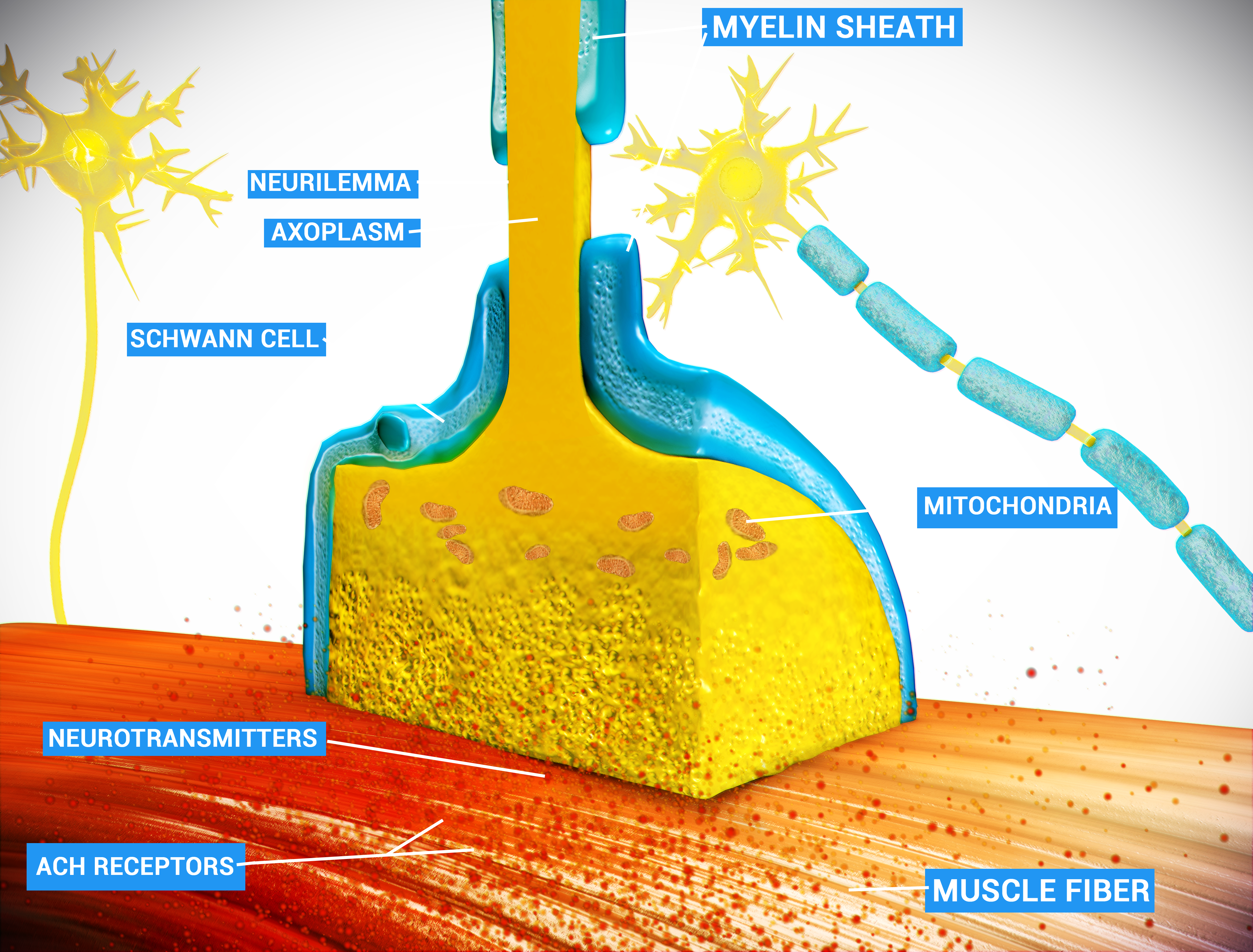
Excess acetylcholine at nicotinic receptors at the neuromuscular junction initially produces fasciculations — visible, involuntary muscle twitching from repetitive firing of motor endplate potentials. As inhibition deepens, sustained depolarization of the endplate produces a depolarizing neuromuscular block, manifesting as flaccid weakness and, in severe poisoning, paralysis of the diaphragm and intercostal muscles. This paralysis cannot be reversed by atropine, which acts only at muscarinic receptors, and combined with the concurrent bronchospasm and bronchorrhea, it creates a situation where mechanical ventilation becomes the only intervention that can ensure adequate oxygenation.

Educational diagram of the neuromuscular junction showing the motor nerve terminal, synaptic vesicles, synaptic cleft, and postsynaptic nicotinic receptors on the muscle fiber
Anatomy of the neuromuscular junction. Organophosphate-induced accumulation of acetylcholine at this site produces fasciculations followed by depolarizing neuromuscular blockade and flaccid paralysis of respiratory muscles in severe poisoning. Doctor Jana, CC BY 4.0, via Wikimedia Commons.
The Nicotinic Component at Autonomic Ganglia

Acetylcholine accumulation at nicotinic receptors in autonomic ganglia stimulates both sympathetic and parasympathetic postganglionic neurons simultaneously. Because sympathetic activation releases norepinephrine and adrenal medullary activation releases epinephrine, mild or early organophosphate poisoning may initially present with tachycardia and elevated blood pressure rather than the bradycardia expected from the muscarinic component.

As poisoning progresses and parasympathetic muscarinic effects become dominant, the cardiovascular picture shifts to bradycardia and hypotension. This biphasic pattern — initial sympathomimetic followed by parasympathomimetic predominance — can complicate early assessment and explains why tachycardia at presentation does not rule out organophosphate toxicity.

The Central Nervous System Component and Seizures

Central nervous system acetylcholinesterase inhibition produces anxiety, agitation, and cognitive impairment at moderate degrees of toxicity, progressing to seizures, loss of consciousness, and central respiratory depression in severe poisoning. Organophosphate-induced seizures are initially cholinergic in origin, but they rapidly evolve into a self-sustaining pattern driven by glutamate excitotoxicity that no longer depends on ongoing cholinergic stimulation.

This transition explains why benzodiazepines remain effective anticonvulsants even after full muscarinic blockade with atropine has been achieved — they suppress seizures by enhancing gamma-aminobutyric acid activity, independent of cholinergic tone. It also explains why delay in benzodiazepine administration makes seizures harder to control.

Four Mechanisms of Respiratory Failure in Severe Organophosphate Poisoning

All four can operate simultaneously in severe poisoning. Intubation and mechanical ventilation are the only interventions that address all four at once. Atropine does not reverse neuromuscular junction paralysis or central respiratory depression.

1. Neuromuscular junction depolarizing blockade: flaccid paralysis of the diaphragm and intercostal muscles.

2. Bronchospasm: muscarinic receptor-mediated airway smooth muscle contraction increasing airway resistance.

3. Bronchorrhea: muscarinic receptor-mediated gland hypersecretion flooding the airways with secretions.

4. Central respiratory depression: central nervous system acetylcholinesterase inhibition suppressing medullary respiratory centers.


Section 4

Antidote Management — Atropine, Pralidoxime, and Benzodiazepines

Three antidotes targeting three different pathophysiological mechanisms: muscarinic blockade with atropine, enzyme reactivation with pralidoxime before aging, and seizure control with benzodiazepines

Effective management of organophosphate poisoning requires simultaneous use of three pharmacological interventions, each targeting a distinct mechanism of toxicity. Atropine blocks muscarinic receptors to reduce secretions, bronchospasm, and bradycardia. Pralidoxime reactivates inhibited acetylcholinesterase before aging renders it permanently inactive. Benzodiazepines suppress seizures by enhancing gamma-aminobutyric acid receptor activity. Understanding what each antidote can and cannot do is as important as knowing when to use it.

Antidote 1
Atropine
  • Competitive antagonist at muscarinic receptors (all subtypes)
  • Blocks muscarinic effects: reduces bronchospasm, bronchorrhea, bradycardia, secretions
  • Does NOT reverse neuromuscular junction paralysis
  • Does NOT reactivate acetylcholinesterase
  • Crosses the blood-brain barrier: treats central nervous system muscarinic effects
  • Titration endpoint: drying of bronchial secretions, NOT normalization of heart rate
  • Large doses required in severe poisoning—far exceeding typical clinical doses
Antidote 2
Pralidoxime (2-PAM)
  • Oxime nucleophile that attacks and removes the phosphoryl group from inhibited acetylcholinesterase
  • Restores enzyme activity if given before aging is complete
  • Addresses neuromuscular junction paralysis and ganglionic effects that atropine cannot treat
  • Does NOT cross the blood-brain barrier (permanently charged quaternary compound)
  • Does NOT treat the central nervous system component of toxicity
  • Administer as early as possible: aging eliminates the window for reactivation
Antidote 3
Benzodiazepines
  • Enhance gamma-aminobutyric acid type A receptor chloride conductance, hyperpolarizing neurons
  • Suppress seizures by raising the seizure threshold
  • Effective even after full muscarinic blockade, because late-phase seizures are glutamate-driven
  • Diazepam preferred in mass-casualty settings; lorazepam or midazolam acceptable in hospital
  • Treat seizures early: delay allows transition to self-sustaining status epilepticus
Atropine: Why the Endpoint Is Secretion Drying, Not Heart Rate

Atropine blocks all muscarinic receptor subtypes, making it effective against the full spectrum of parasympathetic overstimulation. The correct titration endpoint is cessation of bronchorrhea and reduction of bronchospasm — not normalization of heart rate. Using heart rate as the endpoint leads to systematic undertreatment because nicotinic ganglionic stimulation can sustain tachycardia even when muscarinic blockade is inadequate.

Atropine does not address the nicotinic component of the toxidrome — it cannot reverse neuromuscular junction paralysis. However, atropine does cross the blood-brain barrier as a tertiary amine, making it effective against the central nervous system muscarinic component of toxicity in addition to its peripheral effects.

Pralidoxime: Mechanism, Timing, and the Blood-Brain Barrier Limitation

Pralidoxime reactivates phosphorylated acetylcholinesterase by attacking the phosphorus atom of the enzyme adduct and regenerating the free active serine. This reactivation is specific for the pre-aging adduct — once aging is complete, pralidoxime cannot restore the enzyme. The treatment window is therefore determined by the aging kinetics of the specific organophosphate involved, and pralidoxime should always be given as early as possible. Its particular value is addressing the nicotinic component of toxicity, especially neuromuscular junction blockade, that atropine cannot reach.

Pralidoxime is a permanently charged quaternary compound that does not cross the blood-brain barrier in clinically meaningful amounts. It therefore does not treat the central nervous system component of organophosphate toxicity — which is why atropine remains essential even when pralidoxime is given promptly. The two antidotes address different anatomical compartments and are not interchangeable.

Why Atropine Treats the Brain and Pralidoxime Does Not

Atropine is a tertiary amine—it carries no permanent positive charge—and freely crosses the blood-brain barrier. This makes atropine effective against both peripheral muscarinic symptoms and the central nervous system muscarinic component of organophosphate toxicity.

Pralidoxime is a permanently charged quaternary ammonium compound. Charged molecules do not cross the blood-brain barrier. Pralidoxime cannot reach acetylcholinesterase in the brain, and central nervous system acetylcholinesterase inhibition continues even when pralidoxime has successfully reactivated peripheral enzyme. Atropine is therefore essential in addition to pralidoxime, not as a substitute for it.


Section 5

Carbamates vs. Organophosphates — Key Pharmacological Differences

Why carbamate insecticides produce a similar toxidrome with a shorter and more self-limited course, why pralidoxime is not used for carbamate poisoning, and the central importance of decontamination before medical treatment

Carbamate insecticides and organophosphate compounds both inhibit acetylcholinesterase and produce the cholinergic toxidrome, but fundamental differences in their chemistry lead to important clinical management distinctions. Understanding these differences is straightforward once the mechanism of inhibition is clear: carbamates carbamylate the active site serine rather than phosphorylating it, and the carbamyl-enzyme adduct spontaneously hydrolyzes with a half-life of thirty to one hundred twenty minutes. This means that carbamate-inhibited acetylcholinesterase recovers on its own, without any antidote for the enzyme itself.

The Critical Difference: Aging Does Not Occur with Carbamates

The single most important pharmacological distinction between carbamate and organophosphate acetylcholinesterase inhibitors is that carbamate-inhibited enzyme does not age. The carbamyl-enzyme adduct spontaneously hydrolyzes and regenerates active enzyme over thirty to one hundred twenty minutes.

This spontaneous reactivation has two direct clinical consequences. First, pralidoxime is not indicated for carbamate poisoning. The enzyme will reactivate on its own, and some experimental evidence suggests pralidoxime may even paradoxically inhibit acetylcholinesterase in the context of carbamate poisoning, though the human clinical data are not definitive enough to draw firm conclusions. Second, the clinical course of carbamate poisoning is generally shorter and more self-limited than organophosphate poisoning of equivalent initial severity, with symptoms typically resolving within six to twenty-four hours in the absence of massive ongoing exposure.

Common carbamate insecticides include carbofuran, aldicarb, methomyl, and carbaryl. The therapeutic carbamate acetylcholinesterase inhibitors neostigmine and pyridostigmine work by the same carbamylation mechanism, which is why they produce similar but short-lived, controllable cholinergic effects at therapeutic doses. The distinction between therapeutic and toxic carbamate exposure is primarily one of dose and selectivity of receptor access.

Management of Carbamate Poisoning

Atropine remains the mainstay of treatment for the muscarinic component of carbamate toxicity, titrated to secretion drying by the same principle as in organophosphate poisoning. Dose requirements are typically lower and the duration shorter because the enzyme is recovering spontaneously. Pralidoxime is omitted or used only with caution if the agent cannot be confirmed. Benzodiazepines are used for seizures if they occur, and airway management follows the same priorities as for organophosphate poisoning.

The shorter clinical course should not lead to premature relaxation of monitoring — massive carbamate exposures can still be life-threatening during the active inhibition phase.

Decontamination: The First Priority in Any Organophosphate or Carbamate Exposure

Decontamination must precede or occur simultaneously with medical treatment when skin contamination is present, because ongoing dermal absorption perpetuates toxicity even while antidotes are being given. The sequence is straightforward: remove the patient from the contaminated environment, remove all clothing and personal effects (this alone eliminates the majority of the contaminant burden), irrigate the skin with copious water, and irrigate the eyes with water or saline if conjunctival exposure occurred. Personnel performing decontamination require appropriate protective equipment.

The relative priority of decontamination versus antidote administration depends on the agent. For volatile compounds such as sarin, the agent disperses rapidly and decontamination of residual skin contamination is less critical than immediate antidote administration. For liquid agents with persistent dermal absorption such as VX, decontamination takes relatively higher priority.

Comparison
Organophosphates
  • Phosphorylation of active site serine
  • Aging: adduct becomes permanently resistant to pralidoxime over time
  • Treatment window for pralidoxime is time-dependent and compound-specific
  • Pralidoxime indicated: give as early as possible
  • Clinical course: potentially prolonged; redistribution from tissue stores can cause relapse
Comparison
Carbamates
  • Carbamylation of active site serine
  • No aging: spontaneous hydrolysis with half-life of 30 to 120 minutes
  • Enzyme recovers without antidote for the enzyme
  • Pralidoxime NOT indicated (may be harmful; unnecessary)
  • Clinical course: shorter, more self-limited; symptoms typically resolve in 6 to 24 hours
What Both Organophosphates and Carbamates Have in Common

Both produce the full cholinergic toxidrome through acetylcholinesterase inhibition. Both require atropine for muscarinic symptom management, titrated to secretion drying. Both may require benzodiazepines for seizure control. Both require decontamination if dermal exposure has occurred. Both require airway management as the top priority in severe poisoning. The only management difference is that pralidoxime is used in organophosphate poisoning but withheld or avoided in confirmed carbamate poisoning.


Suggested References
Author / Organization Title Source
Colovic MB, Krstic DZ, Lazarevic-Pasti TD, Bondzic AM, Vasic VM Acetylcholinesterase inhibitors: pharmacology and toxicology Current Neuropharmacology. 2013;11(3):315–335
Munoz-Quezada MT, Lucero BA, Iglesias VP, et al. Chronic exposure to organophosphate pesticides and neuropsychological functioning in farm workers: a review International Journal of Occupational and Environmental Health. 2016;22(1):68–79
Eddleston M, Buckley NA, Eyer P, Dawson AH Management of acute organophosphorus pesticide poisoning Lancet. 2008;371(9612):597–607
Karalliedde L, Senanayake N Organophosphorus insecticide poisoning British Journal of Anaesthesia. 1989;63(6):736–750
Jokanovic M Medical treatment of acute poisoning with organophosphorus and carbamate pesticides Toxicology Letters. 2009;190(2):107–115
Holstege CP, Kirk M, Sidell FR Chemical warfare: nerve agent poisoning Critical Care Clinics. 1997;13(4):923–942
Eddleston M, Eyer P, Worek F, et al. Differences between organophosphorus insecticides in human self-poisoning: a prospective cohort study Lancet. 2005;366(9495):1452–1459
Treiman DM GABAergic mechanisms in epilepsy Epilepsia. 2001;42(Suppl 3):8–12
Reigart JR, Roberts JR Recognition and Management of Pesticide Poisonings. 6th ed. US Environmental Protection Agency; 2013. EPA 735-R-13-001
Bradberry SM, Cage SA, Proudfoot AT, Vale JA Poisoning due to pyrethroids Toxicological Reviews. 2005;24(2):93–106
Thiermann H, Worek F, Kehe K Limitations and challenges in treatment of acute chemical warfare agent poisoning Chemico-Biological Interactions. 2013;206(3):435–443
Worek F, Thiermann H, Wille T Oximes in organophosphate poisoning: 60 years of hope and despair Chemico-Biological Interactions. 2016;259(Pt B):93–98