The adrenal cortex produces three classes of steroid hormones from a shared cholesterol precursor. Each class originates in a distinct cortical zone under different regulatory control. This zonal and enzymatic organization explains how drugs that inhibit specific biosynthetic enzymes produce selective effects on steroid output.
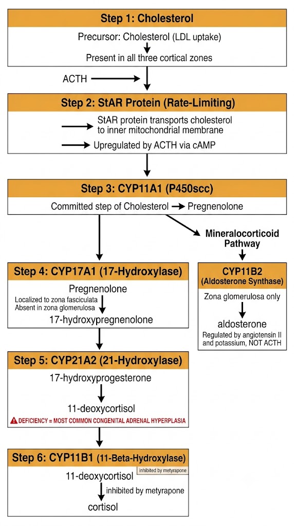
The adrenal cortex is organized into three concentric zones. The outermost zona glomerulosa produces aldosterone and is regulated primarily by the renin-angiotensin-aldosterone system and serum potassium. The middle zona fasciculata is the largest zone and produces cortisol under the control of adrenocorticotropic hormone from the anterior pituitary. The innermost zona reticularis produces adrenal androgens, principally dehydroepiandrosterone and its sulfate ester, also under adrenocorticotropic hormone control.
Zone specificity is determined by enzyme expression. The zona glomerulosa expresses aldosterone synthase (CYP11B2) but not the 17-alpha-hydroxylase activity of CYP17A1, making it capable of producing aldosterone but not cortisol. The zona fasciculata expresses CYP17A1 and CYP11B1 but not CYP11B2, producing cortisol but not aldosterone. The zona reticularis expresses CYP17A1 with high lyase activity to produce dehydroepiandrosterone.
All steroid synthesis begins with cholesterol, derived primarily from low-density lipoprotein uptake. The rate-limiting step is transport of cholesterol from the outer to the inner mitochondrial membrane, mediated by the steroidogenic acute regulatory (StAR) protein. Adrenocorticotropic hormone acutely upregulates StAR expression through a cyclic adenosine monophosphate-dependent mechanism, which is how adrenocorticotropic hormone stimulation rapidly increases cortisol output.
Once at the inner membrane, CYP11A1 (the cholesterol side-chain cleavage enzyme) converts cholesterol to pregnenolone, the committed step. Pregnenolone then enters parallel pathways. In the zona fasciculata, CYP17A1 hydroxylates pregnenolone to 17-hydroxypregnenolone, which is further processed to 17-hydroxyprogesterone. CYP21A2 (21-hydroxylase) then converts 17-hydroxyprogesterone to 11-deoxycortisol, and CYP11B1 (11-beta-hydroxylase) completes cortisol synthesis.
CYP21A2 deficiency blocks the conversion of 17-hydroxyprogesterone to 11-deoxycortisol. Because cortisol cannot be made, adrenocorticotropic hormone remains elevated, driving substrate accumulation proximal to the block. The accumulated 17-hydroxyprogesterone is shunted into androgen synthesis, producing cortisol deficiency, variable aldosterone deficiency, and androgen excess. The degree of virilization in females and salt-wasting depends on mutation severity. Elevated 17-hydroxyprogesterone is the diagnostic marker.
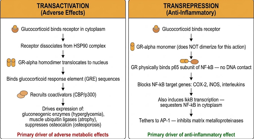
Glucocorticoids act primarily through the glucocorticoid receptor, a cytoplasmic ligand-activated transcription factor. Two distinct signaling modes account for the therapeutic and adverse effect profiles of glucocorticoid therapy: transactivation of metabolic genes drives most adverse effects, while transrepression of inflammatory transcription factors drives most anti-inflammatory benefit.
The unliganded glucocorticoid receptor resides in the cytoplasm bound to a chaperone complex that includes heat shock protein 90 (HSP90), which holds the receptor in a conformation competent for ligand binding. When a glucocorticoid binds the receptor, a conformational change releases the receptor from the complex and triggers translocation to the nucleus, where it acts as a transcription factor.
In the nucleus, activated glucocorticoid receptor homodimers bind glucocorticoid response element sequences in the promoters of target genes and recruit coactivator complexes that enhance transcription. This transactivation drives expression of genes that produce most adverse metabolic effects: gluconeogenic enzymes (causing hyperglycemia), muscle-specific ubiquitin ligases (causing muscle atrophy), and suppression of osteocalcin synthesis (contributing to osteoporosis).
The anti-inflammatory actions of glucocorticoids depend on a mechanistically distinct process. Rather than binding directly to DNA, glucocorticoid receptor monomers physically interact with and inhibit pro-inflammatory transcription factors, principally nuclear factor kappa-B and activator protein-1. Nuclear factor kappa-B normally drives transcription of cyclooxygenase-2, inducible nitric oxide synthase, multiple interleukins, and adhesion molecules. By binding the p65 subunit of nuclear factor kappa-B, the glucocorticoid receptor blocks transcription of these pro-inflammatory targets without requiring glucocorticoid response element binding.
Glucocorticoids also induce transcription of the inhibitory protein I-kappa-B, which sequesters nuclear factor kappa-B in the cytoplasm, providing a second layer of nuclear factor kappa-B suppression. Similarly, glucocorticoid receptor binding to activator protein-1 (a heterodimer of c-Fos and c-Jun) reduces transcription of matrix metalloproteinases and additional cytokines.
Adverse metabolic effects (hyperglycemia, osteoporosis, muscle atrophy, skin thinning) are driven primarily by glucocorticoid response element-dependent transactivation. Anti-inflammatory effects are driven primarily by tethered transrepression of nuclear factor kappa-B and activator protein-1. This distinction has motivated decades of research into agents that favor transrepression over transactivation, though no currently available drug achieves this separation in patients. Budesonide achieves a partial pharmacokinetic separation by limiting systemic transactivation through high first-pass hepatic metabolism while maintaining local anti-inflammatory transrepression in the gut or airway.
The hypothalamic-pituitary-adrenal axis regulates cortisol secretion and is suppressed by exogenous glucocorticoids in a dose- and duration-dependent manner. Understanding this suppression is inseparable from understanding the risks of steroid therapy and the rationale for tapering and stress-dose supplementation.
The hypothalamus releases corticotropin-releasing hormone into the portal circulation, stimulating the anterior pituitary to secrete adrenocorticotropic hormone, which in turn drives cortisol synthesis in the zona fasciculata. Cortisol feeds back negatively to suppress both corticotropin-releasing hormone and adrenocorticotropic hormone secretion, completing the regulatory loop.
Cortisol secretion follows a circadian rhythm driven by the suprachiasmatic nucleus. Levels peak 30 to 60 minutes after awakening (the cortisol awakening response) and fall through the day, reaching their nadir around midnight. This rhythm has a direct clinical implication: once-daily glucocorticoid doses given in the morning produce less total hypothalamic-pituitary-adrenal axis suppression than the same dose given in the evening, because morning dosing adds to an already-elevated cortisol level that has partially suppressed the axis, whereas evening dosing suppresses the nighttime adrenocorticotropic hormone surge that drives the next morning peak.
Once-daily oral glucocorticoids should be taken in the morning (7:00 to 8:00 AM). This exploits the normal circadian pattern: the axis is already partially suppressed by endogenous cortisol at this time, so the incremental suppression added by an exogenous morning dose is minimized. Evening dosing suppresses the nighttime adrenocorticotropic hormone surge with substantially greater cumulative effect on hypothalamic-pituitary-adrenal axis function. Patients must be counseled specifically about timing, not simply told to take the medication once daily.
Persistently elevated exogenous glucocorticoid levels suppress corticotropin-releasing hormone and adrenocorticotropic hormone transcription through glucocorticoid receptor-mediated repression of the respective gene promoters. Prolonged suppression leads to adrenocortical atrophy. The axis retains basal function at lower suppression, but the stress response is blunted.
As a general guide: doses below a prednisone equivalent of 5 mg per day rarely cause clinically significant suppression at any duration. Doses above 20 mg per day for more than three weeks cause substantial suppression. Duration matters as much as dose — the same cumulative amount given over a longer period causes more suppression than the same amount given over a shorter course.
The choice among hydrocortisone, prednisone, methylprednisolone, dexamethasone, and budesonide is grounded in their distinct potency ratios, biologic durations of action, mineralocorticoid activity, and routes of metabolism. These properties determine which agent is appropriate for which clinical context.
All glucocorticoids share a critical pharmacokinetic feature: their biologic duration of action substantially exceeds their plasma half-life. This dissociation occurs because glucocorticoid receptor-mediated changes in gene transcription persist after plasma concentrations have fallen. Hydrocortisone has a plasma half-life of 60 to 90 minutes but produces effects lasting 8 to 12 hours. Prednisone has a plasma half-life of 2 to 3 hours but a biologic duration of 18 to 36 hours. This means once-daily dosing is pharmacologically sufficient even for short-acting agents.
Prednisone is an inactive prodrug converted to prednisolone by 11-beta-hydroxysteroid dehydrogenase type 1 in the liver. In patients with severe hepatic insufficiency, this conversion is impaired and prednisolone is preferred because it requires no activation. For patients with normal liver function, prednisone and prednisolone are clinically interchangeable.
All systemic glucocorticoids are metabolized primarily by cytochrome P450 3A4. Cytochrome P450 3A4 inducers (rifampin, phenytoin, carbamazepine, phenobarbital) accelerate glucocorticoid metabolism and can reduce plasma concentrations enough to cause loss of therapeutic effect or, in dependent patients, adrenal crisis. Cytochrome P450 3A4 inhibitors (ketoconazole, ritonavir and other protease inhibitors, clarithromycin) increase glucocorticoid concentrations and can cause iatrogenic Cushing syndrome even at standard doses. Patients on ritonavir-boosted antiretroviral regimens who receive fluticasone-containing inhaled corticosteroids are at particular risk for this interaction; beclomethasone is the preferred alternative in this population because it is not a cytochrome P450 3A4 substrate.
| Author / Organization | Title | Source |
|---|---|---|
| Miller WL, Auchus RJ | The molecular biology, biochemistry, and physiology of human steroidogenesis and its disorders | Endocr Rev. 2011;32(1):81–151 |
| Stocco DM | StAR protein and the regulation of steroid hormone biosynthesis | Annu Rev Physiol. 2001;63:193–213 |
| Chrousos GP | Adrenocorticosteroids and adrenocortical antagonists. In: Katzung BG, ed. Basic and Clinical Pharmacology, 14th ed. | McGraw-Hill; 2018:687–710 |
| Barnes PJ | Glucocorticosteroids: current and future directions | Br J Pharmacol. 2011;163(1):29–43 |
| Stahn C, Buttgereit F | Genomic and nongenomic effects of glucocorticoids | Nat Clin Pract Rheumatol. 2008;4(10):525–533 |
| Rhen T, Cidlowski JA | Antiinflammatory action of glucocorticoids — new mechanisms for old drugs | N Engl J Med. 2005;353(16):1711–1723 |
| Debono M et al. | Modified-release hydrocortisone to provide circadian cortisol profiles | J Clin Endocrinol Metab. 2009;94(5):1548–1554 |
| Crowley RK et al. | Clinical insights into adrenal insufficiency: low-dose ACTH stimulation test versus insulin tolerance test | Clin Endocrinol (Oxf). 2010;73(4):461–466 |
| Liu D et al. | A practical guide to the monitoring and management of the complications of systemic corticosteroid therapy | Allergy Asthma Clin Immunol. 2013;9(1):30 |
| Czock D et al. | Pharmacokinetics and pharmacodynamics of systemically administered glucocorticoids | Clin Pharmacokinet. 2005;44(1):61–98 |
| Henzen C et al. | Suppression and recovery of adrenal response after short-term, high-dose glucocorticoid treatment | Lancet. 2000;355(9203):542–545 |