CHAPTER 28  ·  ADRENOCORTICOSTEROID PHARMACOLOGY
Section 01
Molecular Basis of Glucocorticoid Anti-Inflammatory Action
Annexin-A1 and phospholipase A2 inhibition, nuclear factor kappa-B and activator protein-1 transrepression, cyclooxygenase-2 and inducible nitric oxide synthase suppression, and the cytokine network targeted by glucocorticoids

The anti-inflammatory potency of glucocorticoids derives from simultaneous suppression of multiple arms of the inflammatory cascade at the transcriptional level. Rather than blocking a single mediator, the glucocorticoid receptor inhibits the master transcription factors that drive the entire inflammatory gene program, while also inducing proteins that further suppress inflammation post-translationally.

Upstream Eicosanoid Suppression

Glucocorticoids suppress eicosanoid synthesis through two convergent mechanisms. First, they induce annexin-A1 (lipocortin-1), an endogenous inhibitor of cytosolic phospholipase A2. Because phospholipase A2 releases arachidonic acid from membrane phospholipids — the substrate for both cyclooxygenase and lipoxygenase pathways — annexin-A1 induction simultaneously reduces prostaglandins, thromboxanes, and leukotrienes. This explains why glucocorticoids suppress a broader range of eicosanoids than nonsteroidal anti-inflammatory drugs, which block only cyclooxygenase. Second, glucocorticoids transcriptionally repress cyclooxygenase-2 expression through nuclear factor kappa-B inhibition, preventing inducible cyclooxygenase-2 upregulation at inflamed sites.

Nuclear Factor Kappa-B Suppression

Nuclear factor kappa-B is the master regulator of the acute inflammatory transcriptome. Glucocorticoids suppress it through three parallel mechanisms: (1) glucocorticoid receptor monomers tether directly to the p65 subunit of nuclear factor kappa-B, blocking coactivator binding; (2) glucocorticoids induce transcription of I-kappa-B-alpha, the inhibitory protein that sequesters nuclear factor kappa-B in the cytoplasm; (3) the glucocorticoid receptor recruits histone deacetylase 2 to promoters of nuclear factor kappa-B target genes, reversing histone acetylation and closing chromatin. The histone deacetylase 2 mechanism is clinically relevant to glucocorticoid resistance in chronic obstructive pulmonary disease: oxidative stress from cigarette smoke inactivates histone deacetylase 2 in airway macrophages, impairing this arm of glucocorticoid action.

Cytokine Suppression Profile

The cytokine targets of glucocorticoids span both initiating signals and amplification circuits. At the initiating level, interleukin-1 beta and tumor necrosis factor-alpha (the principal macrophage-derived proximal cytokines driving fever, acute-phase protein synthesis, and immune cell recruitment) are suppressed. At the amplification level, interleukin-2 suppression impairs T cell clonal expansion; interleukin-6 suppression blunts the hepatic acute-phase response; suppression of interferon-gamma reduces macrophage activation; and suppression of interleukin-8 reduces neutrophil chemotaxis. Glucocorticoids also suppress inducible nitric oxide synthase and the adhesion molecules intercellular adhesion molecule-1 and E-selectin through nuclear factor kappa-B inhibition, reducing vascular permeability and leukocyte transmigration — contributing to the rapid reduction in tissue edema that is among the first clinically visible effects.

Post-Transcriptional Suppression: MKP-1

Glucocorticoids induce mitogen-activated protein kinase phosphatase-1, which inactivates the p38 mitogen-activated protein kinase and c-Jun N-terminal kinase pathways. These kinases stabilize messenger RNA of pro-inflammatory cytokines including tumor necrosis factor-alpha and interleukin-6 by phosphorylating proteins that protect cytokine messenger RNA from degradation. Mitogen-activated protein kinase phosphatase-1 induction accelerates cytokine messenger RNA decay, reducing protein output at the post-transcriptional level independently of transcriptional repression. This dual action — transcriptional repression plus post-transcriptional destabilization — explains why glucocorticoids reduce cytokine output more completely than agents targeting only one mechanism.


Section 02
Glucocorticoid Effects on Immune Cells
Neutrophil demargination, T lymphocyte redistribution and interleukin-2 suppression, macrophage and dendritic cell impairment, eosinophil apoptosis, and interpreting the complete blood count in steroid-treated patients

Glucocorticoids produce predictable, characteristic changes in circulating leukocyte populations within hours of administration. Understanding the mechanism behind each change is necessary for correctly interpreting blood counts in steroid-treated patients and for understanding both the therapeutic immunosuppression and infectious complications of chronic therapy.

Neutrophilia: Redistribution, Not Increased Function

Glucocorticoids cause a 2- to 3-fold rise in circulating neutrophil count within 4 to 6 hours, driven by two redistribution phenomena: suppression of adhesion molecule expression (L-selectin, Mac-1) on the neutrophil surface reduces margination to the vascular endothelium, and accelerated release of mature neutrophils from bone marrow storage pools expands the circulating pool. Antimicrobial function is not enhanced. An elevated neutrophil count in a steroid-treated patient therefore cannot be used as a reliable marker of bacterial infection.

Lymphopenia: Redistribution and Interleukin-2 Suppression

Circulating T cells fall markedly (70 to 90% at pharmacological doses) within hours of glucocorticoid administration, primarily by redistribution to lymphoid tissues (spleen, lymph nodes, bone marrow) driven by altered homing receptor expression. This redistribution reverses within 24 hours of a single dose. The therapeutic immunosuppression in ongoing immune responses depends primarily on suppression of interleukin-2 production by T helper cells: without interleukin-2, antigen-activated T cells cannot undergo clonal expansion to mount an adaptive immune response. At high concentrations or with prolonged exposure, glucocorticoids also induce apoptosis in immature thymocytes.

Macrophage and Dendritic Cell Suppression

In macrophages, glucocorticoids suppress the classical activation phenotype: the respiratory burst, phagocytic activity, antigen presentation via reduced major histocompatibility complex class II expression, and production of interleukin-1 beta, tumor necrosis factor-alpha, and interleukin-12 are all impaired. The capacity to kill intracellular pathogens is reduced, explaining the increased susceptibility to organisms normally controlled by cell-mediated immunity. In dendritic cells, glucocorticoids impair maturation, reduce costimulatory molecule expression (CD80, CD86), and shift the cytokine profile away from T helper type 1-promoting patterns, reducing the efficiency of naive T cell priming.

Eosinophil Apoptosis

Glucocorticoids promote eosinophil apoptosis, rapidly and profoundly reducing tissue eosinophil numbers. The fall in blood eosinophil count after a single systemic glucocorticoid dose is one of the most sensitive indicators of glucocorticoid receptor engagement. Mast cell numbers in tissues are also reduced with prolonged use through suppression of stem cell factor. These effects account for the prominent efficacy of glucocorticoids in allergic diseases including asthma and allergic rhinitis, where eosinophils and mast cells are the dominant effectors of type 2 inflammation.

Interpreting the Complete Blood Count in Glucocorticoid-Treated Patients

Expected steroid effect on the complete blood count: neutrophilia (2- to 3-fold rise from demargination, not infection), lymphopenia (T cells predominantly, from redistribution), monocytopenia (redistribution), eosinopenia (indicates active glucocorticoid receptor engagement). A rising eosinophil count in a patient who should be therapeutically suppressed warrants consideration of non-compliance or inadequate dosing. A left-shifted neutrophilia with toxic granulations on peripheral smear suggests superimposed infection despite background steroid-induced neutrophilia.
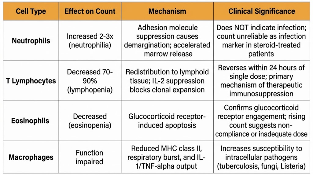
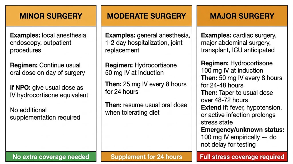
Table showing glucocorticoid effects on neutrophils, lymphocytes, eosinophils, and macrophages
Figure 1. Glucocorticoid effects on circulating leukocyte populations. Neutrophilia results from redistribution, not enhanced antimicrobial function. Lymphopenia and eosinopenia are the primary mechanisms of therapeutic immunosuppression.

Section 03
Glucocorticoids in Critical Illness and Organ Transplantation
Septic shock dosing and landmark trials, dexamethasone in COVID-19 acute respiratory distress syndrome (RECOVERY trial), cerebral edema and bacterial meningitis, and transplant immunosuppression

Glucocorticoids have a contested but clearly evidence-based role in critical illness. Large randomized trials have defined the specific patient populations who benefit and the appropriate agents, doses, and durations. In organ transplantation, glucocorticoids remain integral to both induction and maintenance immunosuppression, though steroid-minimization protocols reflect both their adverse effect burden and the efficacy of newer immunosuppressants.

Septic Shock

The rationale for glucocorticoids in septic shock is that severe sepsis produces relative adrenal insufficiency in some patients through suppression of hypothalamic-pituitary-adrenal axis responsiveness by inflammatory mediators and impaired cortisol synthesis from adrenal ischemia. The ADRENAL trial (2018) found no mortality benefit from hydrocortisone 200 mg per day in mechanically ventilated septic shock patients, though the glucocorticoid group achieved faster shock reversal and faster liberation from mechanical ventilation. The APROCCHSS trial (2018) found a significant mortality benefit with hydrocortisone plus fludrocortisone in patients with more severe septic shock requiring higher vasopressor doses.

The current clinical synthesis: low-dose hydrocortisone (200 mg per day by continuous infusion or 50 mg intravenous every 6 hours) is appropriate for patients with septic shock refractory to adequate fluid resuscitation and vasopressors at doses equivalent to norepinephrine greater than 0.25 micrograms per kilogram per minute. It should not be used in patients who respond to initial resuscitation. Dexamethasone is not appropriate in this setting because of its long duration and absence of mineralocorticoid activity needed to complement hydrocortisone's fluid-retaining effect.

COVID-19 Acute Respiratory Distress Syndrome: RECOVERY Trial

The RECOVERY trial demonstrated that dexamethasone 6 mg once daily for up to 10 days reduced 28-day mortality in hospitalized COVID-19 patients requiring respiratory support. Benefit was concentrated in patients requiring supplemental oxygen or mechanical ventilation (number needed to treat approximately 8 for ventilated patients). Patients not requiring oxygen showed no benefit and a trend toward harm — underscoring the principle that glucocorticoid benefit in infectious respiratory failure is relevant only when dysregulated host inflammation, rather than active viral replication, is the dominant driver of injury. Dexamethasone was selected for once-daily dosing, absence of mineralocorticoid activity, and high potency at a small dose.

Cerebral Edema and Bacterial Meningitis

Dexamethasone is the standard agent for managing cerebral edema from primary or metastatic brain tumors: high potency, no mineralocorticoid activity, and long duration make it preferred over other glucocorticoids in this setting. In bacterial meningitis, dexamethasone 0.15 mg per kilogram every 6 hours for 4 days, given with or immediately before the first antibiotic dose, reduces the risk of severe neurological sequelae (sensorineural hearing loss, particularly in Haemophilus influenzae type b meningitis) by suppressing the intense subarachnoid inflammatory response triggered by bacterial lysis from antibiotics.

Organ Transplantation

Glucocorticoids contribute to transplant immunosuppression primarily by suppressing interleukin-2 production and reducing major histocompatibility complex class II expression on antigen-presenting cells, impairing alloreactive T cell activation. They are synergistic with calcineurin inhibitors (cyclosporine, tacrolimus), which block the calcineurin-nuclear factor of activated T cells pathway of interleukin-2 gene transcription; the combination suppresses interleukin-2 at both the transcriptional and upstream cytokine-signaling levels. An important drug interaction: cytochrome P450 3A4 induction by rifampin or anticonvulsants can reduce glucocorticoid levels enough to precipitate acute rejection. Steroid-minimization protocols that taper prednisone to discontinuation within 3 to 12 months post-transplant are now standard in many kidney transplant centers for low-risk recipients.


Section 04
Glucocorticoids in Rheumatology, Pulmonary Disease, and Dermatology
Bridge therapy in rheumatoid arthritis and systemic lupus erythematosus, high-dose therapy in polymyalgia rheumatica and giant cell arteritis, inhaled corticosteroid pharmacology in asthma and chronic obstructive pulmonary disease, and topical potency classification

Glucocorticoids have disease-specific roles that differ markedly in dose, duration, and intent across specialties. Understanding which dose and agent is appropriate for which indication is high-yield at the second-year level.

Rheumatoid Arthritis and Systemic Lupus Erythematosus

In rheumatoid arthritis, glucocorticoids serve as bridge therapy: low-dose prednisone (5 to 10 mg per day) controls synovial inflammation while awaiting the 8- to 12-week onset of disease-modifying antirheumatic drugs such as methotrexate. Short courses at higher doses (20 to 40 mg for 5 to 10 days) rapidly suppress acute flares. Intra-articular triamcinolone or methylprednisolone is preferred over systemic therapy when only one or two joints are actively inflamed, concentrating drug at the target site while minimizing systemic exposure.

In systemic lupus erythematosus, dosing spans an enormous range depending on organ involvement. Mild flares may be managed with prednisone 20 to 30 mg per day for a short course. Severe organ-threatening manifestations — lupus nephritis, neuropsychiatric lupus, lupus myocarditis — require high-dose intravenous methylprednisolone pulse (500 to 1000 mg per day for 3 days), followed by oral prednisone 1 mg per kilogram per day tapered over months, combined with mycophenolate mofetil or cyclophosphamide for sustained organ protection.

Polymyalgia Rheumatica and Giant Cell Arteritis

Polymyalgia rheumatica requires prednisone 15 to 25 mg per day maintained until symptoms resolve and inflammatory markers normalize, then slowly tapered over 12 to 24 months — relapse rates are high with premature dose reduction. Giant cell arteritis, a large-vessel vasculitis affecting cranial arteries in patients over age 50, requires prednisone 40 to 60 mg per day to prevent irreversible ischemic vision loss from anterior ischemic optic neuropathy. When visual symptoms or transient monocular blindness are present, intravenous methylprednisolone pulse is initiated immediately before starting oral prednisone — even 24 hours of delay can cause permanent blindness. Tocilizumab (interleukin-6 receptor antagonist) is approved as adjunctive therapy in giant cell arteritis and enables a faster prednisone taper with fewer relapses than prednisone monotherapy.

Inhaled Corticosteroids

Inhaled corticosteroids achieve high local airway concentrations with minimal systemic bioavailability through two mechanisms: poor gastrointestinal absorption of swallowed drug, and extensive first-pass hepatic metabolism of the absorbed fraction. Fluticasone propionate has the highest topical potency and lowest gastrointestinal bioavailability among commonly used agents. Budesonide undergoes approximately 85 to 90% first-pass hepatic metabolism. Ciclesonide is an inactive prodrug activated by esterases in airway epithelium. At standard adult doses, inhaled corticosteroids have a favorable safety profile; at high doses (fluticasone propionate greater than 500 micrograms per day), measurable hypothalamic-pituitary-adrenal axis suppression, posterior subcapsular cataracts, and skin thinning can occur.

Topical Glucocorticoid Potency Classification

Topical glucocorticoids are classified into seven potency classes (Class I, superpotent, through Class VII, lowest potency) based on a vasoconstrictor assay measuring skin blanching. Class I agents (clobetasol propionate 0.05%) should be limited to 2 consecutive weeks on restricted body surface area and avoided on the face, axillae, and groin. Class VI to VII agents (hydrocortisone 1%, desonide) are safe for facial and intertriginous use and in children. Vehicle matters: ointments have the highest penetration (occlusive effect), creams are intermediate, and lotions are preferred for scalp application. Skin atrophy, striae, and telangiectasia result from glucocorticoid receptor-dependent suppression of collagen synthesis and represent irreversible changes with prolonged high-potency use.


Section 05
Perioperative Glucocorticoid Management and Stress-Dose Protocols
The surgical cortisol stress response, risk stratification of hypothalamic-pituitary-adrenal axis suppression, stress-dose supplementation by surgical category, and recognition of perioperative adrenal crisis

The perioperative period is a high-stakes context for glucocorticoid management. Major surgery normally drives a large cortisol surge essential for hemodynamic stability; patients with a suppressed hypothalamic-pituitary-adrenal axis cannot mount this response independently. Unrecognized perioperative adrenal crisis is entirely preventable with appropriate prophylaxis.

Risk Stratification

Patients on prednisone equivalent less than 5 mg per day, or any glucocorticoid for less than 3 weeks, have minimal suppression and require no supplementation — continue their usual dose through the perioperative period. Patients on prednisone 5 to 20 mg per day for 3 or more weeks have partial suppression and uncertain stress response capacity; supplementation is proportional to surgical stress. Patients on prednisone greater than 20 mg per day for more than 3 weeks, or any patient with Cushingoid features, are presumed to have substantial suppression and receive full stress-dose supplementation for major procedures. Patients off glucocorticoids for more than 3 months generally have recovered hypothalamic-pituitary-adrenal axis function and require no special management.

Perioperative Protocol
Stress-Dose Supplementation by Surgical Category
  • Minor surgery (local or regional anesthesia, same-day discharge) — no supplementation beyond usual morning dose; if nothing by mouth, give usual dose as intravenous hydrocortisone equivalent
  • Moderate surgery (general anesthesia, 1 to 2 day hospitalization) — hydrocortisone 50 mg intravenous at induction, then 25 mg intravenous every 8 hours for 24 hours; resume usual oral dose when tolerating diet
  • Major surgery (prolonged general anesthesia, intensive care unit care anticipated) — hydrocortisone 100 mg intravenous at induction, then 50 mg intravenous every 8 hours for 24 to 48 hours, tapering to usual dose over 48 to 72 hours; extend if fever, hypotension, or active infection prolongs the stress state
  • Emergency surgery with unknown hypothalamic-pituitary-adrenal axis status — give hydrocortisone 100 mg intravenous empirically; do not delay surgery for cortisol testing; obtain random cortisol and adrenocorticotropic hormone sample before the dose if timing allows
Recognizing Perioperative Adrenal Crisis

Clinical clues suggesting adrenal crisis rather than hypovolemia or cardiac dysfunction: hypotension refractory to fluid resuscitation and vasopressors, hemodynamic instability disproportionate to estimated blood loss, hyponatremia without hyperkalemia (secondary adrenal insufficiency spares mineralocorticoid function), and a history of chronic glucocorticoid use. In any hemodynamically unstable postoperative patient on or recently discontinuing glucocorticoids, give hydrocortisone 100 mg intravenous empirically without waiting for cortisol testing. Hemodynamic improvement within 30 to 60 minutes supports the diagnosis.

Three-panel diagram showing perioperative glucocorticoid stress-dose protocol by surgical category
Figure 2. Perioperative glucocorticoid stress-dose protocol stratified by surgical category. Patients on prednisone equivalent less than 5 mg per day or steroids for less than 3 weeks require no supplementation beyond their usual dose.

Suggested References
Author / Organization Title Source
Rhen T, Cidlowski JA Antiinflammatory action of glucocorticoids — new mechanisms for old drugs N Engl J Med. 2005;353(16):1711–1723
Barnes PJ Glucocorticosteroids: current and future directions Br J Pharmacol. 2011;163(1):29–43
Chrousos GP Adrenocorticosteroids and adrenocortical antagonists. In: Katzung BG, ed. Basic and Clinical Pharmacology, 14th ed. McGraw-Hill; 2018:687–710
Liu D et al. A practical guide to the monitoring and management of the complications of systemic corticosteroid therapy Allergy Asthma Clin Immunol. 2013;9(1):30
Stahn C, Buttgereit F Genomic and nongenomic effects of glucocorticoids Nat Clin Pract Rheumatol. 2008;4(10):525–533
Barnes PJ Inhaled corticosteroids Pharmaceuticals (Basel). 2010;3(3):514–540
Annane D et al. Hydrocortisone plus fludrocortisone for adults with septic shock (APROCCHSS) N Engl J Med. 2018;378(9):809–818
RECOVERY Collaborative Group; Horby P et al. Dexamethasone in hospitalized patients with COVID-19 N Engl J Med. 2021;384(8):693–704
Czock D et al. Pharmacokinetics and pharmacodynamics of systemically administered glucocorticoids Clin Pharmacokinet. 2005;44(1):61–98
Stone JH et al. Trial of tocilizumab in giant-cell arteritis N Engl J Med. 2017;377(4):317–328
Marik PE, Varon J Requirement of perioperative stress doses of corticosteroids: a systematic review Arch Surg. 2008;143(12):1222–1226
Venkatesh B et al. Adjunctive glucocorticoid therapy in patients with septic shock (ADRENAL) N Engl J Med. 2018;378(9):797–808