CHAPTER 29  ·  DIABETES PHARMACOLOGY
Section 01
Insulin Physiology: Synthesis, Secretion, and Receptor Signaling
How beta cells sense glucose, the regulated secretory pathway, biphasic insulin release, and the downstream signaling cascade that drives glucose uptake into peripheral tissues

Insulin is the principal anabolic hormone of the body, produced exclusively by the beta cells of the pancreatic islets of Langerhans. Understanding endogenous insulin biology is the essential foundation for rational therapeutic use of exogenous insulin preparations, because the pharmacological goal of insulin therapy is to replicate the time course and magnitude of normal insulin secretion as closely as the available preparations permit.

Insulin is synthesized as preproinsulin and processed in the endoplasmic reticulum to proinsulin, then to mature insulin in secretory granules. A connecting peptide (C-peptide) is released equimolarly with insulin. C-peptide is absent from all commercial insulin preparations, so measuring it clinically distinguishes endogenous secretion from injected insulin and can help identify factitious hypoglycemia.

Glucose-stimulated insulin secretion is triggered when glucose enters the beta cell via glucose transporter 2 and is metabolized by glucokinase, raising the ratio of adenosine triphosphate to adenosine diphosphate inside the cell. This closes adenosine triphosphate-sensitive potassium channels, depolarizes the cell membrane, opens voltage-gated calcium channels, and triggers exocytosis of insulin granules. This potassium channel is the molecular target of sulfonylureas, which close it independent of glucose — explaining both their efficacy and their hypoglycemia risk.

Normal insulin secretion is biphasic. The first phase is a rapid spike within minutes of a glucose load, representing release of granules already docked at the membrane. The second phase is sustained and involves reserve granule recruitment and amplification by incretin hormones (glucagon-like peptide-1 from intestinal L cells and glucose-dependent insulinotropic polypeptide from intestinal K cells). Loss of first-phase secretion is one of the earliest defects in type 2 diabetes mellitus.

Insulin Receptor Signaling

Insulin binds its receptor tyrosine kinase, activating a cascade through insulin receptor substrate proteins and phosphatidylinositol 3-kinase to Akt. In muscle and adipose tissue, Akt triggers translocation of glucose transporter type 4 vesicles to the plasma membrane, dramatically increasing glucose uptake. In the liver, Akt promotes glycogen synthesis and suppresses gluconeogenesis. These are the key metabolic effects: glucose uptake in periphery, glycogen storage in liver, inhibition of lipolysis in fat.
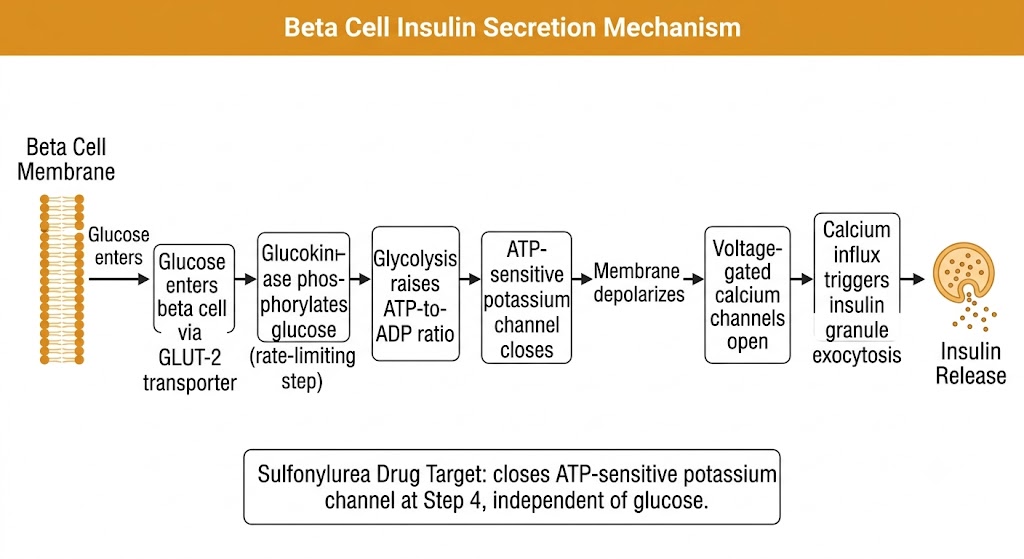
Flow diagram showing glucose-stimulated insulin secretion in the pancreatic beta cell, from glucose entry via GLUT-2 through glucokinase metabolism, ATP-sensitive potassium channel closure, membrane depolarization, calcium influx, and insulin granule exocytosis, with a note indicating the sulfonylurea drug target at the potassium channel step.
Beta cell glucose-stimulated insulin secretion pathway. Generated with Gemini AI for educational use.
Section 02
Insulin Preparations: Rapid-, Short-, Intermediate-, and Long-Acting
Structural modifications that determine pharmacokinetic profiles, and the clinical rationale for each preparation class

Commercially available insulin preparations span a range from ultra-rapid analogs designed to mimic mealtime secretion to near-peakless basal analogs that provide 24 to 42 hours of background coverage. Each reflects specific amino acid substitutions or formulation changes that alter the speed of subcutaneous absorption.

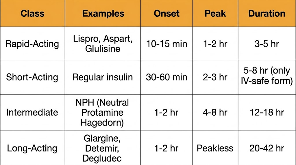
Regular insulin (human insulin) forms hexamers in the vial and must dissociate to monomers before absorption, producing an onset of 30 to 60 minutes and duration of 5 to 8 hours. It must be injected 30 minutes before meals. Regular insulin is the only preparation safe for intravenous use and remains important for intravenous infusions in the intensive care unit and perioperative setting.

Rapid-acting analogs (lispro, aspart, glulisine) carry amino acid substitutions that disrupt hexamer formation, yielding onset within 10 to 15 minutes, peak at 1 to 2 hours, and duration of 3 to 5 hours. They can be injected immediately before or at the start of a meal, substantially improving postprandial glucose control and convenience compared with regular insulin.

Neutral protamine Hagedorn insulin is an intermediate-acting preparation with onset 1 to 2 hours, peak 4 to 8 hours, and duration 12 to 18 hours. Its pronounced peak causes nocturnal hypoglycemia when injected at bedtime and produces significant day-to-day variability. It remains widely used where cost is a concern.

Long-acting basal analogs are designed to provide a flat, peakless background insulin level. Glargine (precipitates at subcutaneous pH, dissolving slowly) provides approximately 20 to 24 hours of coverage with the standard formulation and approximately 36 hours with the concentrated formulation. Detemir (fatty acid chain causes albumin binding in subcutaneous tissue) provides 18 to 22 hours, often requiring twice-daily dosing. Degludec forms multi-hexamer complexes that dissociate slowly, providing more than 42 hours of coverage with the least day-to-day variability of any basal analog.

Memorizing the Classes

Rapid: lispro, aspart, glulisine — inject at mealtime, peak in 1–2 hours.
Short: regular insulin — inject 30 minutes before meals, only preparation for intravenous use.
Intermediate: neutral protamine Hagedorn — twice daily, nocturnal hypoglycemia risk.
Long: glargine, detemir, degludec — once daily basal, peakless profile.

Comparison table of insulin preparation classes showing rapid-acting (lispro, aspart, glulisine: onset 10-15 min, peak 1-2 hr, duration 3-5 hr), short-acting (regular insulin: onset 30-60 min, peak 2-3 hr, duration 5-8 hr, only IV-safe form), intermediate (NPH: onset 1-2 hr, peak 4-8 hr, duration 12-18 hr), and long-acting (glargine, detemir, degludec: onset 1-2 hr, peakless, duration 20-42 hr).
Insulin preparation classes: onset, peak, and duration profiles. Generated with Gemini AI for educational use.
Section 03
Clinical Use: Basal-Bolus Dosing and Titration
Physiological rationale for basal-bolus therapy, starting doses, titration principles, and insulin management in special contexts

Effective insulin therapy matches pharmacokinetics to physiology. In type 1 diabetes mellitus, where endogenous secretion is absent, the goal is complete replacement: a basal insulin suppresses hepatic glucose output between meals and overnight, while bolus insulin covers each meal. In type 2 diabetes mellitus, insulin is usually added after oral agents fail, often starting with basal-only therapy.

Starting total daily doses are typically 0.4 to 0.5 units per kilogram per day in type 1 diabetes mellitus, split approximately 50 percent basal and 50 percent bolus. In type 2 diabetes mellitus beginning basal-only therapy, 0.1 to 0.2 units per kilogram per day is a common starting point. Basal insulin is titrated by increasing the dose by 2 units every 3 days when fasting glucose exceeds the target (typically 80 to 130 mg/dL), holding if any fasting glucose falls below 80 mg/dL.

Bolus dosing uses a carbohydrate-to-insulin ratio (how many grams of carbohydrate one unit covers) and a correction factor (how much one unit lowers blood glucose in mg/dL). A common starting estimate for the correction factor is 1800 divided by the total daily dose. Continuous glucose monitoring has substantially improved bolus titration by providing real-time postprandial glucose curves.

Perioperative and Intensive Care Unit Insulin

Target blood glucose in surgical and critically ill patients is 140 to 180 mg/dL. Long-acting basal insulin is continued at 75 to 80 percent of the usual dose; all bolus insulin is held while the patient is fasting. Intravenous regular insulin infusion is used for major surgery and critical illness. The NICE-SUGAR trial showed that intensive glucose control targeting 81 to 108 mg/dL increased mortality compared with conventional control (140 to 180 mg/dL), primarily due to severe hypoglycemia.

Section 04
Adverse Effects, Drug Interactions, and Special Populations
Hypoglycemia mechanisms and management, weight gain, injection site complications, drug interactions, and insulin use in pregnancy

Hypoglycemia is the most clinically consequential adverse effect of insulin and the primary barrier to achieving optimal glycemic control. The adverse effect profile also includes weight gain and injection site complications. Insulin is the only antihyperglycemic agent with an established safety record in pregnancy.

Hypoglycemia is defined by the Whipple triad: symptoms consistent with hypoglycemia, a low measured glucose (below 70 mg/dL for level 1, below 54 mg/dL for clinically significant level 2), and relief with glucose administration. Neurogenic symptoms (sweating, tremor, palpitations, hunger) appear at approximately 65 to 70 mg/dL. Neuroglycopenic symptoms (confusion, visual changes, seizure) appear at approximately 55 to 60 mg/dL and reflect direct neuronal glucose deprivation. Common precipitants include excess dose, missed meals, unplanned exercise, and alcohol (which suppresses hepatic gluconeogenesis).

Hypoglycemia unawareness develops in some patients with longstanding type 1 diabetes mellitus: repeated hypoglycemic episodes lower the glucose threshold at which counter-regulatory responses are triggered, so the first sign of hypoglycemia may be loss of consciousness without any warning symptoms. Continuous glucose monitoring with low-glucose alerts is recommended for these patients.

Treatment of mild hypoglycemia follows the rule of 15: 15 grams of fast-acting glucose orally, recheck in 15 minutes, repeat if still below 70 mg/dL. For severe hypoglycemia with inability to take oral glucose, glucagon (intramuscular or intranasal) is the standard out-of-hospital treatment; intravenous dextrose is given in hospital. Glucagon mobilizes hepatic glycogen and is ineffective in patients with depleted stores (prolonged fasting, alcoholism, hepatic failure).

Key Drug Interactions

Agents that increase insulin requirements: glucocorticoids (dose- and timing-dependent; postprandial and afternoon glucose most affected), atypical antipsychotics (olanzapine, clozapine), thiazide diuretics, calcineurin inhibitors (tacrolimus especially).
Agents that mask hypoglycemia or potentiate it: non-selective beta-blockers blunt tachycardia and tremor warning symptoms (sweating is preserved); alcohol inhibits gluconeogenesis and can cause delayed hypoglycemia 4 to 8 hours after ingestion.

Weight gain of 2 to 6 kg is predictable with insulin initiation, driven by reversal of glycosuria, enhanced lipogenesis, and increased appetite from hypoglycemia episodes. Adding a glucagon-like peptide-1 receptor agonist or sodium-glucose cotransporter-2 inhibitor to insulin attenuates weight gain and provides cardiovascular benefit in type 2 diabetes mellitus.

Insulin is the standard of care for both type 1 diabetes mellitus and gestational diabetes mellitus in pregnancy. Insulin does not cross the placenta in clinically significant amounts. Requirements typically double by the third trimester due to placental hormone-driven insulin resistance, then fall abruptly after delivery. Lispro and aspart are the preferred rapid-acting analogs in pregnancy. Regular insulin, neutral protamine Hagedorn, and glargine are also used with established safety records.

Visual Summary
Insulin Pharmacology: Mechanisms, Preparations, and Clinical Framework
Key mechanisms, preparation classes, dosing principles, and adverse effect management
Suggested References
Author / Organization Title Source
Saltiel AR, Kahn CR Insulin signalling and the regulation of glucose and lipid metabolism Nature. 2001;414(6865):799–806
Rorsman P, Braun M Regulation of insulin secretion in human pancreatic islets Annu Rev Physiol. 2013;75:155–179
Nauck MA, Meier JJ Incretin hormones: their role in health and disease Diabetes Obes Metab. 2018;20(Suppl 1):5–21
Korenblat KM et al Liver, muscle, and adipose tissue insulin action is directly related to intrahepatic triglyceride content in obese subjects Gastroenterology. 2008;134(5):1369–1375
Hirsch IB Insulin analogues N Engl J Med. 2005;352(2):174–183
Havelund S et al The mechanism of protraction of insulin detemir, a long-acting, acylated analog of human insulin Pharm Res. 2004;21(8):1498–1504
Becker RHA et al New insulin glargine 300 Units/mL provides a more even activity profile and prolonged glycemic control at steady state Diabetes Care. 2015;38(4):637–643
Frid AH et al New insulin delivery recommendations Mayo Clin Proc. 2016;91(9):1231–1255
Duckworth WC, Bennett RG, Hamel FG Insulin degradation: progress and potential Endocr Rev. 1998;19(5):608–624
Riddle MC et al The treat-to-target trial: randomized addition of glargine or human NPH insulin to oral therapy of type 2 diabetic patients Diabetes Care. 2003;26(11):3080–3086
American Diabetes Association Standards of Care in Diabetes—2024 Diabetes Care. 2024;47(Suppl 1):S1–S321
NICE-SUGAR Study Investigators Intensive versus conventional glucose control in critically ill patients N Engl J Med. 2009;360(13):1283–1297
Cryer PE Mechanisms of hypoglycemia-associated autonomic failure in diabetes N Engl J Med. 2013;369(4):362–372
Heise T et al Ultra-long-acting insulin degludec has a flat and stable glucose-lowering effect in type 2 diabetes Diabetes Obes Metab. 2012;14(10):944–950
Blumer I et al Diabetes and pregnancy: an Endocrine Society clinical practice guideline J Clin Endocrinol Metab. 2013;98(11):4227–4249
↑ Back to top