Immunopharmacology is concerned with how drugs modify immune function, whether to suppress pathological inflammation, restore deficient responses, or redirect immune activity against cancer. A working understanding of the cellular architecture of innate and adaptive immunity is essential for predicting drug effects, anticipating toxicities, and making sense of the rapidly expanding landscape of biologic and small-molecule immune therapies.
The innate immune system provides immediate, non-antigen-specific defense through neutrophils, monocytes, macrophages, dendritic cells, natural killer cells, mast cells, basophils, and eosinophils. Neutrophils are the most abundant circulating leukocytes and the first cells recruited to acute infection sites; they kill pathogens through phagocytosis, oxidative burst, and degranulation. Granulocyte colony-stimulating factor and granulocyte-macrophage colony-stimulating factor are the pharmacological regulators of neutrophil production, used clinically to reverse chemotherapy-induced neutropenia.
Macrophages are the central mediators of chronic inflammation, secreting tumor necrosis factor-alpha, interleukin-1 beta, and interleukin-6 in response to pathogen encounter. They are the primary target of tumor necrosis factor inhibitors and interleukin-1 antagonists. Dendritic cells are the most potent antigen-presenting cells and the critical link between innate sensing and adaptive activation. Abatacept and belatacept exploit the dendritic cell-to-T-cell interface by blocking the co-stimulatory ligands needed for full T-cell activation.
Natural killer cells kill virus-infected and tumor cells without prior sensitization. They also mediate antibody-dependent cellular cytotoxicity through their Fc receptor, which is how monoclonal antibodies such as rituximab and trastuzumab recruit natural killer cells to destroy opsonized targets. Calcineurin inhibitors and corticosteroids impair natural killer cell function, contributing to viral vulnerability in transplant recipients.
T lymphocytes develop in the thymus and are divided by surface markers and function into helper subsets and cytotoxic cells. CD4-positive helper T cells direct immune responses by secreting cytokines that activate macrophages, stimulate B-cell antibody production, and recruit effector cells. The major CD4 subsets are Th1 cells (producing interferon-gamma, driving macrophage activation against intracellular pathogens), Th2 cells (producing interleukin-4, interleukin-5, and interleukin-13, driving eosinophilic inflammation and IgE production), Th17 cells (producing interleukin-17A, driving neutrophilic mucosal inflammation), and regulatory T cells (producing interleukin-10 and transforming growth factor-beta, suppressing responses and maintaining self-tolerance). Drugs targeting these subsets include dupilumab (blocking the interleukin-4/interleukin-13 receptor used by Th2 cells) and secukinumab (blocking interleukin-17A produced by Th17 cells).
CD8-positive cytotoxic T lymphocytes kill target cells through perforin-granzyme pathways and Fas-mediated apoptosis. In tumor microenvironments, these cells enter functional exhaustion, upregulating checkpoint receptors including programmed death-1 and cytotoxic T-lymphocyte-associated protein 4. Checkpoint inhibitor drugs reverse this exhaustion to restore anti-tumor killing; these agents are covered in depth in Chapter 34.
B lymphocytes are the effectors of humoral immunity. Upon antigen encounter with T-cell help through the CD40 ligand-CD40 interaction, B cells proliferate, undergo class-switch recombination, and differentiate into antibody-secreting plasma cells. IgG mediates opsonization and complement activation; IgE drives mast cell and basophil degranulation in allergy. Pharmacological targeting of B cells includes rituximab and obinutuzumab (depleting B cells via anti-CD20), belimumab (reducing B-cell survival factor), and intravenous immunoglobulin (modulating Fc receptor signaling).
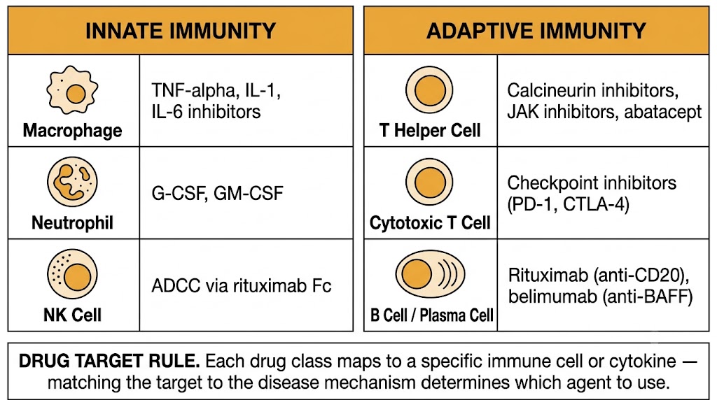
Neutrophils: granulocyte colony-stimulating factor (filgrastim), granulocyte-macrophage colony-stimulating factor (sargramostim). Macrophages: tumor necrosis factor inhibitors, interleukin-1 antagonists, interleukin-6 inhibitors, corticosteroids. Dendritic cells and co-stimulation: abatacept, belatacept. T cells broadly: calcineurin inhibitors, mammalian target of rapamycin inhibitors, corticosteroids, azathioprine, mycophenolate. Th2 and eosinophils: dupilumab (interleukin-4 receptor), mepolizumab and benralizumab (interleukin-5 axis). Th17: secukinumab and ixekizumab (interleukin-17A), guselkumab and risankizumab (interleukin-23). B cells: rituximab (anti-CD20), belimumab (anti-B-cell activating factor), intravenous immunoglobulin.
The innate immune system detects pathogens and tissue damage through germline-encoded pattern recognition receptors that trigger immediate inflammatory signaling. These receptors and their downstream cascades are direct or indirect targets of several pharmacological agents, from vaccine adjuvants that exploit pattern recognition to interleukin-1 antagonists that block the cytokine product of inflammasome activation.
Toll-like receptors are transmembrane pattern recognition receptors expressed on innate immune cells, endothelial cells, and epithelial surfaces. They recognize conserved microbial structures: toll-like receptor 4 recognizes lipopolysaccharide from gram-negative bacteria; toll-like receptor 2 recognizes bacterial lipopeptides; toll-like receptor 3 recognizes double-stranded RNA from viral replication; toll-like receptor 7 and toll-like receptor 8 recognize single-stranded RNA; toll-like receptor 9 recognizes unmethylated CpG motifs in bacterial and viral DNA. All toll-like receptors signal through MyD88, activating nuclear factor kappa B and driving transcription of tumor necrosis factor-alpha, interleukin-1 beta, and interleukin-6.
Toll-like receptor agonists are exploited as vaccine adjuvants to strengthen adaptive immune responses. Monophosphoryl lipid A, a detoxified toll-like receptor 4 agonist, is the adjuvant in the recombinant zoster vaccine (Shingrix) and the malaria vaccine. CpG-1018, a synthetic toll-like receptor 9 agonist, is the adjuvant in the hepatitis B vaccine Heplisav-B and produces substantially higher seroconversion rates than aluminum-only adjuvanted hepatitis B vaccines, particularly in older adults.
The NLRP3 inflammasome is a cytoplasmic multiprotein complex that senses intracellular danger signals. It is activated by extracellular adenosine triphosphate released from damaged cells, monosodium urate crystals in gout, calcium pyrophosphate crystals in pseudogout, cholesterol crystals in atherosclerotic plaques, and silica fibers. When activated, NLRP3 recruits the adaptor protein ASC and caspase-1, which cleaves pro-interleukin-1 beta and pro-interleukin-18 into their biologically active secreted forms. NLRP3 is thus the mechanistic link between metabolic damage signals and interleukin-1 beta-driven inflammation.
Three drugs target the interleukin-1 pathway. Anakinra is recombinant interleukin-1 receptor antagonist, blocking both interleukin-1 alpha and interleukin-1 beta at their shared receptor; its short half-life requires daily subcutaneous injection. Canakinumab is a monoclonal antibody selective for interleukin-1 beta with a half-life of approximately 26 days, allowing monthly or quarterly dosing; it is approved for cryopyrin-associated periodic syndromes, systemic juvenile idiopathic arthritis, and gouty arthritis. Rilonacept is a soluble decoy receptor fusion protein that binds both interleukin-1 alpha and interleukin-1 beta and is approved for recurrent pericarditis and cryopyrin-associated periodic syndromes.
Gout: monosodium urate crystals activate NLRP3, generating interleukin-1 beta that drives acute articular inflammation. Canakinumab is approved for flares refractory to colchicine, nonsteroidal anti-inflammatory drugs, and corticosteroids. Cryopyrin-associated periodic syndromes: gain-of-function NLRP3 mutations cause constitutive interleukin-1 beta secretion; canakinumab or rilonacept produces near-complete suppression of fever, rash, and systemic inflammation. Anakinra has the broadest clinical flexibility due to its short half-life, making it useful for acute flares (gout, pericarditis, cytokine storm) where rapid offset is desirable.
Cytokines are low-molecular-weight proteins that coordinate immune responses through specific cell-surface receptors. Pharmacological targeting of individual cytokines or their receptors now constitutes one of the largest classes of therapeutic agents in medicine, spanning rheumatology, dermatology, gastroenterology, pulmonology, and oncology.
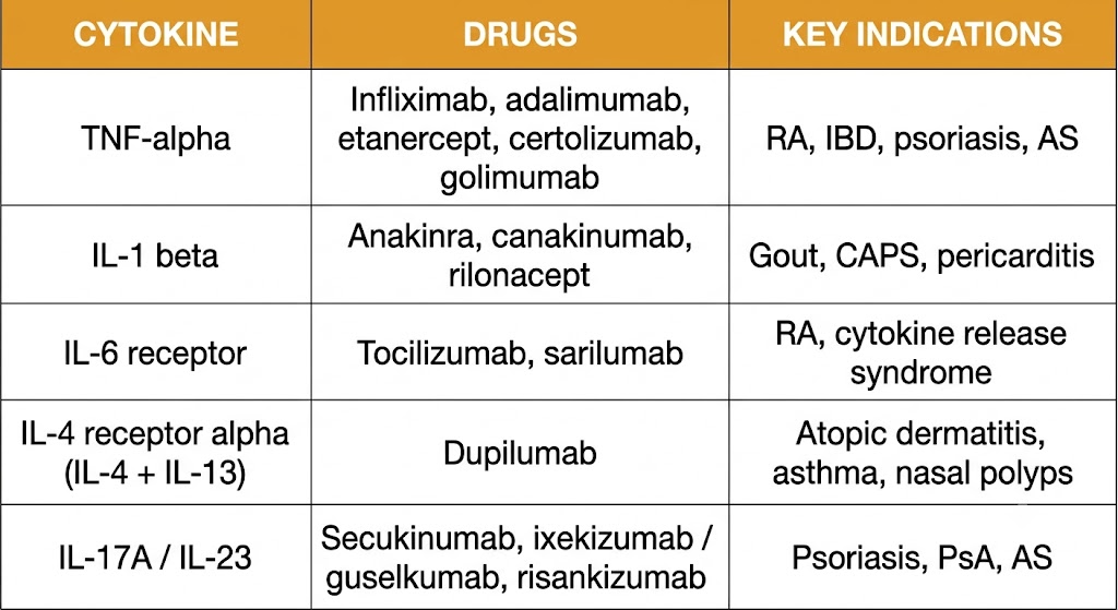
Tumor necrosis factor-alpha is produced primarily by activated macrophages and T cells and drives transcription of additional inflammatory cytokines, adhesion molecules, and acute-phase proteins through nuclear factor kappa B. It is central to the pathogenesis of rheumatoid arthritis, inflammatory bowel disease, psoriasis, ankylosing spondylitis, and psoriatic arthritis.
Five inhibitors are approved. Infliximab is a chimeric immunoglobulin G1 monoclonal antibody. Adalimumab and golimumab are fully human immunoglobulin G1 antibodies. Certolizumab pegol is a pegylated Fab fragment lacking the Fc region, which means it does not cross the placenta via neonatal Fc receptor-mediated transport and is the preferred tumor necrosis factor inhibitor in pregnancy. Etanercept is a dimeric fusion protein of the tumor necrosis factor receptor 2 extracellular domain with immunoglobulin G1 Fc; it binds both tumor necrosis factor-alpha and lymphotoxin-alpha and differs from the monoclonal antibodies in being ineffective in granulomatous diseases such as Crohn's disease and sarcoidosis.
Interleukin-6 is produced by macrophages, T cells, and fibroblasts and drives hepatic synthesis of acute-phase reactants, fever, Th17 differentiation, and thrombocytosis. It signals through a receptor complex including the ligand-binding interleukin-6 receptor alpha chain and the signal-transducing gp130 subunit. Tocilizumab and sarilumab are monoclonal antibodies blocking the interleukin-6 receptor alpha chain, suppressing C-reactive protein so effectively that C-reactive protein can no longer serve as an infection biomarker in patients receiving these agents. This is a critical clinical caveat: use procalcitonin and clinical assessment to detect concurrent infection in patients on interleukin-6 receptor inhibitors.
The type 2 immune response is driven by Th2 cells producing interleukin-4, interleukin-5, and interleukin-13. Interleukin-4 promotes IgE class switching and Th2 differentiation. Interleukin-5 governs eosinophil production, maturation, and survival. Interleukin-13 drives mucus hypersecretion and airway smooth muscle hyperreactivity. Dupilumab blocks the interleukin-4 receptor alpha chain shared by the receptors for both interleukin-4 and interleukin-13, producing simultaneous blockade of both cytokines; it is approved for atopic dermatitis, asthma, chronic rhinosinusitis with nasal polyposis, eosinophilic esophagitis, and prurigo nodularis. Mepolizumab and reslizumab (anti-interleukin-5) and benralizumab (anti-interleukin-5 receptor alpha) are approved for severe eosinophilic asthma.
Interleukin-23 shares its p40 subunit with interleukin-12 but pairs it with a unique p19 subunit. Interleukin-23 drives expansion of Th17 cells, which secrete interleukin-17A and interleukin-17F to stimulate neutrophilic inflammation at epithelial surfaces, driving the pathogenesis of psoriasis, psoriatic arthritis, and ankylosing spondylitis.
Ustekinumab targets the shared p40 subunit, blocking both interleukin-12 and interleukin-23, and is approved for psoriasis, psoriatic arthritis, Crohn's disease, and ulcerative colitis. Selective interleukin-23 p19 inhibitors (guselkumab, risankizumab, tildrakizumab) are more specific, preserving interleukin-12-dependent Th1 immunity while suppressing the interleukin-23/Th17 axis. Secukinumab and ixekizumab target interleukin-17A directly and carry a class-specific risk of new-onset or worsening inflammatory bowel disease; they are contraindicated in patients with active inflammatory bowel disease.
Tumor necrosis factor-alpha: infliximab, adalimumab, etanercept, certolizumab, golimumab. Interleukin-1 alpha and beta: anakinra; interleukin-1 beta only: canakinumab; interleukin-1 trap: rilonacept. Interleukin-6 receptor: tocilizumab, sarilumab. Interleukin-4 receptor alpha (blocks interleukin-4 and interleukin-13): dupilumab. Interleukin-5: mepolizumab, reslizumab; interleukin-5 receptor alpha: benralizumab. Interleukin-12/23 p40: ustekinumab. Interleukin-23 p19: guselkumab, risankizumab, tildrakizumab. Interleukin-17A: secukinumab, ixekizumab; interleukin-17A and interleukin-17F: bimekizumab.
Two additional intracellular and plasma-based signaling systems are central to immunopharmacology. The Janus kinase-signal transducer and activator of transcription pathway is the shared intracellular signal for more than fifty cytokines, making Janus kinase inhibitors uniquely broad-spectrum oral immunosuppressants. The complement cascade provides immediate effector defense against pathogens but, when dysregulated, drives severe hematological and renal diseases that are now treatable with specific complement inhibitors.
Four Janus kinase family members exist: Janus kinase 1, Janus kinase 2, Janus kinase 3, and tyrosine kinase 2. Each isoform associates constitutively with specific cytokine receptor subunits. Janus kinase 3 pairs exclusively with the common gamma chain shared by the receptors for interleukin-2, interleukin-4, interleukin-7, interleukin-9, interleukin-15, and interleukin-21. Janus kinase 2 transduces signals from erythropoietin, thrombopoietin, growth hormone, and granulocyte colony-stimulating factor receptors, as well as from interferon-gamma. Janus kinase 1 participates in signaling by interleukin-6 (through gp130) and by type I and type II interferons. Tyrosine kinase 2 couples with the receptors for interleukin-12, interleukin-23, and type I interferons.
Upon cytokine binding, receptor dimerization brings the associated Janus kinases into proximity, triggering mutual transphosphorylation and activation, followed by phosphorylation of signal transducer and activator of transcription proteins that dimerize and translocate to the nucleus to drive target gene transcription. All approved Janus kinase inhibitors are adenosine triphosphate-competitive kinase inhibitors. Tofacitinib preferentially inhibits Janus kinase 1 and Janus kinase 3; baricitinib inhibits Janus kinase 1 and Janus kinase 2; upadacitinib is a more selective Janus kinase 1 inhibitor. Inhibition of Janus kinase 2 at clinical doses suppresses erythropoietin and granulocyte colony-stimulating factor signaling, causing anemia and neutropenia. Janus kinase inhibitors carry a class-wide black box warning for serious infections, major adverse cardiovascular events, venous thromboembolism, malignancy, and mortality, based on the ORAL Surveillance safety trial of tofacitinib.
The complement cascade is activated through three pathways converging at the central cleavage of complement component C3. The classical pathway is triggered by antibody-antigen complexes binding C1q. The lectin pathway is activated by mannose-binding lectin or ficolins binding microbial carbohydrates. The alternative pathway is constitutively active at low level through spontaneous C3 hydrolysis and is amplified on pathogen surfaces lacking host regulatory proteins. All three pathways generate C3 convertases that cleave C3 into C3a (an anaphylatoxin) and C3b (an opsonin). C5 convertases then cleave C5 into C5a (a potent anaphylatoxin and chemotactic factor) and C5b, which nucleates the membrane attack complex causing cell lysis.
Eculizumab and ravulizumab are monoclonal antibodies that bind C5 and prevent its cleavage, blocking both C5a generation and membrane attack complex formation while preserving upstream C3b-mediated opsonization. Eculizumab requires intravenous infusions every two weeks; ravulizumab has an engineered extended half-life allowing dosing every eight weeks with equivalent efficacy. Both are approved for paroxysmal nocturnal hemoglobinuria, atypical hemolytic uremic syndrome, generalized myasthenia gravis with anti-acetylcholine receptor antibodies, and neuromyelitis optica spectrum disorder with anti-aquaporin-4 antibodies. Because terminal complement is the primary defense against encapsulated bacteria, meningococcal vaccination against serogroups A, C, W, Y, and B is mandatory at least two weeks before initiating any complement inhibitor, with antibiotic prophylaxis recommended throughout therapy.
Based on the ORAL Surveillance trial comparing tofacitinib to tumor necrosis factor inhibitors in high-cardiovascular-risk rheumatoid arthritis patients: serious infections including tuberculosis reactivation; malignancy including lung cancer and lymphoma; major adverse cardiovascular events; thrombosis including deep vein thrombosis and pulmonary embolism; mortality. Use restricted to patients who have had inadequate response to tumor necrosis factor inhibitors in rheumatic indications. Mandatory pre-treatment screening: tuberculin skin test or interferon-gamma release assay for latent tuberculosis; hepatitis B surface antigen and core antibody; complete blood count; fasting lipids. Herpes zoster reactivation is the most common infectious complication; recombinant zoster vaccine (Shingrix) is strongly recommended before initiating any Janus kinase inhibitor.
| Author / Organization | Title | Source |
|---|---|---|
| Janeway CA Jr, Travers P, Walport M, Shlomchik MJ | Immunobiology: The Immune System in Health and Disease, 9th ed. | Garland Science, New York; 2017. ISBN 9780815345053 |
| Bluestone JA, St Clair EW, Turka LA | CTLA4Ig: bridging the basic immunology with clinical application | Immunity. 2006;24(3):233-238 |
| Ridker PM, Everett BM, Thuren T, et al | Antiinflammatory therapy with canakinumab for atherosclerotic disease | N Engl J Med. 2017;377(12):1119-1131 |
| Sanz I, Lund FE | B cell responses to pathogens and vaccines in health and disease | J Immunol. 2020;205(2):293-301 |
| Kawai T, Akira S | The role of pattern-recognition receptors in innate immunity: update on Toll-like receptors | Nat Immunol. 2010;11(5):373-384 |
| Martinon F, Mayor A, Tschopp J | The inflammasomes: guardians of the body | Annu Rev Immunol. 2009;27:229-265 |
| Tracey D, Klareskog L, Sasso EH, Salfeld JG, Tak PP | Tumor necrosis factor antagonist mechanisms of action: a comprehensive review | Pharmacol Ther. 2008;117(2):244-279 |
| Tanaka T, Narazaki M, Kishimoto T | IL-6 in inflammation, immunity, and disease | Cold Spring Harb Perspect Biol. 2014;6(10):a016295 |
| Gandhi NA, Bennett BL, Graham NMH, et al | Targeting key proximal drivers of type 2 inflammation in disease | Nat Rev Drug Discov. 2016;15(1):35-50 |
| Miossec P, Kolls JK | Targeting IL-17 and Th17 cells in chronic inflammation | Nat Rev Drug Discov. 2012;11(10):763-776 |
| O'Shea JJ, Schwartz DM, Villarino AV, et al | The JAK-STAT pathway: impact on human disease and therapeutic intervention | Annu Rev Med. 2015;66:311-328 |
| Ytterberg SR, Bhatt DL, Mikuls TR, et al | Cardiovascular and cancer risk with tofacitinib in rheumatoid arthritis | N Engl J Med. 2022;386(4):316-326 |
| Ricklin D, Hajishengallis G, Yang K, Lambris JD | Complement: a key system for immune surveillance and homeostasis | Nat Immunol. 2010;11(9):785-797 |
| Lee JW, Sicre de Fontbrune F, Wong Lee Lee L, et al | Ravulizumab (ALXN1210) vs eculizumab in adult patients with PNH naive to complement inhibitors | Blood. 2019;133(6):530-539 |