Transplant Immunosuppression: Calcineurin Inhibitors, mTOR Inhibitors, and Antimetabolites
Mechanism, pharmacokinetics, toxicity, drug interactions, and transplant protocol pharmacology
Chapter 40 · Module 2 of 5
Section 1
Calcineurin Inhibitors
Cyclosporine and tacrolimus: the calcineurin-NFAT pathway, immunophilin binding, pharmacokinetics, and therapeutic drug monitoring
Calcineurin inhibitors are the cornerstone of solid organ transplant immunosuppression. Both cyclosporine and tacrolimus block the same downstream target, calcineurin, but bind distinct intracellular proteins, differ substantially in potency, and require therapeutic drug monitoring to navigate a narrow window between rejection and toxicity.
Mechanism of Action
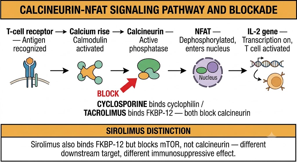
T-cell activation begins with antigen recognition through the T-cell receptor, which raises intracellular calcium and activates the calcium-calmodulin complex. This complex stimulates calcineurin, a serine-threonine phosphatase that dephosphorylates nuclear factor of activated T cells (NFAT), allowing it to translocate into the nucleus and drive transcription of interleukin-2 and other T-cell activation genes.
Cyclosporine binds cyclophilin, a cytoplasmic immunophilin, forming a cyclosporine-cyclophilin complex that inhibits calcineurin. Tacrolimus (also called FK-506) binds a different immunophilin, FK-binding protein 12 (FKBP-12), forming a tacrolimus-FKBP-12 complex that likewise inhibits calcineurin. Both complexes prevent NFAT dephosphorylation, blocking interleukin-2 transcription and halting T-cell activation and proliferation. Tacrolimus is approximately 100-fold more potent than cyclosporine on a weight basis and has largely replaced cyclosporine as the calcineurin inhibitor of choice in kidney, liver, heart, and lung transplantation.
Pharmacokinetics and Therapeutic Drug Monitoring
Both calcineurin inhibitors have highly variable oral bioavailability — cyclosporine averages 20 to 50% depending on formulation, and tacrolimus averages 25% with a wide range. Both are extensively metabolized by cytochrome P450 3A4 (CYP3A4) and the related CYP3A5 isoform in the liver and intestinal wall, with P-glycoprotein serving as an important efflux transporter. CYP3A5 genetic polymorphisms are clinically relevant for tacrolimus: patients who express functional CYP3A5 (more common in patients of African ancestry) require substantially higher doses to reach target trough concentrations.
Both drugs distribute extensively into erythrocytes — tacrolimus approximately 75 to 80%, cyclosporine approximately 60% — so therapeutic drug monitoring samples must be collected in ethylenediaminetetraacetic acid-anticoagulated whole blood, not plasma. Trough concentrations drawn immediately before the morning dose (C0) are the standard monitoring parameter. For tacrolimus in kidney transplant, target troughs are typically 8 to 12 nanograms per milliliter in the early post-transplant period and 5 to 8 nanograms per milliliter during maintenance. Both drugs are eliminated primarily through biliary excretion; dose adjustment for renal impairment is not required on a pharmacokinetic basis, though both agents cause nephrotoxicity that limits their use as renal function declines.
The calcineurin-NFAT signaling pathway and the site of calcineurin inhibitor blockade. Cyclosporine and tacrolimus bind different immunophilins but inhibit the same target. Sirolimus binds FKBP-12 but blocks mTOR downstream, not calcineurin. Abbreviations: FKBP-12 = FK-binding protein 12; IL-2 = interleukin-2; mTOR = mammalian target of rapamycin; NFAT = nuclear factor of activated T cells.
Calcineurin Inhibitor Mechanism Summary
Cyclosporine binds cyclophilin → cyclosporine-cyclophilin complex inhibits calcineurin → NFAT remains phosphorylated in cytoplasm → interleukin-2 gene not transcribed → T-cell activation blocked. Tacrolimus binds FKBP-12 → tacrolimus-FKBP-12 complex inhibits calcineurin by the same mechanism. Both immunophilins are cytoplasmic peptidyl-prolyl isomerases; the drug-immunophilin complex, not the drug alone, is the active inhibitor of calcineurin. Note: sirolimus also binds FKBP-12 but the sirolimus-FKBP-12 complex inhibits mTOR, not calcineurin — a critical mechanistic distinction.
Section 2
Calcineurin Inhibitor Toxicity and Drug Interactions
Nephrotoxicity mechanisms, metabolic and neurological adverse effects, and clinically significant CYP3A4 and P-glycoprotein interactions
The toxicity profiles of cyclosporine and tacrolimus overlap substantially because both inhibit calcineurin, but they differ in ways that influence drug selection and clinical monitoring. Both cause nephrotoxicity and increase infection risk. Cyclosporine has a more pronounced metabolic and cosmetic profile; tacrolimus causes more neurotoxicity and diabetes. Both share a dense web of drug interactions through CYP3A4 and P-glycoprotein.
Nephrotoxicity
Calcineurin inhibitor nephrotoxicity occurs through two distinct mechanisms. The first is acute functional nephrotoxicity: calcineurin inhibitors cause afferent arteriolar vasoconstriction in the kidney, reducing the glomerular filtration rate in a dose-dependent, reversible manner. This accounts for the acute rise in serum creatinine seen within days to weeks of initiation and responds to dose reduction. The second is chronic structural nephrotoxicity: prolonged exposure causes irreversible interstitial fibrosis, tubular atrophy, and arteriolar hyalinosis — collectively called calcineurin inhibitor nephropathy — which does not reverse with dose reduction and contributes to progressive chronic kidney disease in long-term transplant recipients.
Comparing Cyclosporine and Tacrolimus Toxicity
Cyclosporine produces more predictable hypertension, hyperlipidemia with elevated low-density lipoprotein, and hyperuricemia. Its cosmetic effects — hirsutism and gingival hyperplasia — are prominent and influence drug selection in some patients. Tacrolimus is more diabetogenic: new-onset diabetes after transplantation occurs in 10 to 20% of patients receiving tacrolimus versus 5 to 10% with cyclosporine, reflecting more potent impairment of pancreatic beta-cell insulin secretion through the FKBP-12 pathway. Tacrolimus causes significantly more neurotoxicity: tremor occurs in up to 40% of patients, and supratherapeutic levels can cause seizures and posterior reversible encephalopathy syndrome (PRES). Both drugs cause hypomagnesemia through renal tubular magnesium wasting, and both can rarely cause thrombotic microangiopathy resembling hemolytic uremic syndrome through endothelial injury.
Drug Interactions via CYP3A4
Calcineurin inhibitors have among the most clinically significant drug interaction profiles of any therapeutic class. Strong CYP3A4 inhibitors raise calcineurin inhibitor concentrations and increase toxicity risk. The most important interactions are with azole antifungals (fluconazole, voriconazole, itraconazole, posaconazole), which can raise tacrolimus or cyclosporine levels several-fold within days and require pre-emptive dose reduction with intensive trough monitoring. Clarithromycin and erythromycin are also potent CYP3A4 inhibitors and raise calcineurin inhibitor levels; azithromycin has minimal CYP3A4 inhibitory activity and is safer in transplant recipients.
Strong CYP3A4 inducers dramatically reduce calcineurin inhibitor exposure. Rifampin is the most clinically important: coadministration reduces tacrolimus or cyclosporine area under the curve by 70 to 90%, risking acute rejection within days. Calcineurin inhibitor doses must be increased three- to five-fold when rifampin is initiated and must be reduced rapidly when it is discontinued. Other important inducers include carbamazepine, phenytoin, phenobarbital, and St. John's wort. Grapefruit juice inhibits intestinal CYP3A4 and should be avoided.
Calcineurin Inhibitor Toxicity Comparison
Shared: Nephrotoxicity (functional and structural), hypertension, hypomagnesemia, hyperkalemia, hyperuricemia, increased infection risk, thrombotic microangiopathy (rare). Cyclosporine predominant: Hyperlipidemia (elevated low-density lipoprotein), hirsutism, gingival hyperplasia. Tacrolimus predominant: New-onset diabetes after transplantation (10 to 20%), neurotoxicity (tremor, posterior reversible encephalopathy syndrome at toxic levels), alopecia. Key interaction rule: Any CYP3A4 or P-glycoprotein inhibitor or inducer requires immediate trough monitoring and dose adjustment — rifampin plus a calcineurin inhibitor requires a three- to five-fold dose increase and twice-weekly trough monitoring.
Section 3
mTOR Inhibitors and Antimetabolites
Sirolimus and everolimus (mTORC1 inhibition), azathioprine (thiopurine metabolism, TPMT pharmacogenomics), and mycophenolate mofetil (IMPDH inhibition, teratogenicity)
Two additional drug classes complete the backbone of transplant immunosuppression. Mammalian target of rapamycin inhibitors block the proliferative response to interleukin-2 downstream of calcineurin, offering a mechanistically complementary approach that enables calcineurin inhibitor dose reduction. Antimetabolites deplete the nucleotide precursors required for lymphocyte DNA synthesis and have been used in transplantation for decades, with mycophenolate mofetil now largely replacing azathioprine in most protocols.
mTOR Inhibitors: Sirolimus and Everolimus
Sirolimus and everolimus are macrolide compounds that, like tacrolimus, bind FKBP-12. However, the sirolimus-FKBP-12 complex does not inhibit calcineurin; instead it inhibits mammalian target of rapamycin complex 1 (mTORC1), a serine-threonine kinase that integrates nutrient, energy, and growth factor signals to regulate protein synthesis and cell cycle progression from G1 to S phase. In the context of T-cell immunosuppression, mammalian target of rapamycin inhibitors block the proliferative response to interleukin-2 downstream of calcineurin — which is why the combination of a calcineurin inhibitor (suppressing interleukin-2 production) with a mammalian target of rapamycin inhibitor (blocking the response to interleukin-2) produces synergistic immunosuppression and allows lower doses of each agent.
Both sirolimus and everolimus are metabolized by CYP3A4 and P-glycoprotein, creating drug interaction profiles identical to those of the calcineurin inhibitors. Sirolimus has a half-life of approximately 60 hours, allowing once-daily dosing; everolimus has a shorter half-life of approximately 28 to 30 hours and is dosed twice daily. Therapeutic drug monitoring is required for both. The principal toxicities unique to this class are pulmonary toxicity (sirolimus-induced pneumonitis occurs in 3 to 11% of patients and can range from asymptomatic infiltrates to organizing pneumonia requiring drug discontinuation) and impaired wound healing (mammalian target of rapamycin inhibitors inhibit fibroblast proliferation, increasing the risk of wound dehiscence, and are typically avoided in the first 4 to 12 weeks after transplant surgery). Hypertriglyceridemia is prominent, peripheral edema is common, and oral mucositis is a frequent complaint.
Azathioprine and Thiopurine Metabolism
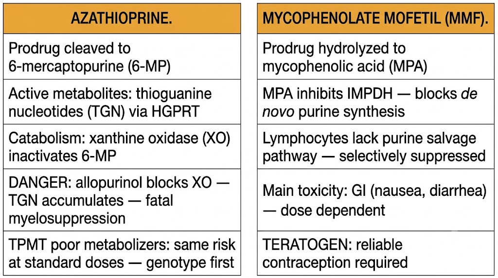
Azathioprine is a prodrug cleaved to 6-mercaptopurine, which is converted along competing metabolic pathways. The anabolic pathway via hypoxanthine-guanine phosphoribosyltransferase generates thioguanine nucleotides, the active immunosuppressive metabolites that incorporate into DNA and inhibit purine synthesis. The catabolic pathway via xanthine oxidase generates inactive thiouric acid. Thiopurine methyltransferase (TPMT) methylates 6-mercaptopurine to an inactive metabolite, shunting drug away from the active pathway.
The most dangerous drug interaction with azathioprine is coadministration with allopurinol or febuxostat, both xanthine oxidase inhibitors used for gout. Blocking xanthine oxidase causes four-fold accumulation of thioguanine nucleotides, producing severe, potentially fatal myelosuppression (pancytopenia). This combination is absolutely contraindicated unless the azathioprine dose is reduced by 67 to 75%, with weekly complete blood count monitoring.
TPMT activity is governed by common pharmacogenomic polymorphisms. Approximately 89 to 94% of individuals are homozygous for the high-activity wild-type allele and metabolize azathioprine normally. Approximately 6 to 11% are heterozygous carriers of a low-activity variant (TPMT*3A is most common in patients of European ancestry; TPMT*3C is more prevalent in patients of Asian and African ancestry) and require dose reduction of 30 to 50%. Approximately 0.3% are homozygous for low-activity alleles and develop profound myelotoxicity at standard doses. TPMT genotyping or phenotyping before initiating azathioprine is recommended. NUDT15 polymorphisms, more prevalent in patients of East Asian ancestry, also predict thiopurine myelotoxicity and should be tested in patients of relevant ancestry.
Mycophenolate Mofetil
Mycophenolate mofetil is a prodrug hydrolyzed to mycophenolic acid (MPA), which inhibits inosine monophosphate dehydrogenase (IMPDH), the rate-limiting enzyme in the de novo purine synthesis pathway. Lymphocytes are uniquely dependent on de novo purine synthesis and cannot effectively use the salvage pathway, making IMPDH inhibition selectively immunosuppressive for lymphocytes. Mycophenolic acid undergoes extensive enterohepatic recirculation, producing a secondary plasma peak at 6 to 12 hours after dosing.
Mycophenolate mofetil has largely replaced azathioprine in transplant protocols because of superior efficacy and a more favorable hematological safety profile. Its principal toxicities are gastrointestinal — nausea, vomiting, diarrhea, and abdominal cramping in 30 to 45% of patients — and dose-dependent. Gastrointestinal symptoms often improve with dose reduction or switching to enteric-coated mycophenolate sodium. Mycophenolate mofetil is a potent human teratogen, causing cleft palate, ear abnormalities, limb hypoplasia, and cardiac defects in up to 25% of exposed pregnancies. All women of childbearing potential must use reliable contraception during therapy. Azathioprine is the preferred antimetabolite when maintenance immunosuppression is required during pregnancy.
Antimetabolite mechanisms and key toxicities. Azathioprine requires pharmacogenomic screening (TPMT) and carries a fatal interaction with xanthine oxidase inhibitors. Mycophenolate mofetil is preferred in most transplant protocols but is an absolute teratogen requiring contraception. Abbreviations: GI = gastrointestinal; HGPRT = hypoxanthine-guanine phosphoribosyltransferase; IMPDH = inosine monophosphate dehydrogenase; MMF = mycophenolate mofetil; MPA = mycophenolic acid; TGN = thioguanine nucleotide; TPMT = thiopurine methyltransferase; XO = xanthine oxidase.
Allopurinol and febuxostat inhibit xanthine oxidase, the primary catabolic pathway for 6-mercaptopurine. Coadministration with azathioprine at standard doses causes four-fold accumulation of thioguanine nucleotides, producing severe pancytopenia that can be fatal. If gout management requires xanthine oxidase inhibitor use in a patient on azathioprine: reduce azathioprine dose by 67 to 75% and monitor complete blood count weekly for the first four weeks, then monthly. Safer alternatives: switch immunosuppressant to mycophenolate mofetil, or use a uricosuric agent (probenecid) for gout management. Colchicine and nonsteroidal anti-inflammatory drugs do not interact with azathioprine.
Section 4
Transplant Protocols, Rejection, and Induction Agents
Triple therapy maintenance, acute cellular versus antibody-mediated rejection, basiliximab, and anti-thymocyte globulin
Effective transplant immunosuppression integrates multiple drug classes with complementary mechanisms across three phases: induction at the time of transplant, maintenance to prevent ongoing rejection, and treatment of established rejection episodes. Understanding the pharmacological rationale of each phase and the distinction between cellular and antibody-mediated rejection is essential for clinical practice.
Triple Therapy Maintenance
The backbone of maintenance immunosuppression in solid organ transplantation is three mechanistically complementary agents: a calcineurin inhibitor (almost always tacrolimus), an antimetabolite (almost always mycophenolate mofetil), and a corticosteroid (prednisone at low maintenance doses). Each targets a distinct step in T-cell activation: the calcineurin inhibitor blocks NFAT activation and interleukin-2 transcription; mycophenolate mofetil blocks the proliferative response by depleting purine precursors; and corticosteroids suppress cytokine production and co-stimulatory signals. The combination provides synergistic immunosuppression at doses lower than any single agent alone, reducing the cumulative toxicity of each component. Calcineurin inhibitor-sparing protocols substitute a mammalian target of rapamycin inhibitor to preserve renal function.
Acute Cellular Rejection
Acute cellular rejection is mediated by recipient T cells recognizing donor alloantigens directly on donor antigen-presenting cells (direct allorecognition) or as processed peptides on recipient antigen-presenting cells (indirect allorecognition). In kidney transplantation it typically presents in the first 3 to 12 months with rising serum creatinine and reduced urine output, though many cases are subclinical and detected only on surveillance biopsy. Diagnosis requires allograft biopsy classified according to the Banff criteria, grading tubulointerstitial rejection (Banff I), vascular rejection (Banff II), and severe rejection (Banff III). Treatment is pulse intravenous methylprednisolone 250 to 1,000 milligrams daily for 3 to 5 days; steroid-resistant rejection is treated with anti-thymocyte globulin.
Antibody-Mediated Rejection
Antibody-mediated rejection is mediated by recipient immunoglobulin G antibodies against donor human leukocyte antigens (donor-specific antibodies). These bind to endothelial cells in the transplanted organ, activate complement, and recruit natural killer cells via antibody-dependent cellular cytotoxicity. It is more difficult to treat than acute cellular rejection and is the leading cause of late kidney allograft failure. Treatment of acute antibody-mediated rejection includes plasmapheresis to remove circulating donor-specific antibodies, high-dose intravenous immunoglobulin at 2 grams per kilogram to modulate anti-donor immune responses, and rituximab to deplete donor-specific antibody-producing B cells. Outcomes are significantly worse than for acute cellular rejection.
Induction Agents
Basiliximab is a chimeric anti-CD25 monoclonal antibody (CD25 is the interleukin-2 receptor alpha chain) administered as two fixed doses on day 0 and day 4 post-transplant. It saturates the high-affinity interleukin-2 receptor for approximately 4 to 6 weeks, blocking interleukin-2-driven T-cell proliferation in the critical early post-transplant period. It is well tolerated with minimal adverse effects and is the standard induction agent in standard-immunological-risk recipients.
Anti-thymocyte globulin, available as a rabbit preparation (thymoglobulin), is a polyclonal antibody directed against multiple T-cell surface antigens including CD3, CD4, CD8, CD25, and CD28. It causes profound and prolonged lymphocyte depletion through complement-dependent cytotoxicity and antibody-dependent cellular cytotoxicity, and is used for high-immunological-risk recipients and for steroid-resistant acute rejection. Adverse effects include cytokine release syndrome during infusion (managed by pre-medication with corticosteroids, antihistamines, and acetaminophen), leukopenia, thrombocytopenia, and increased risk of cytomegalovirus infection and post-transplant lymphoproliferative disorder.
Transplant Protocol Summary
Standard-risk induction: Basiliximab 20 milligrams intravenously on day 0 and day 4. High-risk induction: Anti-thymocyte globulin (thymoglobulin) 1.5 milligrams per kilogram per day intravenously for 3 to 7 days; pre-medicate for cytokine release syndrome. Maintenance triple therapy: Tacrolimus (target trough 8 to 12 nanograms per milliliter early, 5 to 8 nanograms per milliliter maintenance) plus mycophenolate mofetil 1 to 1.5 grams twice daily plus prednisone tapered to 5 to 10 milligrams per day by 3 to 6 months. Acute cellular rejection: Pulse methylprednisolone 250 to 1,000 milligrams intravenously daily for 3 to 5 days; steroid-resistant: anti-thymocyte globulin. Acute antibody-mediated rejection: Plasmapheresis plus intravenous immunoglobulin plus rituximab. Prophylaxis required in all transplant recipients: Trimethoprim-sulfamethoxazole (Pneumocystis jirovecii), valganciclovir (cytomegalovirus in high-risk donor-positive/recipient-negative pairs), antifungal prophylaxis per protocol.
Suggested References
Author / Organization
Title
Source
Halloran PF
Immunosuppressive drugs for kidney transplantation
N Engl J Med. 2004;351(26):2715-2729
Vanhove T, Annaert P, Kuypers DRJ
Clinical determinants of calcineurin inhibitor disposition: a mechanistic review
Drug Metab Rev. 2016;48(1):88-112
Staatz CE, Tett SE
Clinical pharmacokinetics and pharmacodynamics of tacrolimus in solid organ transplantation
Clin Pharmacokinet. 2004;43(10):623-653
Naesens M, Kuypers DRJ, Sarwal M
Calcineurin inhibitor nephrotoxicity
Clin J Am Soc Nephrol. 2009;4(2):481-508
Ekberg H, Tedesco-Silva H, Demirbas A, et al
Reduced exposure to calcineurin inhibitors in renal transplantation
N Engl J Med. 2007;357(25):2562-2575
Sehgal SN
Sirolimus: its discovery, biological properties, and mechanism of action
Transplant Proc. 2003;35(3 Suppl):7S-14S
Relling MV, Schwab M, Whirl-Carrillo M, et al
Clinical pharmacogenomics implementation consortium guideline for thiopurine dosing based on TPMT and NUDT15 genotypes
Clin Pharmacol Ther. 2019;105(5):1095-1105
Allison AC, Eugui EM
Mycophenolate mofetil and its mechanisms of action
Immunopharmacology. 2000;47(2-3):85-118
Rhen T, Cidlowski JA
Antiinflammatory action of glucocorticoids — new mechanisms for old drugs
N Engl J Med. 2005;353(16):1711-1723
Kidney Disease: Improving Global Outcomes Transplant Work Group
KDIGO clinical practice guideline for the care of kidney transplant recipients
Am J Transplant. 2009;9(Suppl 3):S1-S155
Haas M, Loupy A, Lefaucheur C, et al
The Banff 2017 kidney meeting report: revised diagnostic criteria for chronic active T cell-mediated rejection and antibody-mediated rejection