CHAPTER 40  ·  IMMUNOPHARMACOLOGY
Section 1
Complement System Pharmacology
Classical, lectin, and alternative pathway activation; C5 inhibitors (eculizumab, ravulizumab); C3 inhibition (pegcetacoplan); alternative pathway blockade (iptacopan); and the meningococcal infection mandate

The complement cascade is activated through three pathways converging at C3 (complement component 3) cleavage, generating opsonins, anaphylatoxins, and the membrane attack complex that lyses target cells. When host regulatory proteins are genetically absent or overwhelmed, complement drives severe hematological, renal, and neurological disease. Pharmacological complement inhibition has become a transformative strategy for several rare but life-threatening conditions.

Complement Pathways and Disease Mechanisms

The classical pathway is triggered by C1q binding to antibody-antigen complexes on cell surfaces, sequentially activating C4 (complement component 4) and C2 (complement component 2) to form the classical pathway C3 convertase (C4b2a). The lectin pathway is activated by mannose-binding lectin or ficolins recognizing carbohydrate patterns on microbial surfaces, activating MBL-associated serine proteases to form the same C4b2a convertase. The alternative pathway operates as a continuous low-level surveillance mechanism through spontaneous C3 hydrolysis (tick-over), amplified on pathogen or damaged cell surfaces lacking host regulatory proteins — primarily CD55 (decay-accelerating factor, which accelerates convertase decay), CD59 (protectin, which blocks membrane attack complex assembly), and factor H (which promotes C3b cleavage by factor I). All three pathways converge on C3 convertases that cleave C3 into C3a (an anaphylatoxin promoting mast cell degranulation and vascular permeability) and C3b (an opsonin that deposits on target surfaces). C5 (complement component 5) convertases then cleave C5 into C5a (the most potent complement-derived inflammatory mediator, driving neutrophil chemotaxis and activation) and C5b, which nucleates the membrane attack complex (C5b-9) causing osmotic lysis.

Paroxysmal nocturnal hemoglobinuria (PNH) is caused by somatic mutations in the PIGA gene (phosphatidylinositol glycan biosynthesis class A) in hematopoietic stem cells, impairing GPI (glycosylphosphatidylinositol) anchor biosynthesis and depleting CD55 and CD59 from erythrocyte and platelet surfaces. Without these regulators, PNH erythrocytes undergo spontaneous complement-mediated intravascular hemolysis, and platelets are activated, causing the thrombotic complications that are the leading cause of mortality in PNH. Atypical hemolytic uremic syndrome (aHUS) results from mutations or autoantibodies affecting alternative pathway regulatory proteins (factor H, factor I, or CD46 known as membrane cofactor protein), producing uncontrolled alternative pathway activation in the renal microvasculature causing thrombotic microangiopathy with hemolytic anemia, thrombocytopenia, and acute kidney injury. In neurology, neuromyelitis optica spectrum disorder (NMOSD) with anti-aquaporin-4 (anti-AQP4) antibodies activates complement at astrocyte foot processes causing astrocyte destruction, and generalized myasthenia gravis (gMG) with anti-acetylcholine receptor antibodies activates terminal complement at the neuromuscular junction.

C5 Inhibitors: Eculizumab and Ravulizumab

Eculizumab is a humanized monoclonal antibody that binds C5 with high affinity, preventing its cleavage into C5a and C5b by the C5 convertase. This blocks both C5a-mediated inflammation and membrane attack complex formation while preserving upstream C3b-mediated opsonization, which is important for ongoing host defense. Eculizumab is approved for PNH, aHUS, gMG, and NMOSD with anti-AQP4 antibodies. In PNH it dramatically reduces intravascular hemolysis, transfusion requirements, and thrombotic events. It is administered intravenously every two weeks after a loading phase.

Ravulizumab is an engineered successor to eculizumab with four amino acid substitutions that extend its plasma half-life approximately four-fold through increased affinity for the neonatal Fc receptor, allowing maintenance dosing every eight weeks rather than every two weeks. It has the same mechanism, the same approved indications, and the same safety requirements as eculizumab. The clinical decision between the two agents is primarily based on dosing convenience.

Because the terminal complement complex is the primary defense mechanism against encapsulated bacteria — particularly Neisseria meningitidis (meningococcus) — all complement inhibitors carry a mandatory meningococcal vaccination requirement. Both the meningococcal ACWY conjugate vaccine and the meningococcal type B vaccine (Bexsero or Trumenba) must be administered at least two weeks before the first dose. Prophylactic antibiotics (penicillin V or amoxicillin) are recommended throughout therapy. Patients must be educated about the symptoms of meningococcal disease — sudden severe headache, fever, stiff neck, petechial rash — and the need for immediate emergency evaluation, because complement inhibitor-related meningococcal disease can be fulminant and fatal within hours.

Proximal and Alternative Pathway Inhibitors

Approximately 30% of PNH patients treated with anti-C5 therapy experience residual hemolysis through extravascular mechanisms: C5 inhibition does not prevent C3b deposition on PNH erythrocytes, and C3b-opsonized erythrocytes are removed by splenic and hepatic macrophages. Pegcetacoplan is a pegylated cyclic peptide that binds C3 and C3b directly, blocking all three complement pathways upstream of C5 and preventing both intravascular hemolysis (through downstream MAC blockade) and extravascular hemolysis (through prevention of C3b opsonization). It is approved for PNH in adults inadequately controlled on eculizumab or ravulizumab and is administered subcutaneously twice weekly.

Iptacopan is an oral factor B inhibitor; factor B is the serine protease that associates with C3b to form the alternative pathway C3 convertase (C3bBb). By selectively inhibiting factor B, iptacopan blocks only the alternative pathway amplification loop while leaving the classical and lectin pathways intact, preserving a broader component of innate immune defense than upstream pan-complement inhibitors. Iptacopan is approved as oral monotherapy for PNH and demonstrated superiority over anti-C5 therapy in reducing extravascular hemolysis and improving hemoglobin levels in clinical trials. Avacopan is an oral small-molecule antagonist of the C5a receptor 1 (C5aR1, also called CD88), approved for anti-neutrophil cytoplasmic antibody (ANCA)-associated vasculitis — specifically granulomatosis with polyangiitis and microscopic polyangiitis — where it serves as a corticosteroid-sparing strategy by blocking C5a-driven neutrophil activation in the renal glomerulus without requiring meningococcal vaccination because MAC formation is unaffected.
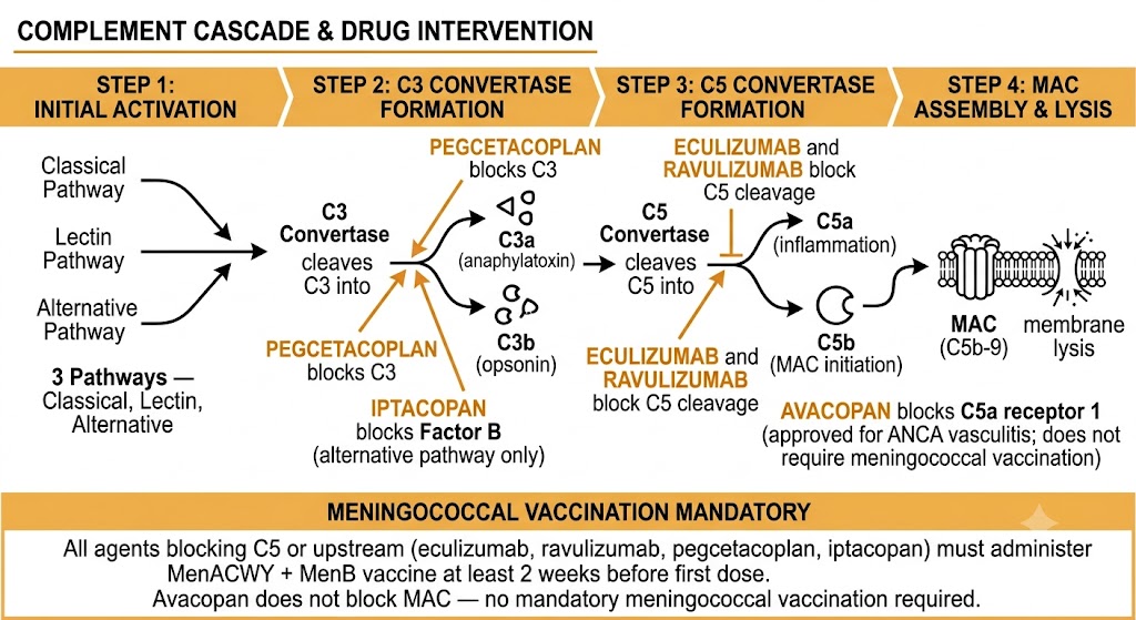
Flow diagram of the complement cascade showing four steps: Step 1 (three activation pathways — classical, lectin, alternative), Step 2 (C3 convertase cleaving C3 into C3a anaphylatoxin and C3b opsonin, with pegcetacoplan blocking C3 and iptacopan blocking factor B of the alternative pathway), Step 3 (C5 convertase cleaving C5 into C5a driving inflammation and C5b initiating MAC assembly, with eculizumab and ravulizumab blocking C5 cleavage, and avacopan blocking C5a receptor 1 for ANCA vasculitis without requiring meningococcal vaccination), and Step 4 (MAC C5b-9 causing membrane lysis), with a meningococcal vaccination mandatory warning box below stating all agents blocking C5 or upstream require MenACWY plus MenB vaccine at least two weeks before first dose while avacopan does not block MAC and does not require mandatory meningococcal vaccination.
The complement cascade and approved drug intervention points. Pegcetacoplan and iptacopan act upstream at C3; eculizumab and ravulizumab act at C5; avacopan blocks the C5a receptor without affecting MAC formation and does not require meningococcal vaccination. Abbreviations: ANCA = anti-neutrophil cytoplasmic antibody; MAC = membrane attack complex; MenACWY = meningococcal serogroups A, C, W, Y; MenB = meningococcal type B.
Meningococcal Infection Risk — Mandatory for All Complement Inhibitors

Terminal complement is the primary defense against Neisseria meningitidis. All complement inhibitors — eculizumab, ravulizumab, pegcetacoplan, iptacopan — increase the risk of fulminant meningococcal disease. Required before first dose: meningococcal ACWY conjugate vaccine AND meningococcal type B vaccine at least two weeks before initiation. If urgent treatment is needed, start prophylactic antibiotics (penicillin V 250 milligrams twice daily) immediately and continue until at least two weeks after vaccination. Continue antibiotic prophylaxis throughout therapy. Educate patients: sudden headache, fever, neck stiffness, or petechial rash requires immediate emergency evaluation — meningococcal disease in complement-inhibited patients can be fatal within hours. Note: avacopan (C5a receptor 1 antagonist) does not block MAC formation and does not require meningococcal vaccination.

Section 2
IVIG, Co-stimulation Blockade, and Plasma Cell-Directed Therapy
Intravenous immunoglobulin mechanisms and indications, abatacept and belatacept (CD28 co-stimulation blockade), BTK inhibition, and plasma cell targeting with daratumumab

The final dimension of immunopharmacology covered in this chapter encompasses agents that modulate immune function through three additional mechanisms: polyspecific immunoglobulin replacement and immunomodulation via intravenous immunoglobulin; selective interruption of the T-cell co-stimulatory signal required for full activation; and direct depletion of long-lived plasma cells that perpetuate pathogenic autoantibody production beyond the reach of B-cell-depleting agents.

Intravenous Immunoglobulin: Mechanisms and Indications

Intravenous immunoglobulin (IVIG) is a pooled polyspecific IgG preparation derived from thousands of healthy donors. At high immunomodulatory doses (typically 1 to 2 grams per kilogram), IVIG acts through several converging mechanisms. First, it saturates the neonatal Fc receptor (FcRn) responsible for IgG recycling and extended plasma half-life, accelerating catabolism of endogenous IgG — including pathogenic autoantibodies — reducing their circulating half-life by 10 to 14 days. Second, it blocks activating Fc-gamma receptors on macrophages, dendritic cells, and natural killer cells, preventing phagocytosis of opsonized cells; this is the mechanism most relevant to immune thrombocytopenia (ITP), where it rapidly raises platelet counts by blocking splenic macrophage destruction of antibody-coated platelets. Third, IVIG contains anti-idiotypic antibodies that can directly neutralize pathogenic autoantibodies. Fourth, it modulates complement activation and cytokine production.

IVIG has two broad categories of use. In primary and secondary immunodeficiency states (common variable immunodeficiency, X-linked agammaglobulinemia, hypogammaglobulinemia from chronic lymphocytic leukemia or post-hematopoietic stem cell transplant), IVIG is replacement therapy restoring functional antibody levels at monthly replacement dosing (typically 400 to 600 milligrams per kilogram monthly). At high immunomodulatory doses, IVIG is used for Guillain-Barre syndrome, chronic inflammatory demyelinating polyneuropathy, multifocal motor neuropathy, ITP, Kawasaki disease, dermatomyositis, pemphigus vulgaris, and myasthenia gravis crisis.

Adverse effects include infusion reactions (headache, flushing, chills, fever in 5 to 15% of infusions) managed by slowing the rate and pre-medicating with acetaminophen and antihistamine. Thromboembolic events — deep vein thrombosis, pulmonary embolism, myocardial infarction, and stroke — occur particularly in older patients with cardiovascular risk factors, through IVIG-induced increases in plasma viscosity; adequate hydration and slow infusion rates reduce this risk. Hemolytic anemia can occur from anti-blood group antibodies in the IVIG preparation, particularly in non-O blood group patients receiving large doses. Patients with selective IgA deficiency who have developed anti-IgA antibodies may experience anaphylaxis from IgA in standard IVIG preparations; IgA-depleted IVIG formulations are available for this population.

Co-stimulation Blockade: Abatacept and Belatacept

Full T-cell activation requires two signals: antigen recognition through the T-cell receptor, and a co-stimulatory signal delivered by CD80 (B7-1) or CD86 (B7-2) on antigen-presenting cells engaging CD28 on the T-cell surface. Without the co-stimulatory signal, antigen-specific T cells become anergic rather than activating. Abatacept is a recombinant fusion protein of the extracellular domain of CTLA-4 (cytotoxic T-lymphocyte-associated protein 4) fused to the Fc region of human IgG1. Because CTLA-4 binds CD80 and CD86 with approximately 500 to 2,500-fold higher affinity than CD28, abatacept competitively occupies both co-stimulatory ligands on antigen-presenting cells, delivering an incomplete anergy-inducing signal to antigen-specific T cells. Abatacept is approved for rheumatoid arthritis, psoriatic arthritis, juvenile idiopathic arthritis, and prevention of acute graft-versus-host disease after hematopoietic stem cell transplantation. In rheumatoid arthritis it is particularly effective in seropositive patients (anti-cyclic citrullinated peptide or rheumatoid factor positive), where T-cell-driven pathology is more prominent.

Belatacept is a second-generation CTLA-4-Ig fusion protein with two amino acid substitutions (L104E and A29Y) that confer approximately 10-fold higher affinity for CD80 and CD86 than abatacept. It is approved for prophylaxis of organ rejection in kidney transplant recipients as an alternative to calcineurin inhibitors, providing superior preservation of long-term renal function because it lacks the vasoconstricting and nephrotoxic effects of calcineurin inhibitors. The trade-off is a higher rate of acute rejection in the first year and an increased risk of post-transplant lymphoproliferative disorder in Epstein-Barr virus-seronegative recipients, which is an absolute contraindication to belatacept use. Belatacept is administered intravenously monthly after the peri-transplant period.

A critical conceptual distinction: abatacept and belatacept activate the CTLA-4 inhibitory checkpoint to suppress T-cell responses — the opposite direction from oncology checkpoint inhibitors (pembrolizumab, nivolumab, ipilimumab), which block CTLA-4 and PD-1 to release inhibitory constraints on anti-tumor T cells. The immune-related adverse events of checkpoint inhibitor cancer therapy are autoimmune in nature and can be treated with corticosteroids or abatacept, illustrating the pharmacological reciprocity at these checkpoints.

Plasma Cell-Directed Therapy: Daratumumab and Bortezomib

A fundamental limitation of B-cell-depleting agents such as rituximab is their inability to eliminate long-lived plasma cells, which lack CD20 expression and are maintained in bone marrow survival niches where they continue secreting pathogenic autoantibodies for months to years. Daratumumab is a fully human IgG1 monoclonal antibody directed against CD38 (cluster of differentiation 38), a transmembrane glycoprotein highly expressed on plasma cells (both malignant myeloma cells and non-malignant long-lived plasma cells). Daratumumab depletes CD38-expressing cells through ADCC, complement-dependent cytotoxicity, antibody-dependent cellular phagocytosis, and direct apoptosis induction. Approved for multiple myeloma and light chain amyloidosis, daratumumab is under active investigation in refractory antibody-mediated autoimmune diseases including myasthenia gravis, systemic lupus erythematosus, pemphigus vulgaris, and anti-GBM (glomerular basement membrane) disease, where case series have demonstrated rapid and deep depletion of both short-lived and long-lived plasma cells. A practical safety note: CD38 is expressed on erythrocytes, causing a pan-reactive positive direct antiglobulin test that interferes with blood compatibility testing; specialized pre-transfusion testing is required for daratumumab-treated patients.

Bortezomib is a reversible proteasome inhibitor approved for multiple myeloma and mantle cell lymphoma that selectively depletes plasma cells through a different mechanism. Plasma cells are uniquely vulnerable to proteasome inhibition because their extremely high rate of immunoglobulin synthesis generates a large burden of misfolded protein chains that must be cleared by the proteasome; when proteasome function is blocked, misfolded immunoglobulin accumulates, triggering the terminal unfolded protein response and apoptosis. Bortezomib has been used off-label for antibody-mediated transplant rejection, severe lupus nephritis, and neuromyelitis optica refractory to standard therapy. Its dose-limiting toxicity is peripheral sensory neuropathy, occurring in 30 to 40% of patients.

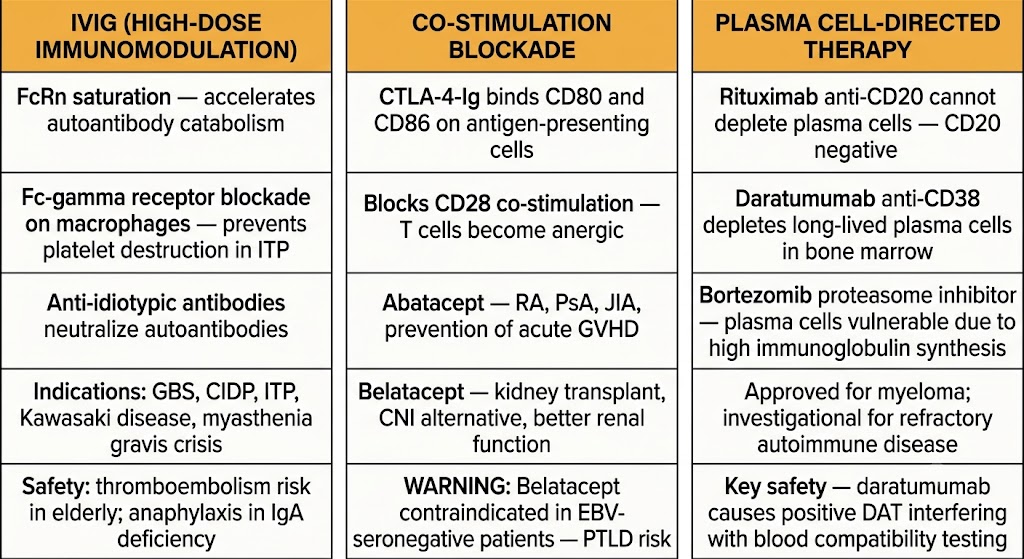
Three-panel comparison diagram: left panel IVIG high-dose immunomodulation showing FcRn saturation accelerating autoantibody catabolism, Fc-gamma receptor blockade on macrophages preventing platelet destruction in ITP, anti-idiotypic antibody neutralization, indications including GBS/CIDP/ITP/Kawasaki/myasthenia gravis crisis, and thromboembolism and IgA deficiency anaphylaxis safety notes; center panel co-stimulation blockade showing CTLA-4-Ig binding CD80 and CD86 to block CD28 co-stimulation causing T-cell anergy, abatacept approved for RA/PsA/JIA/acute GVHD prevention, belatacept as CNI alternative in kidney transplant with better renal function, and belatacept contraindication in EBV-seronegative patients due to PTLD risk; right panel plasma cell-directed therapy showing rituximab cannot deplete CD20-negative plasma cells, daratumumab anti-CD38 depleting long-lived bone marrow plasma cells, bortezomib proteasome inhibitor exploiting plasma cell vulnerability to high immunoglobulin synthesis, indications in myeloma and investigational autoimmune disease, and daratumumab positive DAT safety note.
Three complementary immunotherapy approaches: IVIG (polyspecific immunoglobulin immunomodulation), co-stimulation blockade (abatacept and belatacept), and plasma cell-directed therapy (daratumumab and bortezomib). Plasma cell-directed therapy addresses the limitation of rituximab, which cannot eliminate CD20-negative long-lived plasma cells. Abbreviations: CIDP = chronic inflammatory demyelinating polyneuropathy; CNI = calcineurin inhibitor; CTLA-4 = cytotoxic T-lymphocyte-associated protein 4; DAT = direct antiglobulin test; EBV = Epstein-Barr virus; GBS = Guillain-Barre syndrome; GVHD = graft-versus-host disease; ITP = immune thrombocytopenia; PTLD = post-transplant lymphoproliferative disorder.
Co-stimulation Blockade vs. Checkpoint Inhibition — Key Distinction

Abatacept and belatacept are CTLA-4-Ig fusion proteins that activate the CTLA-4 inhibitory checkpoint, suppressing autoreactive T-cell responses. Oncology checkpoint inhibitors (ipilimumab, pembrolizumab, nivolumab) block CTLA-4 or PD-1 to release inhibitory constraints on anti-tumor T cells — the opposite pharmacological direction. Immune-related adverse events from checkpoint inhibitor cancer therapy are autoimmune in nature (inflammatory arthritis, colitis, thyroiditis, pneumonitis) and can be treated with corticosteroids; abatacept is emerging as a treatment option for checkpoint inhibitor-induced inflammatory arthritis, directly illustrating this pharmacological reciprocity.


Suggested References
Author / Organization Title Source
Ricklin D, Hajishengallis G, Yang K, Lambris JD Complement: a key system for immune surveillance and homeostasis Nat Immunol. 2010;11(9):785-797
Noris M, Remuzzi G Overview of complement activation and regulation Semin Nephrol. 2013;33(6):479-492
Hillmen P, Hall C, Marsh JCW, et al Effect of eculizumab on hemolysis and transfusion requirements in patients with paroxysmal nocturnal hemoglobinuria N Engl J Med. 2004;350(6):552-559
Peffault de Latour R, Brodsky RA, Ortiz S, et al Pharmacokinetic and pharmacodynamic effects of ravulizumab and eculizumab on complement component 5 in adults with paroxysmal nocturnal hemoglobinuria Haematologica. 2020;105(8):2039-2046
Jayne DRW, Merkel PA, Schall TJ, Bekker P; ADVOCATE Study Group Avacopan for the treatment of ANCA-associated vasculitis N Engl J Med. 2021;384(7):599-609
Schwab I, Nimmerjahn F Intravenous immunoglobulin therapy: how does IgG modulate the immune system? Nat Rev Immunol. 2013;13(3):176-189
Vincenti F, Larsen C, Durrbach A, et al Costimulation blockade with belatacept in renal transplantation N Engl J Med. 2005;353(8):770-781
Ponader S, Burger JA Bruton's tyrosine kinase: from X-linked agammaglobulinemia toward targeted therapy for B-cell malignancies J Clin Oncol. 2014;32(17):1830-1839
Nutt SL, Hodgkin PD, Tarlinton DM, Corcoran LM The generation of antibody-secreting plasma cells Nat Rev Immunol. 2015;15(3):160-171
Chung A, Tumlin J, Kooienga L, et al Daratumumab in autoimmune diseases: a mechanism-based review Am J Hematol. 2023;98(6):998-1010
Vande Casteele N, Herfarth H, Katz J, Falck-Ytter Y, Singh S American Gastroenterological Association Institute technical review on the role of therapeutic drug monitoring in the management of inflammatory bowel diseases Gastroenterology. 2017;153(3):835-857