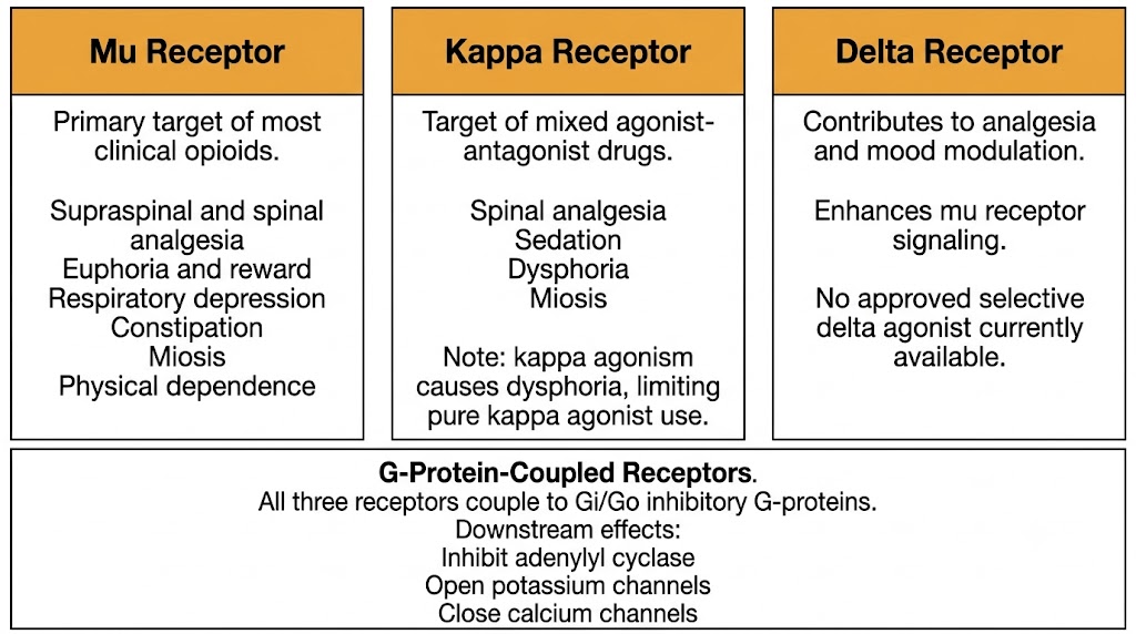
Figure 1. The three classical opioid receptor types and their primary pharmacological effects. All three couple to Gi/Go inhibitory G-proteins. Figure generated by Gemini AI.
Introduction to Medical Pharmacology
Module 1 — Opioid Receptors and the Endogenous Opioid System
Chapter 13 · Module 1 of 5Section 1
Three classical receptor types, one pharmacological family
Opioid drugs produce their effects by binding to specific receptors distributed throughout the nervous system and peripheral tissues. Understanding which receptor does what is the foundation of everything else in opioid pharmacology: why morphine relieves pain and depresses breathing, why certain drugs cause dysphoria instead of euphoria, and why buprenorphine behaves so differently from methadone.
The term opioid refers to any substance, endogenous or synthetic, that binds to opioid receptors and produces effects reversible by the antagonist naloxone. The older term opiate refers specifically to naturally occurring alkaloids from the opium poppy, principally morphine and codeine. Because modern clinical pharmacology includes a vast range of synthetic compounds that act on opioid receptors but bear no structural resemblance to plant alkaloids, the broader term opioid is preferred throughout this chapter.
All three classical opioid receptors belong to the G-protein-coupled receptor superfamily and couple primarily to inhibitory G-proteins of the Gi and Go class. Despite sharing this common signaling mechanism, the three receptor types produce distinct pharmacological profiles depending on where they are expressed and which circuits they modulate.
Receptor Type
Mu Receptor
Receptor Type
Kappa Receptor
Receptor Type
Delta Receptor
Clinical Anchor: Which Receptor Matters Most
The mu receptor drives virtually all of the clinically relevant pharmacology of the opioid analgesics used in practice: analgesia, euphoria, respiratory depression, constipation, miosis, and physical dependence are all primarily mu receptor-mediated effects. The kappa receptor becomes clinically important when understanding mixed agonist-antagonist drugs such as buprenorphine, nalbuphine, and butorphanol, which exploit kappa agonism to produce analgesia while blocking mu-mediated euphoria and dependence.
Figure 1. The three classical opioid receptor types and their primary pharmacological effects. All three couple to Gi/Go inhibitory G-proteins. Figure generated by Gemini AI.
Section 2
Gi and Go coupling produces three coordinated inhibitory effects
When an opioid agonist binds to a mu, kappa, or delta receptor, the receptor activates inhibitory G-proteins (Gi and Go). This produces three distinct downstream effects that together reduce neuronal excitability and decrease the transmission of pain signals. All three mechanisms contribute to analgesia; the same mechanisms in brainstem respiratory neurons account for respiratory depression.
Gi protein activation inhibits adenylyl cyclase, reducing intracellular cyclic adenosine monophosphate levels. This decreases the activity of downstream signaling proteins and reduces gene expression in pain-processing neurons. With chronic opioid use, neurons compensate by upregulating adenylyl cyclase. When opioids are abruptly withdrawn, this upregulated enzyme is suddenly unopposed, causing a surge in cyclic adenosine monophosphate that drives many of the symptoms of opioid withdrawal.
Opioid receptor activation opens inwardly rectifying potassium channels, allowing potassium ions to flow out of the cell. The resulting hyperpolarization makes neurons less likely to fire action potentials. This mechanism contributes to both the analgesic effect (pain-transmitting neurons in the spinal cord fire less) and the sedative and respiratory depressant effects (brainstem neurons controlling wakefulness and breathing are hyperpolarized).
Opioids inhibit voltage-gated calcium channels on presynaptic nerve terminals. Because calcium influx is required for neurotransmitter release, this reduces the release of pain-transmitting neurotransmitters — including glutamate, substance P, and calcitonin gene-related peptide — from primary afferent pain fibers into the spinal cord dorsal horn. This presynaptic inhibition at nociceptive synapses is a major mechanism of spinal opioid analgesia and is the primary reason intrathecal and epidural opioids can produce profound analgesia at very low doses.
Three Mechanisms, One Net Effect
All three downstream effects work together to reduce neuronal excitability: less cyclic adenosine monophosphate signaling, hyperpolarization through potassium channel opening, and reduced neurotransmitter release through calcium channel closure. The same mechanisms that silence pain-processing neurons also act in brainstem respiratory centers — which is why there is no currently available opioid analgesic that is completely free of respiratory depression risk at sufficient doses.
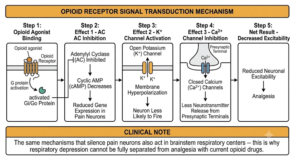
Figure 2. Opioid receptor signal transduction mechanism. The same downstream effects that produce analgesia also act in brainstem respiratory centers, explaining why respiratory depression risk cannot be fully separated from analgesia. Figure generated by Gemini AI.
Section 3
The body's own opioid system: beta-endorphin, enkephalins, and dynorphins
The existence of opioid receptors in the brain implied that the body must produce its own ligands for them. The discovery of the endogenous opioid peptides in the 1970s confirmed this and revealed a natural pain-modulation system that is engaged during stress, exercise, and injury. Three peptide families are the primary endogenous opioids, each derived from a distinct precursor protein and each with a characteristic receptor preference.
Beta-endorphin is derived from the precursor protein pro-opiomelanocortin, which is produced in the pituitary gland and in hypothalamic neurons. It is the most potent endogenous opioid peptide and acts primarily at mu and delta receptors. Beta-endorphin is released during stress, intense exercise, and pain, producing the widespread analgesia and mood elevation associated with these states. It functions both as a neurotransmitter in the brain and as a circulating neuromodulator.
The enkephalins, met-enkephalin and leu-enkephalin, are derived from the precursor protein proenkephalin. They are widely distributed throughout the brain, spinal cord, and peripheral nervous system. Enkephalins act preferentially at delta receptors and also modestly at mu receptors. They function as short-range neurotransmitters in pain-modulating circuits, particularly in the spinal cord dorsal horn where they inhibit incoming pain signals from primary afferent neurons.
Dynorphin A and dynorphin B are derived from the precursor protein prodynorphin. Dynorphins act primarily at kappa receptors. They are found in the hypothalamus, spinal cord, and limbic system. The association of kappa receptor activation with dysphoria means that dynorphin release in limbic circuits contributes to the unpleasant aspects of stress and negative emotional states, in contrast to the rewarding effects of mu receptor-activating beta-endorphin.
Summary Table
Endogenous Peptides at a Glance
Clinical Relevance
Why This Matters for Prescribing
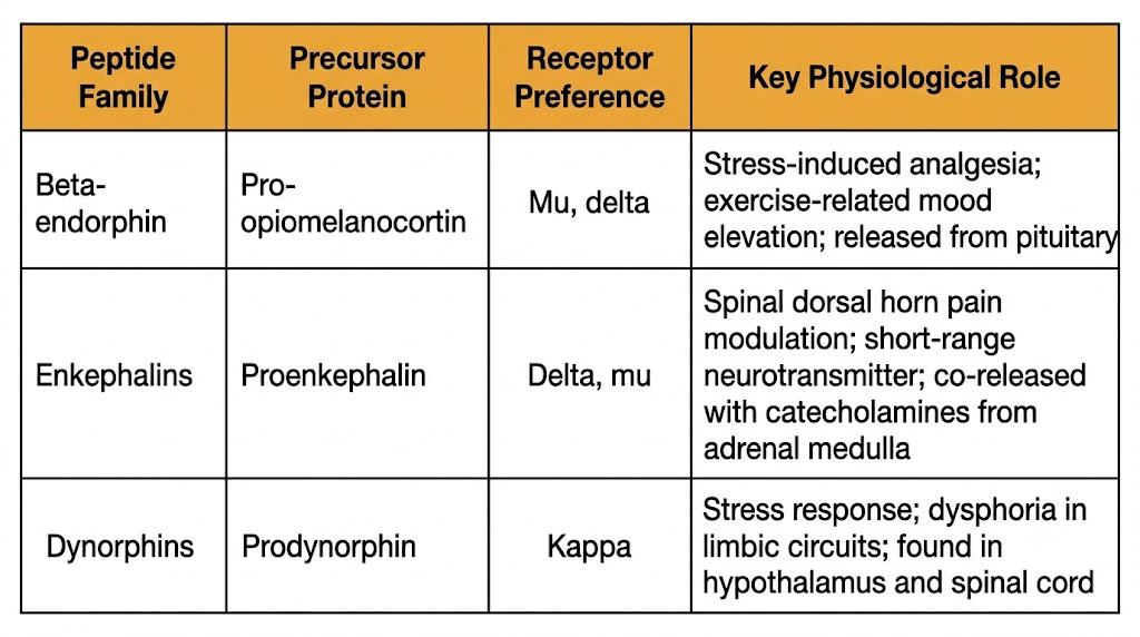
Figure 3. Endogenous opioid peptide families, their precursor proteins, receptor preferences, and key physiological roles. Figure generated by Gemini AI.
Section 4
Supraspinal, spinal, and peripheral mechanisms each contribute
Opioid receptors are distributed at three anatomically distinct levels of the pain pathway, and each level contributes to the analgesic effect. This multilevel architecture has direct clinical implications — it explains why intrathecal and epidural opioids are so potent at such low doses, and it provides a framework for understanding why systemic opioids produce effects in the brain as well as at the site of pain.
In the brain, the periaqueductal gray matter is the most important opioid-sensitive structure for pain modulation. Opioid activation here triggers a cascade of descending inhibitory signals that travel down through the brainstem to the spinal cord, suppressing pain transmission at the dorsal horn. This descending inhibitory system is the mechanism by which stress-induced analgesia occurs naturally and can be amplified pharmacologically by systemically administered opioids. The limbic system is also rich in mu receptors; opioid action here reduces the emotional and affective unpleasantness of pain, separate from the sensory intensity component.
The spinal cord dorsal horn — particularly the superficial layers that receive primary pain fiber input — contains a high density of mu and delta receptors on both presynaptic terminals and postsynaptic neurons. Presynaptically, opioid-mediated closure of calcium channels reduces the release of pain-transmitting substances from incoming nerve fibers. Postsynaptically, potassium channel opening hyperpolarizes dorsal horn neurons, reducing their responsiveness to any remaining input. The clinical consequence of this concentrated action at a single anatomical site is that intrathecal and epidural opioids can produce profound analgesia at doses 100 to 1,000 times lower than are required systemically, with correspondingly reduced sedation and respiratory depression risk compared to high-dose systemic dosing.
Opioid receptors are also expressed on the peripheral terminals of primary afferent pain fibers. Under normal conditions these peripheral receptors play a limited role, but during tissue inflammation they become more active and more accessible. Inflammatory mediators enhance opioid receptor coupling at peripheral terminals, and the disruption of normal tissue barriers allows exogenous opioids to reach these sites more readily. This peripheral mechanism provides a rationale for intra-articular opioid administration in the context of joint inflammation and is an area of active research aimed at developing opioid drugs that act predominantly at peripheral sites to minimize central nervous system adverse effects.
Section 5
Three concepts that are related but clinically distinct
Repeated opioid exposure produces predictable neuroadaptations that alter how the nervous system responds to both the drug and to its absence. Three terms describe different aspects of these adaptations, and they are frequently confused — both in clinical settings and on examinations. Precision in distinguishing them is clinically essential.
Tolerance is a pharmacological adaptation in which repeated exposure to a drug results in a diminished response at the same dose, requiring dose escalation to maintain the original effect. With opioids, tolerance develops to some effects but not others. Tolerance develops to: analgesia, euphoria, sedation, nausea, and respiratory depression. Tolerance does not develop to: constipation and miosis. This is clinically important — a patient on stable high-dose chronic opioid therapy will still have pinpoint pupils and persistent constipation regardless of how long they have been on the medication, even if the analgesic effect has partially diminished.
Physical dependence is a state of neuroadaptation in which the nervous system has adjusted its baseline function to account for the persistent presence of opioids. When the drug is abruptly removed, the compensatory changes are suddenly unopposed, producing a withdrawal syndrome. The molecular basis of this adaptation is upregulation of adenylyl cyclase during chronic opioid exposure. When opioids are removed, cyclic adenosine monophosphate levels surge above normal, driving the autonomic and somatic features of opioid withdrawal: anxiety, restlessness, sweating, gooseflesh, diarrhea, cramping, tachycardia, and hypertension. Physical dependence is a pharmacological phenomenon that can occur in any patient receiving regular opioids — it does not imply addiction.
Addiction (opioid use disorder) is a behavioral syndrome characterized by compulsive drug-seeking and continued use despite harm. It is distinct from physical dependence, though the two can co-occur. The neurobiological substrate of opioid addiction is the mesolimbic dopamine system. Mu receptor activation in the ventral tegmental area suppresses inhibitory interneurons that normally restrain dopamine neurons. With this inhibition removed, dopamine neurons fire more actively and release dopamine in the nucleus accumbens, producing the intense reward signal that drives reinforced drug-seeking behavior. The speed of opioid delivery to the brain strongly influences the magnitude of this dopamine surge — which explains why intravenous and intranasal routes produce greater euphoria and higher addiction potential than oral routes of the same drug.
Key Distinction: Dependence Is Not Addiction
Physical dependence is a predictable physiological consequence of regular opioid use. A surgical patient who receives around-the-clock opioid infusion for ten days will be physically dependent and will experience withdrawal if the drug is stopped abruptly. This patient does not have opioid use disorder. Addiction requires compulsive use despite harm, loss of control over use, and continued use in the face of adverse consequences — behavioral features that are entirely separate from the physiological adaptation of dependence.
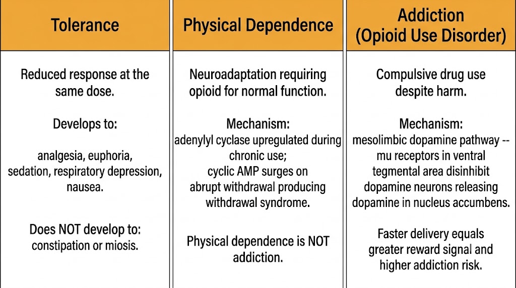
Figure 4. Tolerance, physical dependence, and addiction are distinct phenomena with different mechanisms. Physical dependence is a pharmacological adaptation and does not equal addiction. Figure generated by Gemini AI.
| Author / Organization | Title | Source |
|---|---|---|
| Pasternak GW, Pan YX | Mu opioids and their receptors: evolution of a concept | Pharmacol Rev. 2013;65(4):1257–1317 |
| Brownstein MJ | A brief history of opiates, opioid peptides, and opioid receptors | Proc Natl Acad Sci USA. 1993;90(12):5391–5393 |
| Hughes J, Smith TW, Kosterlitz HW, et al. | Identification of two related pentapeptides from the brain with potent opiate agonist activity | Nature. 1975;258(5536):577–580 |
| Williams JT, Ingram SL, Henderson G, et al. | Regulation of mu-opioid receptors: desensitization, phosphorylation, internalization, and tolerance | Pharmacol Rev. 2013;65(1):223–254 |
| Stein C | Opioid receptors | Annu Rev Med. 2016;67:433–451 |
| Koob GF, Volkow ND | Neurobiology of addiction: a neurocircuitry analysis | Lancet Psychiatry. 2016;3(8):760–773 |
| Pattinson KT | Opioids and the control of respiration | Br J Anaesth. 2008;100(6):747–758 |
| Corbett AD, Henderson G, McKnight AT, Paterson SJ | 75 years of opioid research: the exciting but vain quest for the Holy Grail | Br J Pharmacol. 2006;147(Suppl 1):S153–S162 |