Introduction to Medical Pharmacology
Phase one and two reactions, cytochrome P450 isoforms, hepatic clearance, enzyme interactions, and pharmacogenomics
Chapter 2 · Module 3 of 5 · PKIN-03Section 1
Oxidation, reduction, and hydrolysis — the functionalization reactions that prepare drugs for elimination or conjugation
Drug metabolism transforms lipophilic compounds into more polar, water-soluble products that can be excreted by the kidneys or bile. Phase one reactions introduce or unmask a functional group on the drug molecule, producing metabolites that may be pharmacologically active, inactive, or occasionally toxic. The cytochrome P450 enzyme family is responsible for the majority of phase one oxidative metabolism and is the central determinant of most clinically important drug-drug metabolic interactions.
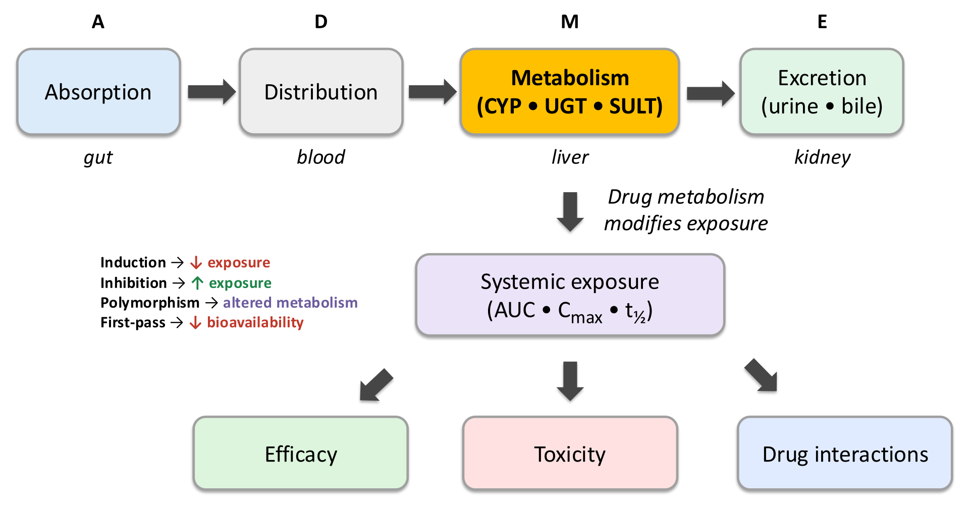
Phase one reactions fall into three categories: oxidation (the most common, accounting for the majority of cytochrome P450-mediated reactions), reduction, and hydrolysis. The cytochrome P450 superfamily — heme-containing monooxygenases located predominantly in the endoplasmic reticulum of hepatocytes — drives the large majority of phase one oxidative drug metabolism. Significant additional cytochrome P450 expression occurs in enterocytes of the small intestinal wall, which accounts for intestinal first-pass metabolism as described in Module 1. The cytochrome P450 reaction uses molecular oxygen and inserts one oxygen atom into the substrate, typically forming a hydroxyl group, converting a lipophilic drug into a more polar hydroxylated product.
Of more than 50 human cytochrome P450 isoforms, six enzymes metabolize the large majority of clinically used drugs: cytochrome P450 3A4, cytochrome P450 2C9, cytochrome P450 2C19, cytochrome P450 2D6, cytochrome P450 1A2, and cytochrome P450 2B6. These isoforms have overlapping but distinct substrate specificities, different tissue expression patterns, and different susceptibilities to induction and inhibition by other drugs — which is why cytochrome P450-mediated drug-drug interactions are so common and clinically consequential.
Non-cytochrome P450 phase one enzymes include monoamine oxidase (which metabolizes catecholamines, serotonin, and tyramine, and is the target of monoamine oxidase inhibitor antidepressants), xanthine oxidase (which metabolizes azathioprine and 6-mercaptopurine, relevant to allopurinol interactions), alcohol dehydrogenase and aldehyde dehydrogenase (which metabolize ethanol and are inhibited by disulfiram), and flavin-containing monooxygenases (which metabolize several drugs including the prodrug clozapine's metabolite pathways).
Cytochrome P450 3A4 is the most abundant hepatic cytochrome P450 and metabolizes approximately 50 percent of clinically used drugs. Its substrates include statins (simvastatin, lovastatin, atorvastatin), calcineurin inhibitors (cyclosporine, tacrolimus), benzodiazepines, most calcium channel blockers, and many other drugs. It is expressed in both the liver and intestinal wall, making it the dominant contributor to both intestinal and hepatic first-pass metabolism for many drugs. Grapefruit juice irreversibly inhibits intestinal cytochrome P450 3A4, increasing bioavailability of sensitive substrates.
Cytochrome P450 2C9 metabolizes warfarin (the active S-enantiomer), many nonsteroidal anti-inflammatory drugs, phenytoin, and sulfonylureas. This isoform is particularly susceptible to competitive inhibition by fluconazole and amiodarone, which can dramatically potentiate warfarin anticoagulation. Cytochrome P450 2C19 metabolizes the proton pump inhibitors (omeprazole, pantoprazole, lansoprazole), clopidogrel (prodrug activation, discussed in Section 6), diazepam, and certain antidepressants. Cytochrome P450 2D6 metabolizes approximately 20 to 25 percent of commonly prescribed drugs, including codeine (prodrug activation to morphine), tramadol, most tricyclic antidepressants, most selective serotonin reuptake inhibitor antidepressants, and beta-blockers including metoprolol and carvedilol. Cytochrome P450 1A2 metabolizes theophylline, caffeine, clozapine, and some antidepressants, and is induced by cigarette smoking — a clinically relevant reason why plasma theophylline and clozapine concentrations fall when patients begin smoking and rise when they stop.
Phase one metabolism does not always inactivate a drug. Metabolites can be pharmacologically active (contributing to the overall drug effect), inactive (the desired outcome for elimination), or toxic (an unwanted and sometimes dangerous consequence of metabolism).
Acetaminophen is the paradigmatic example of toxic metabolite formation. At therapeutic doses, acetaminophen is metabolized primarily by glucuronidation and sulfation (phase two reactions) to non-toxic conjugates that are excreted in urine. A small fraction is oxidized by cytochrome P450 2E1 (and to a lesser extent cytochrome P450 3A4 and 1A2) to a reactive, highly electrophilic metabolite called N-acetyl-para-benzoquinone imine. Under normal circumstances, this toxic metabolite is immediately conjugated with glutathione in the liver, forming a harmless mercapturic acid conjugate. In overdose, or when glutathione stores are depleted (as in chronic alcohol use, malnutrition, or fasting), N-acetyl-para-benzoquinone imine accumulates, covalently binds hepatocyte proteins, and causes hepatocellular necrosis. The antidote N-acetylcysteine works by replenishing glutathione precursors, allowing conjugation of N-acetyl-para-benzoquinone imine before it injures hepatocytes.
Key Cytochrome P450 Isoforms — First Aid Summary
CYP3A4 (~50% of drugs): statins, cyclosporine, tacrolimus, benzodiazepines, calcium channel blockers. Inhibited by azole antifungals, erythromycin, ritonavir, grapefruit. Induced by rifampin, carbamazepine, phenytoin, St. John's wort. CYP2C9: warfarin (S-enantiomer), NSAIDs, phenytoin. Inhibited by fluconazole, amiodarone. CYP2C19: proton pump inhibitors, clopidogrel (activation), diazepam. CYP2D6: codeine (activation), tramadol, tricyclic antidepressants, SSRIs, metoprolol. Inhibited by fluoxetine, paroxetine, bupropion. CYP1A2: theophylline, clozapine, caffeine. Induced by smoking.
Section 2
Glucuronidation, acetylation, sulfation, and the clinical consequences of conjugation enzyme polymorphisms
Phase two reactions attach a polar, water-soluble group to a drug or its phase one metabolite, producing conjugates that are nearly always pharmacologically inactive and readily excreted in urine or bile. Phase two reactions typically follow phase one, but some drugs proceed directly to phase two conjugation without prior oxidative metabolism.
Glucuronidation, catalyzed by uridine diphosphate glucuronosyltransferase enzymes, is the most quantitatively important phase two conjugation pathway. It attaches glucuronic acid to hydroxyl, carboxyl, amino, or thiol groups on the drug or phase one metabolite, producing a glucuronide conjugate that is highly water-soluble and actively secreted into bile or filtered by the kidney. The resulting conjugates are typically pharmacologically inactive, though there are exceptions: morphine-6-glucuronide retains significant opioid receptor agonist activity and contributes substantially to analgesia, particularly in patients with renal failure where the conjugate accumulates.
Glucuronidation capacity is markedly reduced in neonates because uridine diphosphate glucuronosyltransferase enzyme expression is low at birth and does not reach adult levels for several weeks to months. This deficiency was the cause of chloramphenicol gray baby syndrome: when chloramphenicol was given to neonates at doses appropriate for adults, the drug could not be glucuronidated and accumulated to toxic levels, causing cardiovascular collapse, ashen skin color, and death. The lesson is that neonatal drug dosing requires accounting for immature conjugation capacity, not just lower body weight.
Acetylation is catalyzed by N-acetyltransferase enzymes, principally N-acetyltransferase 2 in the liver. It transfers an acetyl group from acetyl-CoA to amino groups on drugs including isoniazid, hydralazine, procainamide, dapsone, and sulfamethazine. Acetylation renders these drugs inactive and more water-soluble for excretion.
N-acetyltransferase 2 is subject to a well-characterized genetic polymorphism that divides people into slow acetylators and rapid (or fast) acetylators. The slow acetylator phenotype is more common in Middle Eastern, North African, and European populations (40 to 70 percent), while the rapid acetylator phenotype predominates in Asian populations (80 to 90 percent). For drugs metabolized by N-acetyltransferase 2, acetylator status has direct clinical consequences. Slow acetylators of isoniazid accumulate higher drug concentrations and are at greater risk of isoniazid-induced peripheral neuropathy (due to drug accumulation) and isoniazid-related drug-induced liver injury. Slow acetylators taking hydralazine or procainamide are at increased risk of developing drug-induced lupus erythematosus, because the parent drug (rather than the acetylated metabolite) is the lupus-inducing agent, and slow acetylators accumulate more of it. Rapid acetylators of isoniazid may have lower drug exposures and potentially reduced bactericidal efficacy when isoniazid is given intermittently rather than daily.
Sulfation transfers sulfate groups to hydroxyl or amino groups, is catalyzed by sulfotransferases, and is an important pathway for some drugs including acetaminophen (at low doses), many steroids, and neurotransmitters. Glutathione conjugation, catalyzed by glutathione S-transferases, is particularly important for detoxifying reactive electrophilic metabolites — including N-acetyl-para-benzoquinone imine from acetaminophen metabolism. When glutathione is depleted, reactive metabolites escape conjugation and cause organ toxicity. Methylation and amino acid conjugation are additional phase two pathways with more limited drug substrates.
Section 3
Capacity-limited and flow-limited metabolism and how they respond differently to enzyme inhibitors and hepatic blood flow changes
Not all drugs are metabolized by the liver in the same way. The distinction between drugs with low hepatic extraction and drugs with high hepatic extraction determines whether a drug's elimination rate is limited by the liver's metabolic capacity or by the rate at which blood delivers drug to the liver. This distinction has direct and sometimes counterintuitive clinical consequences when enzyme inhibitors are added or when hepatic blood flow changes.
A drug with low hepatic extraction is one where the liver's enzyme activity is far greater than needed to metabolize the drug presented to it at typical portal blood flow rates. In this situation, the liver clears only a small fraction of the drug per pass, and plasma concentrations after oral dosing are relatively close to those after parenteral dosing — because first-pass elimination is modest. The key feature of low-extraction drugs is that their clearance depends entirely on how efficiently the metabolizing enzymes can process the drug: enzyme activity (and therefore the free drug concentration available to the enzymes) is the rate-limiting factor. These drugs are therefore called capacity-limited or enzyme-limited.
For low-extraction drugs, enzyme inhibitors substantially reduce clearance and raise plasma concentrations. If an inhibitor halves the activity of the metabolizing cytochrome P450, plasma concentrations of a low-extraction substrate can double. Changes in hepatic blood flow, by contrast, have little effect on clearance of low-extraction drugs — the liver is already far from its capacity limit, and delivering more drug per minute does not change how much gets metabolized.
Examples of low-extraction drugs include warfarin, phenytoin, diazepam, tolbutamide, theophylline, and most proton pump inhibitors. The plasma protein binding of these drugs also influences effective clearance, since only the unbound fraction is accessible to hepatic enzymes.
A drug with high hepatic extraction is one where the liver's enzymes are so efficient that they metabolize nearly all of the drug presented during a single hepatic pass. The liver is not the bottleneck — delivery of drug to the liver is. Hepatic clearance of these drugs therefore approximates hepatic blood flow (approximately 1,500 milliliters per minute in a healthy adult), and their clearance is called flow-limited. Because the liver removes drug so efficiently, oral bioavailability of high-extraction drugs is low, as discussed in Module 1.
For high-extraction drugs, enzyme inhibitors have less effect than might be expected on clearance, because the enzyme was already operating far above the drug concentration needed for near-complete extraction — reducing enzyme activity modestly still leaves capacity for extensive extraction. However, changes in hepatic blood flow substantially affect the clearance of high-extraction drugs. Conditions that reduce hepatic blood flow — propranolol and other drugs that reduce cardiac output, portosystemic shunting in cirrhosis, or hypotension — decrease the delivery rate of drug to the liver and correspondingly reduce clearance, raising plasma concentrations. Conversely, conditions that increase hepatic blood flow, such as meals (which increase splanchnic blood flow), may increase the clearance of high-extraction drugs.
High-extraction drugs include lidocaine, morphine, propranolol, verapamil, most opioids, and many other drugs discussed in Module 1's first-pass section. In cirrhosis with significant portosystemic shunting, blood from the portal circulation bypasses the liver almost entirely before reaching systemic circulation — dramatically increasing the oral bioavailability of high-extraction drugs and requiring dose reduction.
| Property | Low-extraction (capacity-limited) | High-extraction (flow-limited) | Clinical implication |
|---|---|---|---|
| Hepatic extraction ratio | Below ~0.3 | Above ~0.7 | Determines first-pass effect magnitude |
| Rate-limiting factor | Enzyme activity / drug concentration | Hepatic blood flow | Determines which interventions change clearance |
| Effect of enzyme inhibitor | Large increase in plasma concentration | Smaller effect than expected | Enzyme inhibitor drug interactions most dangerous for low-extraction drugs |
| Effect of reduced hepatic blood flow (cirrhosis, propranolol, hypotension) | Minimal effect on clearance | Major reduction in clearance, toxicity risk | High-extraction drugs require dose reduction when hepatic blood flow falls |
| Examples | Warfarin, phenytoin, theophylline, diazepam | Lidocaine, morphine, propranolol, verapamil | Know which category your drug falls in before adjusting dosing |
Section 4
How drugs increase or decrease cytochrome P450 activity, the time course of each effect, and clinically important examples
Drugs can alter the activity of cytochrome P450 enzymes through two fundamentally different mechanisms: induction, which increases enzyme expression and accelerates metabolism of substrates, and inhibition, which reduces enzyme activity and slows metabolism. These two mechanisms produce opposite clinical effects, operate through different mechanisms, and have very different time courses — a distinction that is essential for anticipating and managing drug-drug interactions.
Enzyme induction occurs when a drug increases the transcription of cytochrome P450 genes, leading to higher enzyme protein concentrations in hepatocytes and enterocytes. Because induction requires de novo protein synthesis, its onset is gradual: the full effect typically develops over one to three weeks of continuous exposure to the inducing drug, and the offset after discontinuation is similarly gradual as existing enzyme protein is degraded and new synthesis returns to baseline. This delayed time course is clinically important — a patient stabilized on a drug regimen may not experience the full interaction effect until weeks after starting an inducer, and may experience rebound toxicity weeks after stopping one.
The most clinically important inducers and their principal cytochrome P450 targets include rifampin (cytochrome P450 3A4 and cytochrome P450 2C9 — the most potent enzyme inducer in clinical use, capable of reducing plasma concentrations of sensitive substrates by 80 to 90 percent), carbamazepine (cytochrome P450 3A4 and cytochrome P450 1A2, also auto-induces its own metabolism), phenytoin (cytochrome P450 3A4, cytochrome P450 2C9, and cytochrome P450 2C19), phenobarbital (broad inducer across multiple isoforms), and St. John's wort (cytochrome P450 3A4 — a common source of unexpected interactions when patients do not disclose herbal supplement use). All of these inducers can precipitate treatment failure for co-administered drugs by accelerating their clearance below therapeutic levels.
Clinically consequential induction interactions include rifampin plus oral contraceptives (breakthrough ovulation and contraceptive failure), rifampin plus warfarin (loss of anticoagulation, requiring substantially higher warfarin doses during rifampin therapy and careful dose reduction after discontinuation), carbamazepine plus many antiepileptic drugs and psychiatric medications (complex interactions requiring close therapeutic drug monitoring), and St. John's wort plus cyclosporine (transplant rejection due to dramatically reduced cyclosporine exposure).
Enzyme inhibition reduces cytochrome P450 activity without changing enzyme protein levels, either by competing with the substrate for the active site (competitive inhibition) or by binding irreversibly to the enzyme active site (mechanism-based or irreversible inhibition). Because inhibition acts on existing enzyme protein rather than requiring synthesis of new protein, its onset and offset are rapid — occurring within hours to days of starting or stopping the inhibitor, depending on the inhibitor's half-life and the enzyme's recovery time.
Mechanism-based inhibition, also called suicide inhibition, is a clinically distinct category in which the inhibitor is metabolized by the cytochrome P450 enzyme into a reactive intermediate that covalently inactivates the enzyme. Because the enzyme is irreversibly destroyed, recovery from mechanism-based inhibition requires synthesis of new enzyme protein, which takes days. Erythromycin and clarithromycin, azole antifungals, and grapefruit juice furanocoumarins are mechanism-based inhibitors of cytochrome P450 3A4. Grapefruit's inhibition of intestinal cytochrome P450 3A4 can last 24 to 72 hours after a single exposure, as discussed in Module 1.
Clinically important enzyme inhibitor interactions include azole antifungals (fluconazole, itraconazole, voriconazole, ketoconazole) plus cytochrome P450 3A4 substrates: these combinations can produce severe toxicity with statins (myopathy, rhabdomyolysis), cyclosporine (nephrotoxicity), and other narrow-therapeutic-index drugs. Fluconazole plus warfarin markedly increases anticoagulation by inhibiting both cytochrome P450 2C9 (which metabolizes warfarin's active S-enantiomer) and cytochrome P450 3A4. Cimetidine (an older H2-receptor antagonist that inhibits multiple cytochrome P450 isoforms) has multiple clinically significant interactions including potentiation of warfarin, theophylline, and phenytoin. Selective serotonin reuptake inhibitors, particularly fluoxetine and paroxetine, are potent inhibitors of cytochrome P450 2D6, which explains their ability to dramatically raise plasma concentrations of co-administered tricyclic antidepressants, opioids processed by cytochrome P450 2D6, and other substrates.
Induction vs. Inhibition — Time Course and Clinical Approach
Induction: gradual onset (1–3 weeks), gradual offset after stopping. Key inducers: rifampin, carbamazepine, phenytoin, phenobarbital, St. John's wort. Effect: decreased plasma concentrations of substrates → treatment failure. Inhibition: rapid onset (hours to days). Key inhibitors: azole antifungals, macrolide antibiotics, fluoxetine/paroxetine (CYP2D6), fluconazole/amiodarone (CYP2C9), grapefruit (intestinal CYP3A4 mechanism-based, lasts 24–72 h). Effect: increased plasma concentrations of substrates → toxicity. Always check for cytochrome P450 interactions when adding or stopping any inducer or inhibitor.
Section 5
Metabolizer phenotypes, clinical consequences, and the drugs where genotype most affects outcome
Genetic variation in cytochrome P450 genes produces individuals who metabolize certain drugs at dramatically different rates. The resulting metabolizer phenotypes — poor, intermediate, extensive, and ultra-rapid — can be the difference between therapeutic benefit, treatment failure, and life-threatening toxicity for the same standard dose of a drug.
Genetic polymorphisms in cytochrome P450 genes produce four phenotypic categories. Poor metabolizers have two non-functional alleles and essentially no enzymatic activity for that isoform. Intermediate metabolizers have one functional and one reduced-function or non-functional allele, producing enzyme activity between poor and normal. Extensive metabolizers (sometimes called normal metabolizers) have two functional alleles and represent the population average. Ultra-rapid metabolizers carry gene duplications that produce multiple copies of functional enzyme, metabolizing substrates much faster than the typical individual.
The clinical consequences of metabolizer phenotype depend on whether the drug is a substrate being cleared (in which case poor metabolizers have higher plasma concentrations and more toxicity) or a prodrug being activated (in which case poor metabolizers have less active drug produced and reduced efficacy, while ultra-rapid metabolizers generate more active drug and risk toxicity from it).
Cytochrome P450 2D6 is one of the most polymorphic cytochrome P450 genes, with more than 100 known alleles producing a wide spectrum of enzyme activity. Approximately 7 to 10 percent of European populations are poor metabolizers of cytochrome P450 2D6, while 1 to 2 percent are ultra-rapid metabolizers. The clinical consequences are starkest for codeine, which is a prodrug that requires cytochrome P450 2D6 to convert it to morphine.
In poor metabolizers of cytochrome P450 2D6, codeine is not converted to morphine, producing no meaningful analgesia. These patients receive only the side effects of codeine (constipation, nausea) without the therapeutic benefit and may be inappropriately labeled as opioid non-responders. In ultra-rapid metabolizers, codeine is converted to morphine at an accelerated rate, producing morphine plasma concentrations far above those expected from a standard codeine dose. This can cause respiratory depression, particularly in breastfed infants whose mothers are ultra-rapid metabolizers receiving codeine for postpartum pain — a scenario that led to infant deaths and resulted in the United States Food and Drug Administration issuing a black box warning against codeine use by breastfeeding mothers. The interaction between ultra-rapid cytochrome P450 2D6 metabolizer genotype and codeine use represents one of the most important examples of pharmacogenomics affecting patient safety.
Cytochrome P450 2D6 metabolizer status also affects dosing of many antidepressants (particularly tricyclic antidepressants and certain selective serotonin reuptake inhibitors) and antipsychotics. Poor metabolizers of cytochrome P450 2D6 taking tricyclic antidepressants are at substantially higher risk of cardiac arrhythmias and anticholinergic toxicity from drug accumulation at standard doses. Cytochrome P450 2D6 is also relevant for tamoxifen (discussed in Section 6).
Cytochrome P450 2C19 polymorphisms have two major clinical consequences. Clopidogrel, the antiplatelet prodrug used after coronary stent placement, requires cytochrome P450 2C19 to generate its active thienopyridine metabolite. Poor metabolizers of cytochrome P450 2C19 — approximately 2 to 3 percent of European populations and up to 15 to 20 percent of Asian populations — cannot adequately activate clopidogrel. Clinical studies show that these patients have significantly higher rates of major adverse cardiovascular events after percutaneous coronary intervention with stent placement. The United States Food and Drug Administration added a black box warning to clopidogrel noting reduced effectiveness in poor cytochrome P450 2C19 metabolizers, and the label recommends testing for genotype or considering alternative antiplatelet agents (prasugrel, ticagrelor) in high-risk patients.
For proton pump inhibitors metabolized by cytochrome P450 2C19, poor metabolizers actually have higher drug exposure and potentially greater acid suppression. This is generally clinically beneficial — poor metabolizers have higher eradication rates of Helicobacter pylori when using proton pump inhibitor-based regimens. Ultra-rapid metabolizers of cytochrome P450 2C19 may have reduced proton pump inhibitor efficacy and may require dose escalation for adequate acid suppression.
Warfarin's highly variable dose requirement across patients is partly explained by cytochrome P450 2C9 polymorphisms. The active S-enantiomer of warfarin is primarily metabolized by cytochrome P450 2C9, and variants with reduced enzyme activity (the most important being cytochrome P450 2C9*2 and cytochrome P450 2C9*3 alleles) lead to reduced warfarin clearance and markedly elevated anticoagulation at doses that would be appropriate for patients with normal activity. Poor metabolizers of cytochrome P450 2C9 require substantially lower warfarin doses to maintain the international normalized ratio within the therapeutic range, and they are at higher risk of serious bleeding during warfarin initiation if standard loading approaches are used. Commercial warfarin dosing algorithms now routinely incorporate cytochrome P450 2C9 genotype alongside cytochrome P450 vitamin K epoxide reductase complex subunit 1 (VKORC1) genotype, which determines warfarin's pharmacodynamic target sensitivity.
High-Yield Pharmacogenomics Summary
CYP2D6 poor metabolizer + codeine: no analgesia (no morphine generated). CYP2D6 ultra-rapid metabolizer + codeine: morphine toxicity, respiratory depression — FDA black box warning for breastfeeding mothers. CYP2C19 poor metabolizer + clopidogrel: inadequate platelet inhibition — FDA black box warning, consider prasugrel or ticagrelor. CYP2C9 poor metabolizer + warfarin: elevated bleeding risk at standard doses — use lower doses, incorporate genotype in dosing algorithm. CYP1A2 induction by smoking: lower clozapine and theophylline levels while smoking; levels rise when patient stops smoking.
Section 6
Codeine, clopidogrel, and tamoxifen — when cytochrome P450 metabolism generates the therapeutic effect
While most drugs are pharmacologically active as administered and metabolized to inactive products, some drugs are administered in an inactive or minimally active form that requires cytochrome P450-mediated biotransformation to generate the active therapeutic agent. For these prodrugs, the same genetic variations and drug interactions that would protect against toxicity with an active substrate instead prevent therapeutic efficacy — or, in the case of ultra-rapid metabolizers, cause toxicity from excessive activation.
Codeine is a weak opioid that exerts the large majority of its analgesic effect through demethylation to morphine by cytochrome P450 2D6. Only a small fraction (approximately 10 percent) of an oral codeine dose is converted to morphine under normal circumstances, but this fraction is responsible for the therapeutic effect. Poor metabolizers of cytochrome P450 2D6 convert essentially none of the codeine to morphine and receive no meaningful analgesia. Ultra-rapid metabolizers convert codeine to morphine at a greatly accelerated rate, generating morphine plasma concentrations that can cause respiratory depression at doses that would be safe for extensive metabolizers. This pharmacogenomic consideration led to codeine being contraindicated for pain management in children after tonsillectomy (due to deaths in pediatric patients who were ultra-rapid metabolizers) and to the black box warning against its use in breastfeeding mothers.
Concurrent use of cytochrome P450 2D6 inhibitors — including fluoxetine, paroxetine, bupropion, and quinidine — in a patient taking codeine effectively converts that patient's phenotype to a poor metabolizer, abolishing analgesic efficacy. This is one of the most clinically significant drug-gene-drug interaction patterns in pain management.
Clopidogrel is an antiplatelet prodrug that requires two-step hepatic activation, with the second and rate-limiting step catalyzed by cytochrome P450 2C19. The active metabolite irreversibly blocks the platelet adenosine diphosphate receptor (specifically the purinergic receptor P2Y12), preventing platelet aggregation. Poor metabolizers of cytochrome P450 2C19 generate inadequate amounts of the active metabolite, resulting in insufficient platelet inhibition and higher rates of cardiovascular events after coronary stent placement. This interaction is high-yield for pharmacology examinations and the United States Medical Licensing Examination because it illustrates how a prodrug activation pathway can make a drug variably effective — or entirely ineffective — depending on the patient's genotype.
The concurrent use of omeprazole (a cytochrome P450 2C19 inhibitor that reduces clopidogrel activation) with clopidogrel was a subject of significant regulatory concern and clinical debate. The available evidence suggests that the pharmacokinetic interaction exists but its clinical significance at the population level is uncertain. Pantoprazole, which has less cytochrome P450 2C19 inhibitory activity, is generally preferred over omeprazole when proton pump inhibitor co-administration is necessary in patients on clopidogrel.
Tamoxifen, the selective estrogen receptor modulator used as adjuvant therapy for estrogen receptor-positive breast cancer, undergoes hepatic metabolism to several metabolites. Its most potent active metabolite, endoxifen, is formed by N-demethylation (primarily by cytochrome P450 3A4 and 3A5) to N-desmethyltamoxifen, followed by 4-hydroxylation by cytochrome P450 2D6. Endoxifen has approximately 100-fold greater potency at the estrogen receptor than tamoxifen itself and is responsible for the large majority of tamoxifen's clinical efficacy.
Patients who are poor metabolizers of cytochrome P450 2D6 have substantially lower endoxifen plasma concentrations and, in several observational studies, higher rates of breast cancer recurrence compared to extensive metabolizers receiving the same tamoxifen dose. The concurrent use of potent cytochrome P450 2D6 inhibitors — particularly fluoxetine and paroxetine — with tamoxifen is actively discouraged in oncology practice because these widely used antidepressants can convert an extensive metabolizer to a functionally poor metabolizer phenotype and potentially compromise the effectiveness of adjuvant breast cancer therapy.
Prodrug Activation
Codeine
Prodrug Activation
Clopidogrel
Prodrug Activation
Tamoxifen
| Author / Organization | Title | Source |
|---|---|---|
| Brunton LL, Hilal-Dandan R, Knollmann BC, eds. | Goodman & Gilman’s: The Pharmacological Basis of Therapeutics, 13th ed. | McGraw-Hill, 2018 |
| Zanger UM, Schwab M | Cytochrome P450 enzymes in drug metabolism: regulation of gene expression, enzyme activities, and impact of genetic variation | Pharmacol Ther. 2013;138(1):103-141 |
| Weinshilboum R, Wang L | Pharmacogenomics: precision medicine and drug response | Mayo Clin Proc. 2017;92(11):1711-1722 |
| Cascorbi I | Drug interactions — principles, examples and clinical consequences | Dtsch Arztebl Int. 2012;109(33-34):546-556 |
| Scott SA et al. | Clinical pharmacogenomics implementation consortium guidelines for CYP2C19 genotype and clopidogrel therapy | Clin Pharmacol Ther. 2013;94(3):317-323 |
| Codeine and tramadol products: drug safety communication | FDA safety labeling changes and new black box warning on use of these medicines in children and nursing mothers | US Food and Drug Administration, 2017 |
| Goetz MP et al. | The impact of cytochrome P450 2D6 metabolism in women receiving adjuvant tamoxifen | Breast Cancer Res Treat. 2007;101(1):113-121 |
| Rowland M, Tozer TN | Clinical Pharmacokinetics and Pharmacodynamics: Concepts and Applications, 4th ed. | Lippincott Williams & Wilkins, 2011 |