Calcineurin Inhibitors, Antiproliferatives, mTOR Inhibitors, and Rejection Management
RENL · Module 4 of 5Induction, maintenance, and rejection treatment — three temporal phases with distinct pharmacological strategies
Renal transplantation requires lifelong pharmacological suppression of the recipient's immune response against donor antigens. The fundamental challenge is achieving sufficient immunosuppression to prevent rejection while avoiding the infectious and malignant complications of over-immunosuppression. Three temporal phases structure the pharmacological approach.
Induction immunosuppression is administered during the first days to weeks after transplantation — the period of highest alloimmune risk when the recipient's immune system first encounters donor antigens. The goal is rapid and deep T-cell suppression before maintenance dosing takes full effect. Two strategies are used: interleukin-2 receptor blockade with basiliximab (lower-risk recipients) or lymphocyte depletion with antithymocyte globulin (higher-risk recipients or sensitized patients).
Maintenance immunosuppression is the lifelong triple-drug regimen that prevents chronic rejection. The standard combination is a calcineurin inhibitor plus an antiproliferative agent plus a corticosteroid. Tacrolimus is the preferred calcineurin inhibitor, mycophenolate mofetil the preferred antiproliferative, and low-dose prednisone the corticosteroid component. This triple-drug approach provides complementary immunosuppression at three distinct points in T-cell activation without any single agent needing to carry the full suppressive burden.
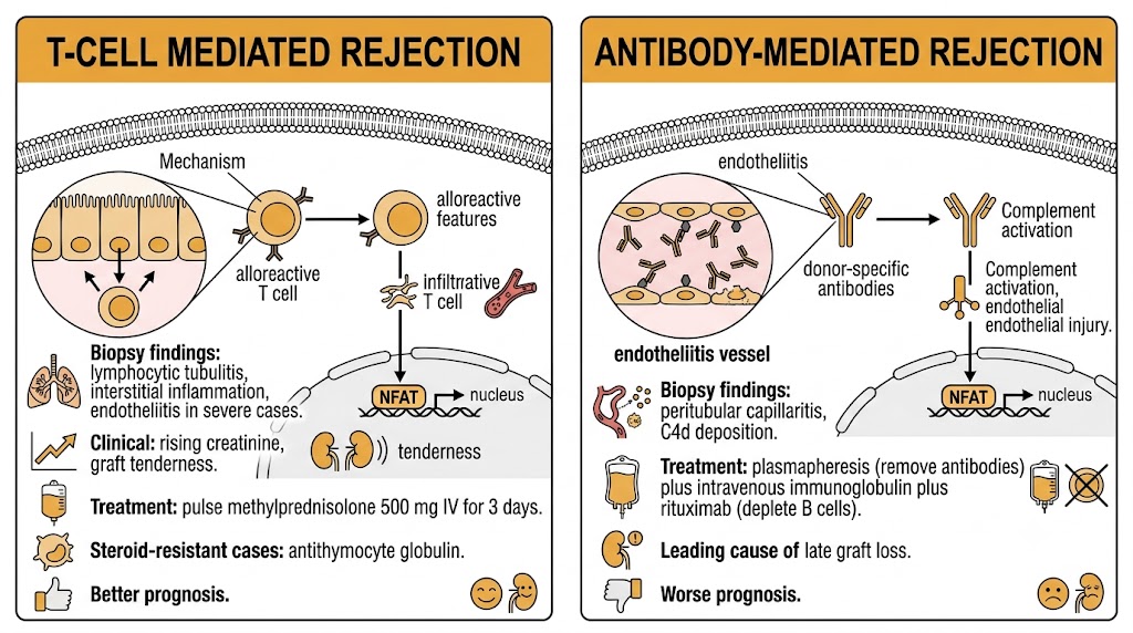
T-cell mediated rejection is treated with pulse intravenous methylprednisolone (500 milligrams intravenously daily for 3 days) as first-line therapy, with antithymocyte globulin reserved for steroid-resistant cases. Antibody-mediated rejection — driven by donor-specific antibodies targeting graft endothelium — is treated with plasmapheresis to remove circulating antibodies, intravenous immunoglobulin to modulate residual antibody effects, and rituximab to deplete antibody-producing B cells. Antibody-mediated rejection carries a substantially worse prognosis than T-cell mediated rejection and often leads to progressive graft loss despite treatment.
Tacrolimus and cyclosporine — convergent downstream targets, divergent adverse effect profiles, and a massive drug interaction network
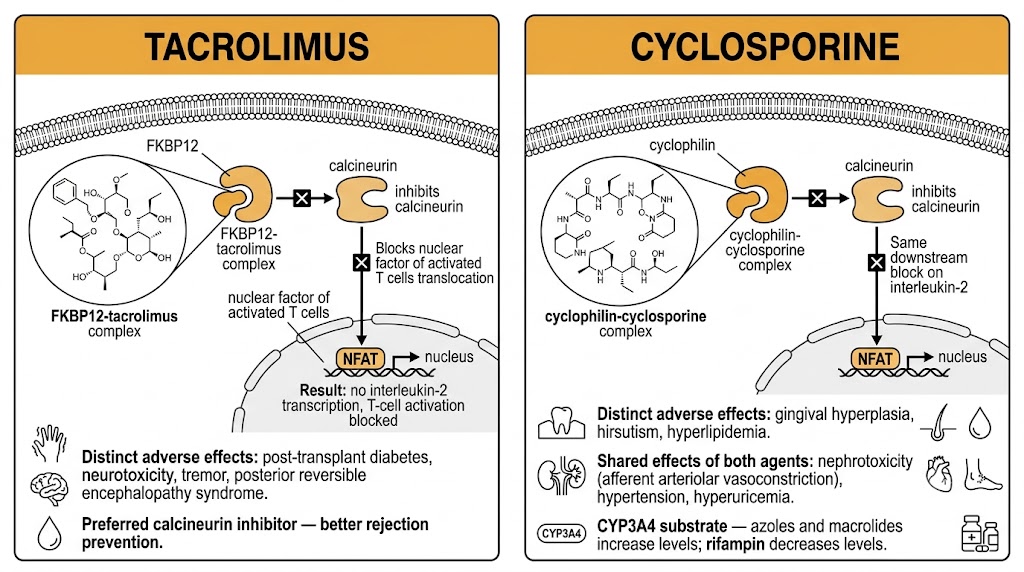
Calcineurin inhibitors are the pharmacological cornerstone of transplant maintenance immunosuppression. Despite binding different intracellular proteins, tacrolimus and cyclosporine converge on the same downstream target — calcineurin — and produce similar degrees of T-cell suppression through the same final pathway.
Tacrolimus binds the immunophilin FKBP12, and the tacrolimus-FKBP12 complex inhibits calcineurin, a phosphatase required for dephosphorylation and nuclear translocation of nuclear factor of activated T cells. Without nuclear factor of activated T cells in the nucleus, interleukin-2 gene transcription is blocked and T-cell activation cannot proceed. Cyclosporine binds cyclophilin instead of FKBP12, but the cyclosporine-cyclophilin complex inhibits the same calcineurin target by the same mechanism. Tacrolimus is approximately 10 to 100 times more potent than cyclosporine on a molar basis, and tacrolimus-based regimens have become the standard because of superior rejection prevention and better long-term graft survival.
Both calcineurin inhibitors are substrates of cytochrome P450 3A4 and the P-glycoprotein efflux transporter, making them targets of a large drug interaction network. Cytochrome P450 3A4 inhibitors — including azole antifungals (fluconazole, voriconazole), macrolide antibiotics (erythromycin, clarithromycin), and diltiazem — dramatically increase calcineurin inhibitor blood levels and risk toxicity. Cytochrome P450 3A4 inducers — rifampin, phenytoin, carbamazepine, and St. John's Wort — reduce levels and risk rejection. Because these drugs have narrow therapeutic indices, therapeutic drug monitoring with trough levels is mandatory and must be repeated after any change to the drug regimen.
Calcineurin inhibitor nephrotoxicity is the most clinically important adverse effect and a leading cause of progressive graft dysfunction over time. Acute nephrotoxicity results from afferent arteriolar vasoconstriction — mediated by thromboxane A2 and endothelin — reducing renal blood flow and glomerular filtration rate in a dose-dependent, reversible manner. Chronic calcineurin inhibitor nephrotoxicity produces irreversible interstitial fibrosis and tubular atrophy from sustained ischemia and direct tubular toxicity, and is a primary driver of calcineurin inhibitor minimization strategies.
Tacrolimus and cyclosporine have divergent adverse effect profiles. Cyclosporine causes gingival hyperplasia, hirsutism, and is more likely to cause hyperlipidemia — effects not seen with tacrolimus. Tacrolimus carries a higher risk of post-transplant diabetes mellitus through pancreatic beta-cell toxicity and peripheral insulin resistance, and a higher risk of neurotoxicity including tremor and posterior reversible encephalopathy syndrome. Both agents elevate blood pressure and uric acid.
Mycophenolate mofetil and azathioprine — blocking lymphocyte proliferation at the nucleotide synthesis level
Antiproliferative agents prevent T- and B-lymphocyte proliferation by targeting purine nucleotide synthesis, which is the rate-limiting metabolic step for lymphocyte clonal expansion after antigen stimulation. Because lymphocytes rely almost exclusively on de novo purine synthesis (lacking the salvage pathway capacity of most other cells), antiproliferative agents provide selective lymphocyte suppression at therapeutic doses.
Mycophenolate mofetil is a prodrug hydrolyzed to mycophenolic acid after oral absorption. Mycophenolic acid uncompetitively inhibits inosine monophosphate dehydrogenase, the enzyme catalyzing the rate-limiting step in de novo guanosine synthesis. Without guanosine nucleotides, T and B cells cannot proliferate. Mycophenolate mofetil has largely replaced azathioprine in transplant regimens because of superior rejection prevention in randomized trials.
The principal adverse effects are gastrointestinal: nausea, vomiting, diarrhea, and abdominal cramping occur in 20 to 30% of patients and are dose-limiting. Switching to the enteric-coated formulation (mycophenolate sodium) may reduce upper gastrointestinal symptoms. Bone marrow suppression producing leukopenia and thrombocytopenia occurs at higher doses. Mycophenolate mofetil is teratogenic and absolutely contraindicated in pregnancy — effective contraception is mandatory in women of childbearing potential.
Azathioprine is a thiopurine prodrug converted to 6-mercaptopurine and then to active thioguanine nucleotides that incorporate into replicating deoxyribonucleic acid, causing strand breaks and cytotoxicity in rapidly dividing cells including lymphocytes. The enzyme thiopurine methyltransferase inactivates 6-mercaptopurine; patients with thiopurine methyltransferase deficiency (approximately 0.3% of the population) accumulate toxic thioguanine nucleotide levels and develop severe myelosuppression at standard doses — thiopurine methyltransferase genotyping before initiation prevents this. The critical drug interaction is with allopurinol, which inhibits xanthine oxidase and dramatically increases 6-mercaptopurine levels; the combination requires an 75% azathioprine dose reduction or substitution with mycophenolate mofetil.
Sirolimus and everolimus — calcineurin-independent immunosuppression with a role in calcineurin inhibitor minimization
The mechanistic target of rapamycin inhibitors sirolimus and everolimus share FKBP12 binding with tacrolimus but act through a completely different downstream mechanism. Rather than inhibiting calcineurin, the drug-FKBP12 complex binds and inhibits mechanistic target of rapamycin complex 1, blocking cytokine-driven T-cell proliferation at the G1-to-S phase transition of the cell cycle.
Because mechanistic target of rapamycin inhibitors suppress T-cell proliferation by a calcineurin-independent pathway, they can replace or reduce calcineurin inhibitor dosing in patients with established calcineurin inhibitor nephrotoxicity. Mechanistic target of rapamycin inhibitors themselves are not nephrotoxic through the vasoconstriction mechanism of calcineurin inhibitors, making them attractive alternatives when renal function is deteriorating from calcineurin inhibitor exposure. However, this advantage must be weighed against their own significant toxicity profile.
Wound healing impairment is a class effect that contraindicates mechanistic target of rapamycin inhibitors in the immediate post-transplant period — they are typically started no earlier than 3 months after surgery when healing is complete. Mouth ulcers (oral aphthous ulceration) are common and can be dose-limiting. Pneumonitis — non-infectious interstitial lung inflammation — is a serious adverse effect requiring drug discontinuation. Dyslipidemia, particularly hypertriglyceridemia, occurs in the majority of patients. Peripheral edema and proteinuria also occur. These toxicities restrict mechanistic target of rapamycin inhibitors largely to a secondary role after calcineurin inhibitor minimization or in specific populations such as patients with post-transplant skin malignancies, where mechanistic target of rapamycin inhibition provides some anti-tumor benefit.
Broad cytokine suppression from corticosteroids; targeted T-cell and lymphocyte depletion from biologics
Corticosteroids provide broad anti-inflammatory immunosuppression at low maintenance doses but accumulate serious toxicity over years of use. Biologic agents — basiliximab and antithymocyte globulin — are used in the induction phase to achieve rapid, deep immune suppression at transplantation.
Corticosteroids suppress transplant immunity through inhibition of nuclear factor kappa B, the transcription factor driving expression of interleukin-1, interleukin-6, tumor necrosis factor-alpha, and other pro-inflammatory cytokines required for T-cell activation and alloimmune responses. At the low maintenance doses used in transplant regimens (prednisone 5 to 10 milligrams daily), rejection prevention is effective but long-term metabolic toxicity accumulates: post-transplant diabetes mellitus, osteoporosis with vertebral fractures, avascular necrosis of the femoral head, adrenal suppression, cataracts, and accelerated cardiovascular disease. These cumulative toxicities drive steroid minimization and steroid-free protocols in lower-immunological-risk recipients.
Basiliximab is a chimeric monoclonal antibody directed against the interleukin-2 receptor alpha chain (CD25), which is expressed on activated T cells. By blocking CD25, basiliximab competitively prevents interleukin-2 from binding its receptor, interrupting the autocrine loop through which activated T cells drive their own clonal expansion. It is administered as two intravenous doses — on the day of transplantation and on post-operative day 4 — and provides T-cell suppression for 4 to 6 weeks. Basiliximab is well tolerated with minimal infusion reactions and is the preferred induction agent for standard-risk recipients.
Antithymocyte globulin is a polyclonal preparation of antibodies against human thymocytes produced in rabbits or horses. Rather than blocking a single receptor, antithymocyte globulin depletes circulating T cells through complement-mediated lysis, antibody-dependent cellular cytotoxicity, and T-cell apoptosis. Rabbit antithymocyte globulin (brand name Thymoglobulin) is more potent than equine antithymocyte globulin and is the standard preparation. Antithymocyte globulin is used in higher-immunological-risk recipients — those who are highly sensitized, repeat transplants, or recipients of extended-criteria donor organs — and for steroid-resistant acute rejection. Adverse effects include infusion reactions (fever, rigors, hypotension), profound lymphopenia, and increased risk of cytomegalovirus infection and post-transplant lymphoproliferative disorder from the sustained immune suppression.
T-cell mediated versus antibody-mediated rejection — distinct mechanisms, diagnostic signatures, and treatment approaches
Acute rejection following renal transplantation is classified by the Banff histopathological criteria into T-cell mediated rejection and antibody-mediated rejection. These represent mechanistically distinct processes with different diagnostic signatures, treatment protocols, and prognoses.
T-cell mediated rejection is characterized histologically by lymphocytic tubulitis — mononuclear cell infiltration of tubular epithelium — and interstitial inflammation. Severe cases show endotheliitis with lymphocytic intimal arteritis. Clinically, it presents as rising creatinine, reduced urine output, and graft tenderness. First-line treatment is pulse methylprednisolone (500 milligrams intravenously daily for 3 consecutive days). Most acute T-cell mediated rejection episodes respond to pulse steroids. Steroid-resistant rejection — defined as failure to improve within 3 to 5 days — is treated with antithymocyte globulin to deplete the alloreactive T-cell population driving the rejection episode.
Antibody-mediated rejection is caused by pre-formed or de novo donor-specific antibodies directed against donor human leukocyte antigens on graft endothelium. These antibodies activate complement, causing endothelial injury and microvascular inflammation detectable on biopsy as peritubular capillary inflammation and complement component 4d deposition. Treatment targets antibody removal and B-cell suppression: plasmapheresis to eliminate circulating donor-specific antibodies, intravenous immunoglobulin to modulate residual antibody-mediated injury, and rituximab (anti-CD20 monoclonal antibody) to deplete antibody-producing B cells and plasma cell precursors. Antibody-mediated rejection is more treatment-resistant than T-cell mediated rejection and is the leading cause of late graft loss.
Tacrolimus and cyclosporine trough levels must be rechecked after starting or stopping any of the following: azole antifungals (increase levels 2 to 5-fold), macrolide antibiotics (increase levels), diltiazem and verapamil (increase levels), rifampin (decreases levels dramatically — risk of rejection), phenytoin and carbamazepine (decrease levels), and St. John's Wort (decreases levels). Failure to recheck levels after these changes is the most common preventable cause of calcineurin inhibitor toxicity or rejection in the outpatient transplant setting.
| Author / Organization | Title | Source |
|---|---|---|
| KDIGO Transplant Work Group | KDIGO clinical practice guideline for the care of kidney transplant recipients | Am J Transplant. 2009;9(Suppl 3):S1–S155 |
| Webster AC, Woodroffe RC, Taylor RS, et al | Tacrolimus versus ciclosporin as primary immunosuppression for kidney transplant recipients | Cochrane Database Syst Rev. 2005;4:CD003961 |
| Halloran PF | Immunosuppressive drugs for kidney transplantation | N Engl J Med. 2004;351(26):2715–2729 |
| Sollinger HW | Mycophenolate mofetil for the prevention of acute rejection in primary cadaveric renal allograft recipients | Transplantation. 1995;60(3):225–232 |
| Lennard L | The clinical pharmacology of 6-mercaptopurine | Eur J Clin Pharmacol. 1992;43(4):329–339 |
| Ekberg H, Tedesco-Silva H, Demirbas A, et al | Reduced exposure to calcineurin inhibitors in renal transplantation | N Engl J Med. 2007;357(25):2562–2575 |
| Solez K, Colvin RB, Racusen LC, et al | Banff 07 classification of renal allograft pathology: updates and future directions | Am J Transplant. 2008;8(4):753–760 |
| Nankivell BJ, Alexander SI | Rejection of the kidney allograft | N Engl J Med. 2010;363(15):1451–1462 |
| Lefaucheur C, Loupy A, Hill GS, et al | Preexisting donor-specific HLA antibodies predict outcome in kidney transplantation | J Am Soc Nephrol. 2010;21(8):1398–1406 |
| Matas AJ, Smith JM, Skeans MA, et al | OPTN/SRTR 2012 annual data report: kidney | Am J Transplant. 2014;14(Suppl 1):11–44 |