Hyperkalemia, Hyponatremia, Metabolic Acidosis and Alkalosis, and Drug-Induced Electrolyte Disorders
RENL · Module 5 of 5Three sequential phases: membrane stabilization, potassium redistribution, and potassium elimination
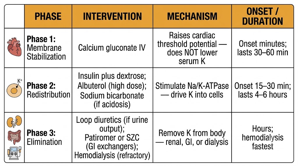
Acute severe hyperkalemia is managed in three mechanistically sequential phases that must be understood as distinct — not interchangeable — interventions. Membrane stabilization prevents fatal arrhythmia but does not lower serum potassium. Redistribution lowers serum potassium temporarily. Elimination actually removes potassium from the body. All three phases may be required simultaneously in severe cases.
Calcium gluconate is the first intervention for hyperkalemia with electrocardiogram changes. Calcium raises the threshold potential of cardiac myocytes, widening the gap between resting membrane potential and threshold and reducing the excitability that produces the peaked T waves, widened QRS complexes, and sine-wave pattern of severe hyperkalemia. The effect begins within minutes and lasts 30 to 60 minutes. Calcium does not lower serum potassium — it buys time for redistribution and elimination measures to work. Calcium chloride delivers three times as much elemental calcium per gram as calcium gluconate and is reserved for patients with central venous access because it causes tissue necrosis if it extravasates from a peripheral vein.
Regular insulin drives potassium into skeletal muscle cells by stimulating sodium-potassium-ATPase activity, shifting approximately 0.5 to 1.5 milliequivalents per liter of serum potassium intracellularly within 15 to 30 minutes. Dextrose is administered concurrently to prevent hypoglycemia, and blood glucose monitoring every 30 to 60 minutes is mandatory. The effect lasts 4 to 6 hours — enough time to initiate elimination measures. Sodium bicarbonate shifts potassium intracellularly through hydrogen-potassium exchange across the cell membrane and is most effective in patients with concurrent metabolic acidosis; its benefit in normokalemic patients is modest and contested.
Albuterol nebulization at 10 to 20 milligrams (four to eight times the standard bronchodilator dose) activates beta-2 adrenergic receptors on skeletal muscle, stimulating cyclic adenosine monophosphate-mediated sodium-potassium-ATPase activation and driving potassium intracellularly. It provides an additive effect when combined with insulin and can lower serum potassium by an additional 0.5 to 1.0 milliequivalents per liter. Tachycardia limits its use in patients with ischemic heart disease or hemodynamic instability.
Loop diuretics increase renal potassium excretion and are appropriate when urine output is adequate. Patiromer and sodium zirconium cyclosilicate are newer gastrointestinal cation exchangers that bind potassium in the gut and remove it in the stool — patiromer in the distal colon, sodium zirconium cyclosilicate throughout the gastrointestinal tract. Sodium polystyrene sulfonate (Kayexalate) is an older resin exchanger whose efficacy data are limited and whose use has been associated with intestinal necrosis, particularly when combined with sorbitol. Hemodialysis is the most effective and fastest method of potassium removal and is the treatment of choice when hyperkalemia is refractory to pharmacological measures or when renal function is absent.
Volume status determines etiology; correction rate determines neurological safety
Hyponatremia is the most common electrolyte abnormality in hospitalized patients. Serum sodium below 135 milliequivalents per liter is classified by volume status — hypovolemic, euvolemic, or hypervolemic — because each category has a distinct pathophysiology and treatment. The correction rate is the dominant safety variable regardless of etiology.
The maximum safe correction rate is 6 to 8 milliequivalents per liter in the first 24 hours, and no more than 10 to 12 milliequivalents per liter in any 24-hour period. Correction faster than these limits — particularly in patients with severe chronic hyponatremia (below 120 milliequivalents per liter present for more than 48 hours) — risks osmotic demyelination syndrome. In osmotic demyelination syndrome, the brain, having adapted to a low-sodium environment by extruding osmoles, is exposed to a rapidly rising osmolality before it can restore intracellular solutes; myelin sheaths in the pons and extrapontine regions are disrupted, causing dysarthria, dysphagia, quadriplegia, and potentially locked-in syndrome. Osmotic demyelination syndrome is largely irreversible and prevented by strict adherence to correction rate limits.
Three percent sodium chloride solution (containing 513 milliequivalents per liter of sodium) is the pharmacological intervention for acute severe hyponatremia with neurological symptoms — seizures, obtundation, or herniation. For symptomatic patients, 100 to 150 milliliters of 3% sodium chloride given as a rapid intravenous infusion over 10 to 20 minutes raises serum sodium by approximately 2 to 3 milliequivalents per liter and can abort active seizures. Serum sodium must be rechecked every 2 hours during active treatment to ensure the correction rate remains within safe limits. Once symptoms resolve, the rate is slowed immediately.
Vasopressin antagonists (vaptans) produce aquaresis — electrolyte-free water excretion — and are specifically suited to euvolemic hyponatremia from syndrome of inappropriate antidiuretic hormone secretion and hypervolemic hyponatremia from heart failure or cirrhosis, where antidiuretic hormone levels are inappropriately elevated. They are contraindicated in hypovolemic hyponatremia, where antidiuretic hormone secretion is appropriate and water excretion would worsen volume depletion. Their overcorrection risk requires free water access and frequent sodium monitoring — the same safety protocol outlined in Module 2. They are not first-line for most hyponatremia and are reserved for cases where fluid restriction and treating the underlying cause are insufficient.
If serum sodium rises faster than the correction limit, relowering is required to prevent osmotic demyelination syndrome. Administer intravenous free water (5% dextrose in water) at 3 to 6 milliliters per kilogram per hour to reduce serum sodium back toward the target range. Desmopressin can be given to reduce ongoing free water excretion. The goal is to keep cumulative 24-hour sodium correction below 10 to 12 milliequivalents per liter — even if this means actively lowering sodium that has already risen too fast.
Established indications, important limitations, and the anion gap as the key diagnostic tool
Metabolic acidosis is defined by a primary fall in serum bicarbonate and arterial pH. The anion gap — calculated as sodium minus the sum of chloride and bicarbonate — separates anion gap metabolic acidosis (lactic acidosis, diabetic ketoacidosis, toxin ingestion, renal failure) from non-anion gap metabolic acidosis (renal tubular acidosis, diarrhea, bicarbonate loss), each with different treatment implications for bicarbonate therapy.
Renal tubular acidosis is the clearest indication for oral sodium bicarbonate supplementation. In type 1 (distal) renal tubular acidosis, the collecting duct fails to secrete hydrogen ions adequately, producing a urine that cannot be acidified below a pH of 5.5 despite systemic acidemia. Alkali therapy corrects the acidosis, prevents nephrocalcinosis from calcium phosphate precipitation, and reduces osteomalacia from bone buffering of chronic acid load. In type 2 (proximal) renal tubular acidosis, bicarbonate is wasted in the urine because the proximal tubule cannot reabsorb it adequately; large supplemental doses are required because any administered bicarbonate is promptly filtered and lost. Chronic kidney disease-related metabolic acidosis responds to bicarbonate supplementation, with evidence from the bicarbonate supplementation study showing slowed chronic kidney disease progression when bicarbonate was maintained in the normal range.
In lactic acidosis and diabetic ketoacidosis, intravenous bicarbonate therapy carries risks that limit its routine use. The buffering reaction — bicarbonate plus hydrogen ion yields carbon dioxide plus water — generates carbon dioxide that freely diffuses into cells and crosses the blood-brain barrier, paradoxically worsening intracellular and cerebrospinal fluid acidosis even as extracellular pH rises. Additionally, rapid alkalization may reduce the drive to hyperventilate, allowing arterial carbon dioxide to rise. In diabetic ketoacidosis, treating the underlying insulin deficiency and restoring volume is the definitive intervention; bicarbonate is generally reserved for pH below 6.9 when cardiac contractility and catecholamine responsiveness are critically impaired by extreme acidemia.
Urine chloride as the diagnostic pivot — chloride-responsive versus chloride-resistant etiologies
Metabolic alkalosis is defined by a primary rise in serum bicarbonate and arterial pH. The urine chloride concentration is the pivotal diagnostic test that divides metabolic alkalosis into two mechanistically distinct categories with different treatments.
A urine chloride below 20 milliequivalents per liter indicates chloride-responsive metabolic alkalosis, in which the kidney is avidly retaining sodium and chloride in response to volume depletion or chloride deficit. Common causes include vomiting (loss of hydrochloric acid), nasogastric suctioning, and prior diuretic use that has since been discontinued. The alkalosis is maintained by the kidney's bicarbonate reabsorption in response to volume contraction and secondary aldosteronism. Treatment is isotonic saline to restore volume and chloride, which allows the kidney to excrete the excess bicarbonate. Potassium repletion is also required because hypokalemia independently perpetuates metabolic alkalosis through intracellular hydrogen-potassium exchange.
A urine chloride above 20 milliequivalents per liter indicates ongoing mineralocorticoid activity driving hydrogen ion and potassium secretion in the collecting duct, independent of volume status. Etiologies include primary hyperaldosteronism, Cushing's syndrome, exogenous mineralocorticoid use, and Bartter or Gitelman syndromes. Saline administration cannot correct this alkalosis because the mineralocorticoid excess continues to drive bicarbonate reabsorption regardless of volume status. Treatment targets the underlying mineralocorticoid excess — spironolactone or eplerenone for primary hyperaldosteronism, surgical resection of an aldosterone-producing adenoma when identified, or potassium-sparing diuretics for the tubular syndromes.
In volume-overloaded patients — decompensated heart failure, cirrhosis with ascites — who develop metabolic alkalosis from loop or thiazide diuretics but cannot receive saline because of their volume overload, acetazolamide provides targeted bicarbonate excretion. By inhibiting carbonic anhydrase in the proximal tubule, acetazolamide promotes bicarbonate loss in the urine, correcting the alkalosis without adding volume. The ADVOR trial demonstrated that adding acetazolamide to standard intravenous furosemide therapy in acute decompensated heart failure produced faster and more complete decongestion than furosemide alone, with the additional benefit of correcting the metabolic alkalosis that loop diuretics induce.
Amphotericin B, cisplatin, lithium, and tenofovir — four distinct nephrotoxic mechanisms with predictable electrolyte consequences
Several widely used drugs cause electrolyte disorders through specific renal tubular mechanisms that are predictable from their pharmacology. Recognizing these drug-electrolyte relationships allows anticipatory monitoring and timely intervention before serious toxicity develops.
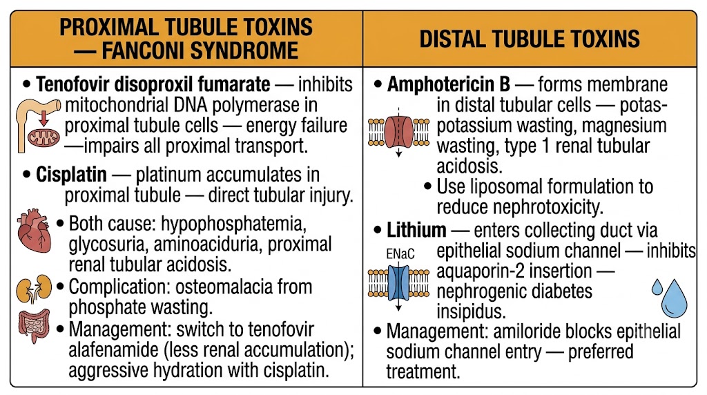
Amphotericin B inserts into ergosterol-rich fungal cell membranes to form pores that disrupt fungal integrity. At renal tubular concentrations, it forms similar pores in tubular cell membranes, producing a distal tubular acidification defect (type 1 renal tubular acidosis pattern), renal potassium wasting (hypokalemia), and renal magnesium wasting (hypomagnesemia). Amphotericin B also causes vasoconstriction of afferent arterioles, reducing glomerular filtration rate. Liposomal amphotericin B formulations dramatically reduce nephrotoxicity by limiting free drug delivery to the renal tubule while maintaining antifungal efficacy — they are the preferred formulation when renal protection is a priority.
Cisplatin causes severe hypomagnesemia through direct damage to the TRPM6 channel — the primary apical magnesium entry channel in the distal convoluted tubule. Platinum accumulation in tubular cells permanently downregulates TRPM6 expression, impairing renal magnesium reabsorption and producing a renal magnesium-wasting state that can persist for months to years after cisplatin discontinuation. Hypokalemia accompanies the hypomagnesemia through the same mechanism described in Module 1 — magnesium depletion prevents suppression of renal outer medullary potassium channel-mediated potassium secretion in the collecting duct. Cisplatin also causes proximal tubular injury producing a Fanconi syndrome pattern with phosphaturia, aminoaciduria, and glycosuria. Aggressive intravenous hydration before and after cisplatin infusion reduces but does not eliminate nephrotoxicity.
Lithium enters collecting duct principal cells via the epithelial sodium channel and accumulates intracellularly, where it inhibits adenylate cyclase-mediated cyclic adenosine monophosphate generation needed for aquaporin-2 insertion in response to antidiuretic hormone. The result is nephrogenic diabetes insipidus — vasopressin-resistant polyuria and polydipsia that can produce severe hypernatremia if fluid intake is restricted. Chronic lithium exposure causes irreversible interstitial fibrosis in some patients. Amiloride is the preferred treatment for lithium-induced nephrogenic diabetes insipidus: it blocks the epithelial sodium channel, reducing lithium entry into principal cells and attenuating the diabetes insipidus without the proximal lithium-reabsorption risk of thiazides.
Tenofovir disoproxil fumarate accumulates in proximal convoluted tubule mitochondria via tubular secretion by organic anion transporters, inhibiting mitochondrial deoxyribonucleic acid polymerase gamma and impairing mitochondrial energy production. Proximal tubular energy failure disrupts all energy-dependent reabsorptive transport at this segment — sodium-coupled glucose transport, phosphate transport, amino acid transport, and bicarbonate reclamation — producing a Fanconi syndrome with hypophosphatemia, glycosuria, aminoaciduria, and proximal renal tubular acidosis. Persistent hypophosphatemia from tubular wasting causes osteomalacia and increased fracture risk. Switching to tenofovir alafenamide, a prodrug formulation with less renal accumulation, substantially reduces the nephrotoxic and bone toxicity profile while maintaining antiviral efficacy.
| Author / Organization | Title | Source |
|---|---|---|
| Kovesdy CP | Management of hyperkalaemia in chronic kidney disease | Nat Rev Nephrol. 2014;10(11):653–662 |
| Elliott MJ, Ronksley PE, Clase CM, et al | Management of patients with acute hyperkalemia | CMAJ. 2010;182(15):1631–1635 |
| Verbalis JG, Goldsmith SR, Greenberg A, et al | Diagnosis, evaluation, and treatment of hyponatremia: expert panel recommendations | Am J Med. 2013;126(10 Suppl 1):S1–S42 |
| Spasovski G, Vanholder R, Allolio B, et al | Clinical practice guideline on diagnosis and treatment of hyponatraemia | Eur J Endocrinol. 2014;170(3):G1–G47 |
| Kraut JA, Madias NE | Metabolic acidosis: pathophysiology, diagnosis and management | Nat Rev Nephrol. 2010;6(5):274–285 |
| de Brito-Ashurst I, Varagunam M, Raftery MJ, Yaqoob MM | Bicarbonate supplementation slows progression of CKD and improves nutritional status | J Am Soc Nephrol. 2009;20(9):2075–2084 |
| Galla JH | Metabolic alkalosis | J Am Soc Nephrol. 2000;11(2):369–375 |
| Mullens W, Dauw J, Martens P, et al | Acetazolamide in acute decompensated heart failure with volume overload | N Engl J Med. 2022;387(13):1185–1195 |
| Perazella MA | Drug-induced nephropathy: an update | Expert Opin Drug Saf. 2005;4(4):689–706 |
| Miller RP, Tadagavadi RK, Ramesh G, Reeves WB | Mechanisms of cisplatin nephrotoxicity | Toxins (Basel). 2010;2(11):2490–2518 |
| Batlle DC, von Riotte AB, Gaviria M, Grupp M | Amelioration of polyuria by amiloride in patients receiving long-term lithium therapy | N Engl J Med. 1985;312(7):408–414 |
| KDIGO CKD Work Group | KDIGO 2024 clinical practice guideline for the evaluation and management of chronic kidney disease | Kidney Int. 2024;105(4S):S117–S314 |