Introduction to Medical Pharmacology
Module 8 — Proarrhythmia, Drug Interactions, and Special Populations
AARR · Module 8 of 9Section 1
The most clinically significant form of antiarrhythmic proarrhythmia -- and how to manage it
Drug-induced QT prolongation is one of the leading causes of drug withdrawal from the market and a major safety concern across multiple drug classes. The mechanism is blockade of the rapid repolarizing potassium channel, which is unusually susceptible to drug interaction across a remarkably broad range of chemical structures -- antibiotics, antifungals, antipsychotics, and antiemetics can all produce the same proarrhythmic effect as the antiarrhythmic drugs themselves.
Blocking the rapid repolarizing potassium channel slows phase 3 repolarization, prolonging the action potential duration and QT interval. A sufficiently prolonged action potential can generate early afterdepolarizations during phase 3, which directly trigger torsades de pointes -- a polymorphic ventricular tachycardia characterized by its twisting QRS axis around the isoelectric baseline. Torsades de pointes typically occurs in runs that may self-terminate or degenerate to ventricular fibrillation.
QT prolongation is necessary but not sufficient for torsades de pointes. The following factors substantially increase risk and must be actively assessed and managed:
Female sex accounts for approximately 70 percent of drug-induced torsades de pointes. Women have a longer baseline corrected QT interval and less repolarization reserve than men.
Hypokalemia and hypomagnesemia directly promote early afterdepolarization formation. These are modifiable risk factors -- electrolytes must be corrected before initiating any QT-prolonging drug and maintained throughout therapy. Target potassium at or above 4.0 to 4.5 mEq/L.
Bradycardia and pauses amplify action potential duration prolongation through reverse use-dependence. Torsades de pointes characteristically occurs after a pause.
Corrected QT interval over 500 milliseconds is the single most important quantitative threshold. If the corrected QT interval exceeds 500 milliseconds during drug therapy, the offending agent must be dose-reduced or discontinued.
Combinations of QT-prolonging drugs produce additive or synergistic QT prolongation. Avoid co-prescribing Class Ia antiarrhythmics with Class III agents, or antiarrhythmics with fluoroquinolone antibiotics, azole antifungals, or antipsychotics.
Many commonly used non-antiarrhythmic drugs carry significant QT prolongation risk by the same mechanism -- blocking the rapid repolarizing potassium channel. Clinically important categories include fluoroquinolone antibiotics (ciprofloxacin, levofloxacin, moxifloxacin), macrolide antibiotics (azithromycin, clarithromycin), azole antifungals (fluconazole, ketoconazole, itraconazole), antipsychotics (haloperidol, quetiapine, ziprasidone), antiemetics (ondansetron, domperidone), and methadone. This list represents categories where the risk is well-established; a complete, continuously updated database is maintained by the CredibleMeds program, which is the clinical standard for QT risk stratification when prescribing decisions require it.
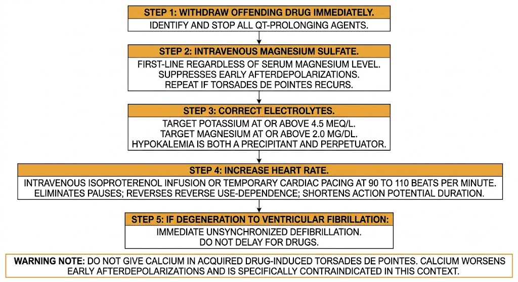
Step 1 -- Withdraw the offending drug immediately. Identify and stop all QT-prolonging agents.
Step 2 -- Intravenous magnesium sulfate is first-line regardless of the serum magnesium level. Magnesium suppresses early afterdepolarizations by blocking the calcium current that sustains them. A repeat dose may be given if torsades de pointes recurs.
Step 3 -- Correct electrolytes. Target potassium at or above 4.5 mEq/L and magnesium at or above 2.0 mg/dL. Hypokalemia is both a precipitant and a perpetuator of torsades de pointes.
Step 4 -- Increase heart rate to eliminate pauses and counteract reverse use-dependence. Intravenous isoproterenol infusion or temporary cardiac pacing at 90 to 110 beats per minute achieves this. Increasing rate shortens the action potential duration and eliminates the pause-dependent trigger for torsades de pointes recurrence.
Step 5 -- Defibrillate immediately if torsades de pointes degenerates to ventricular fibrillation. Do not delay for drug therapy.
Note: Do not give calcium in acquired drug-induced torsades de pointes. Calcium worsens early afterdepolarizations and is specifically indicated only for hyperkalemia and magnesium toxicity, not torsades de pointes.
Section 2
High-risk combinations requiring active dose management or drug avoidance
Antiarrhythmic drugs have extensive interaction potential through enzyme inhibition, transport protein inhibition, and additive effects on QT interval and cardiac conduction. The following interactions are high-yield because they require active management at the time of prescribing.
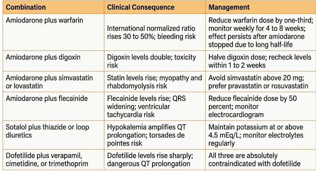
Amiodarone + warfarin: Amiodarone inhibits warfarin metabolism and displaces it from plasma proteins, raising the international normalized ratio by 30 to 50 percent. Reduce the warfarin dose by approximately one-third when amiodarone is added and monitor the international normalized ratio weekly for at least four to eight weeks. The interaction persists for weeks to months after amiodarone is stopped because of its long half-life.
Amiodarone + digoxin: Amiodarone inhibits the transport protein that clears digoxin and reduces its renal clearance, roughly doubling digoxin levels. Halve the digoxin dose when amiodarone is added and recheck levels within one to two weeks.
Amiodarone + simvastatin or lovastatin: Amiodarone inhibits the enzyme responsible for statin metabolism, raising statin levels and markedly increasing the risk of myopathy and rhabdomyolysis. Avoid simvastatin above 20 mg; prefer pravastatin or rosuvastatin in patients on amiodarone.
Amiodarone + flecainide: Amiodarone inhibits flecainide metabolism, raising flecainide levels and causing QRS widening and increased ventricular tachycardia risk. Reduce the flecainide dose by 50 percent and monitor the electrocardiogram when this combination is used.
Sotalol + thiazide or loop diuretics: Diuretic-induced hypokalemia amplifies QT prolongation and the risk of torsades de pointes in patients on sotalol. Maintain potassium at or above 4.5 mEq/L and monitor electrolytes regularly throughout sotalol therapy.
Dofetilide + verapamil, cimetidine, or trimethoprim: These three drugs inhibit the renal tubular transport that clears dofetilide from the body, raising dofetilide levels substantially and producing dangerous QT prolongation. All three are absolutely contraindicated in patients taking dofetilide and cannot be substituted by dose adjustment alone.
Class Ia + Class III antiarrhythmic combination: Combining agents that both block the rapid repolarizing potassium channel produces additive QT prolongation with disproportionate torsades de pointes risk. This combination should be avoided; if unavoidable, intensive QTc monitoring is required.
Section 3
Which drugs require dose adjustment and which are safe regardless of kidney function
Renal impairment affects antiarrhythmic management through two mechanisms: reduced drug clearance leading to accumulation and toxicity, and electrolyte disturbances (particularly hypokalemia in early chronic kidney disease and hyperkalemia in advanced disease) that amplify proarrhythmic risk. Knowing which drugs are renally eliminated is the practical clinical priority.
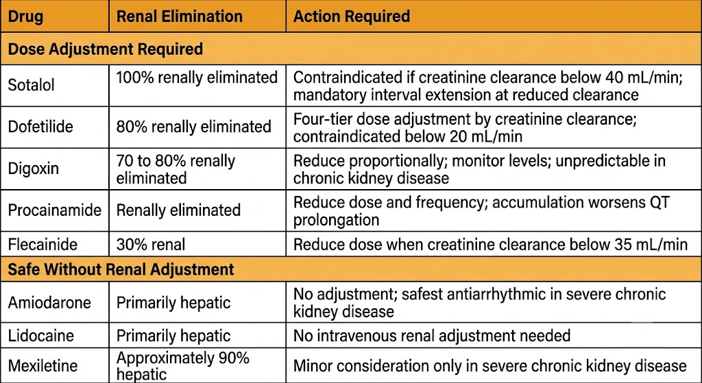
Sotalol is entirely renally eliminated and requires mandatory dose interval extension as creatinine clearance falls. It is contraindicated when creatinine clearance is below 40 mL/min because accumulation produces life-threatening QT prolongation.
Dofetilide is approximately 80 percent renally eliminated. Strict four-tier dose adjustment by creatinine clearance is mandatory; any dose change requires re-initiation with monitored corrected QT interval surveillance. It is contraindicated when creatinine clearance is below 20 mL/min.
Digoxin is 70 to 80 percent renally eliminated with a narrow therapeutic index. Dose reduction proportional to renal function is required; levels are unpredictable in chronic kidney disease and must be monitored regularly.
Procainamide and its active metabolite are both renally eliminated. Accumulation in renal failure prolongs QT interval. Dose and frequency reduction are required, and procainamide should be used with caution in significant renal impairment.
Flecainide is approximately 30 percent renally eliminated; dose reduction is required when creatinine clearance falls below 35 mL/min to prevent QRS widening from accumulation.
Amiodarone is primarily hepatically cleared and requires no dose adjustment in renal impairment, making it the most pharmacokinetically straightforward antiarrhythmic choice in patients with severe chronic kidney disease.
Lidocaine is primarily hepatically metabolized; intravenous dosing requires no renal adjustment, though prolonged infusions can produce central nervous system toxicity from metabolite accumulation.
Mexiletine is approximately 90 percent hepatically eliminated; only minor dose consideration in severe chronic kidney disease. Primarily used for long QT syndrome type 3 and as a ventricular tachycardia adjunct.
Section 4
Balancing maternal arrhythmia control against fetal drug exposure
Arrhythmias during pregnancy range from benign ectopy to life-threatening sustained tachycardias. All antiarrhythmic drugs cross the placenta to some degree. The goal is the lowest effective dose of the agent with the best-established safety record, with drug initiation in the first trimester avoided wherever possible.
Adenosine is the safest antiarrhythmic agent in pregnancy and is first-line for acute supraventricular tachycardia termination. Its elimination half-life of under ten seconds results in negligible fetal exposure. Vagal maneuvers are attempted first; if unsuccessful, adenosine is the agent of choice.
Beta-blockers have the most established safety record among oral antiarrhythmics in pregnancy. Metoprolol is preferred over atenolol, which has been associated with lower birth weight. Concerns include neonatal bradycardia and hypoglycemia; these are manageable with appropriate monitoring. Beta-blockers are used for rate control in atrial fibrillation, supraventricular tachycardia prophylaxis, long QT syndrome, and catecholaminergic polymorphic ventricular tachycardia during pregnancy.
Digoxin has decades of obstetric use and is considered relatively safe. It crosses the placenta; fetal heart rate should be monitored. It is also used for maternal dosing to treat fetal supraventricular tachycardia via placental transfer.
Direct-current cardioversion is safe at any gestational age when hemodynamic compromise requires it. It should be accompanied by fetal monitoring.
Amiodarone should be avoided in pregnancy if at all possible. Its high iodine content produces neonatal hypothyroidism in a significant proportion of exposed infants, and it is also associated with intrauterine growth restriction and premature birth. It is reserved for life-threatening maternal arrhythmias refractory to all other agents.
Flecainide is used for fetal supraventricular tachycardia treatment via maternal dosing and for maternal supraventricular tachycardia in the absence of structural heart disease, but data are limited.
Verapamil intravenous use carries risk of neonatal atrioventricular block and hemodynamic compromise; oral use is relatively safer. It is used for supraventricular tachycardia rate control when beta-blockers fail.
Section 5
Age-related pharmacokinetic changes, polypharmacy risk, and hepatically-cleared drugs
Older patients represent the largest population requiring antiarrhythmic therapy. Age-related reductions in renal function, hepatic blood flow, and lean body mass all alter drug behavior, and polypharmacy dramatically increases interaction and proarrhythmic risk. Hepatic impairment creates distinct challenges for a different subset of antiarrhythmic drugs.
Even without overt chronic kidney disease, kidney function declines with age. Renally-cleared antiarrhythmic drugs -- sotalol, dofetilide, digoxin, procainamide, and flecainide -- all accumulate in elderly patients at standard doses. Renal function should be formally calculated (not estimated from serum creatinine alone, which underestimates impairment in patients with low muscle mass) before initiating any renally-cleared antiarrhythmic.
The practical prescribing principles for elderly patients are: start at approximately half the standard adult dose and uptitrate cautiously; monitor the corrected QT interval at initiation, after any dose change, and after any new interacting drug is added; target digoxin levels in the lower therapeutic range (0.5 to 0.8 nanograms per milliliter); and review all co-medications systematically for QT-prolonging agents, because polypharmacy in elderly patients frequently includes multiple drugs with conditional QT risk (fluoroquinolones, azole antifungals, ondansetron, antidepressants).
For patients already on long-term amiodarone, annual monitoring -- thyroid function tests, liver function tests, chest radiograph, and ophthalmologic evaluation -- is more critical in elderly patients than in younger ones because of cumulative organ toxicity over years of exposure.
Significant hepatic impairment reduces the first-pass metabolism and ongoing clearance of drugs that are primarily hepatically eliminated. The most clinically important antiarrhythmics affected are lidocaine, propafenone, verapamil, and diltiazem. In cirrhosis, all four have substantially increased bioavailability and prolonged half-lives -- lidocaine maintenance infusion should be reduced by approximately half, and propafenone plasma levels can become unpredictable; dose reduction of 50 to 70 percent is required for verapamil and diltiazem in significant hepatic disease.
Amiodarone is primarily hepatically cleared and should be used with caution in hepatic impairment -- it is itself hepatotoxic and can worsen underlying liver disease. Liver function tests must be monitored closely. The renally-cleared drugs (sotalol, dofetilide, digoxin) require minimal adjustment for hepatic impairment alone.
| Author / Organization | Title | Source |
|---|---|---|
| Roden DM | Drug-induced prolongation of the QT interval | N Engl J Med. 2004;350(10):1013–1022 |
| Zipes DP, Camm AJ, Borggrefe M, et al | ACC/AHA/ESC 2006 guidelines for management of patients with ventricular arrhythmias and the prevention of sudden cardiac death | J Am Coll Cardiol. 2006;48(5):e247–e346 |
| Woosley RL, Heise CW, Gallo T, Woosley D, Romero KA | CredibleMeds Combined Risk QTDrugs List | AZCERT, Inc. Available at: www.CredibleMeds.org |
| Tisdale JE, Jayes HA, Kingery JR, et al | Development and validation of a risk score to predict QT-interval prolongation in hospitalized patients | Circ Cardiovasc Qual Outcomes. 2013;6(4):479–487 |
| Matzke GR, Aronoff GR, Atkinson AJ Jr, et al | Drug dosing consideration in patients with acute and chronic kidney disease: a clinical update from Kidney Disease: Improving Global Outcomes (KDIGO) | Kidney Int. 2011;80(11):1122–1137 |
| Page RL, Joglar JA, Caldwell MA, et al | 2015 ACC/AHA/HRS guideline for the management of adult patients with supraventricular tachycardia | J Am Coll Cardiol. 2016;67(13):e27–e115 |
| Fleg JL, Strait J | Age-associated changes in cardiovascular structure and function: a fertile milieu for future disease | Heart Fail Rev. 2012;17(4-5):545–554 |
| Joglar JA, Chung MK, Armbruster AL, et al | 2023 ACC/AHA/ACCP/HRS guideline for the diagnosis and management of atrial fibrillation | J Am Coll Cardiol. 2024;83(1):109–279 |
| Regitz-Zagrosek V, Roos-Hesselink JW, Bauersachs J, et al | 2018 ESC guidelines for the management of cardiovascular diseases during pregnancy | Eur Heart J. 2018;39(34):3165–3241 |
| Goldberger JJ, Cain ME, Hohnloser SH, et al | American Heart Association/American College of Cardiology Foundation/Heart Rhythm Society scientific statement on noninvasive risk stratification techniques for identifying patients at risk for sudden cardiac death | Circulation. 2008;118(14):1497–1518 |
| Hohnloser SH, Kuck KH, Dorian P, et al | Prophylactic use of an implantable cardioverter-defibrillator after acute myocardial infarction (DINAMIT) | N Engl J Med. 2004;351(24):2481–2488 |