CHAPTER 35  ·  ANTIBACTERIAL AGENTS

Section 1

Intrinsic and Acquired Resistance

Natural versus selected resistance, mutation versus horizontal gene transfer, and how resistance spreads

Bacterial resistance to antibiotics is an ancient biological trait. Understanding it requires distinguishing intrinsic resistance — a fixed species-level property present before any antibiotic exposure — from acquired resistance, which emerges through mutation or horizontal gene transfer in organisms that were previously susceptible.

Intrinsic Resistance

Intrinsic resistance is the natural insensitivity of a bacterial species to an antibiotic class, present in every member of the species regardless of prior antibiotic exposure. No selective pressure is required for it to be expressed. Classic examples include the resistance of Gram-negative bacteria to vancomycin (which cannot penetrate the outer membrane), the resistance of Pseudomonas aeruginosa to many penicillins due to limited outer membrane permeability combined with constitutive efflux pump expression, and the resistance of Enterococcus faecalis to all cephalosporins. Intrinsic resistance is predictable and stable; susceptibility testing is not required for drug-organism combinations known to exhibit it.

Acquired Resistance — Mutation

Acquired resistance arises in previously susceptible organisms through spontaneous chromosomal mutation or acquisition of foreign DNA carrying resistance determinants. When antibiotic concentrations kill susceptible bacteria but not a rare pre-existing resistant mutant, the mutant is selectively amplified. Antibiotics therefore select for resistance rather than cause it — a distinction that is mechanistically important. Prolonged subtherapeutic antibiotic concentrations are particularly prone to selecting for resistant mutants, because they kill susceptible organisms while allowing partially resistant variants to survive and undergo further mutational selection.

Acquired Resistance — Horizontal Gene Transfer

Horizontal gene transfer is the primary driver of the modern global resistance crisis. It operates through three mechanisms: conjugation (direct cell-to-cell transfer of plasmids via a pilus), transformation (uptake of free DNA from lysed bacteria), and transduction (bacteriophage-mediated DNA transfer). Conjugation is the most clinically important. Plasmids are extrachromosomal DNA elements that replicate independently and can carry multiple resistance genes, enabling co-transfer of resistance to several antibiotic classes in a single event — which explains why multidrug-resistant organisms frequently emerge with simultaneous resistance to multiple structurally unrelated drug classes.

Mobile genetic elements accelerate gene spread. Transposons carry resistance genes between chromosomes and plasmids. Integrons capture resistance gene cassettes through site-specific recombination and are strongly associated with clinical multidrug resistance. The combined action of these elements explains how resistance genes from environmental bacteria or livestock can appear rapidly in human pathogens worldwide.

Subtherapeutic Dosing and the Mutant Selection Window

When antibiotic concentrations fall below what is required to suppress even partially resistant mutants, those mutants are selectively amplified. Subtherapeutic dosing, missed doses, and early discontinuation are common clinical scenarios that allow concentrations to fall into this window, disproportionately selecting for resistance.


Section 2

Enzymatic Inactivation

Beta-lactamases, extended-spectrum beta-lactamases, carbapenemases, and aminoglycoside-modifying enzymes

Enzymatic inactivation is the most prevalent and clinically consequential category of resistance mechanism worldwide. Beta-lactamases alone account for the majority of resistance to the largest antibiotic class in clinical use, and their ongoing evolution under selective pressure has driven a perpetual cycle of broader-spectrum beta-lactams and new inhibitor combinations.

Beta-Lactamases

Beta-lactamases hydrolyze the beta-lactam ring, destroying antibacterial activity. Narrow-spectrum beta-lactamases hydrolyze penicillins and early cephalosporins but not extended-spectrum cephalosporins or carbapenems. Extended-spectrum beta-lactamases (ESBLs), which arose through mutations, hydrolyze most penicillins, all cephalosporin generations including ceftriaxone and ceftazidime, and aztreonam — but not carbapenems. ESBL-producing organisms, predominantly Escherichia coli and Klebsiella pneumoniae, are defined by susceptibility to carbapenems and inhibition by classical beta-lactamase inhibitors such as clavulanic acid and tazobactam.

AmpC beta-lactamases are inducible cephalosporinases that resist inhibition by classical inhibitors. Organisms with inducible AmpC — including Enterobacter, Serratia, Citrobacter freundii, Acinetobacter, and Pseudomonas aeruginosa — can develop high-level resistance during therapy with third-generation cephalosporins through selection of mutants that constitutively overproduce the enzyme. This is why third-generation cephalosporins are avoided for serious infections caused by these organisms even when initial susceptibility is reported.
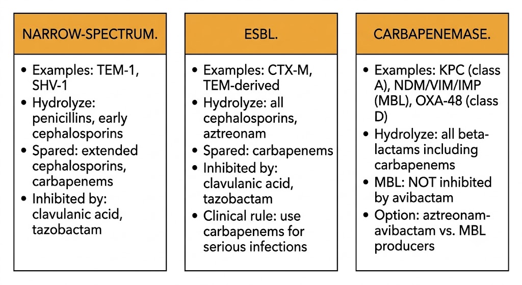
Three-panel diagram comparing beta-lactamase spectrum: narrow-spectrum enzymes hydrolyze penicillins only; extended-spectrum beta-lactamases hydrolyze all cephalosporins and aztreonam but not carbapenems; carbapenemases hydrolyze all beta-lactams including carbapenems.
Figure 1. Beta-lactamase spectrum: from narrow-spectrum hydrolysis through extended-spectrum beta-lactamases to carbapenemases. Generated with Gemini AI.
Carbapenemases

Carbapenemases hydrolyze carbapenems and eliminate the last reliable option for many multidrug-resistant Gram-negative infections. The major families are KPC (Klebsiella pneumoniae carbapenemase, most prevalent in the United States), NDM (New Delhi metallo-beta-lactamase, predominant in South Asia and now global), OXA-48 (prevalent in Europe and the Middle East), and other metallo-beta-lactamases. Metallo-beta-lactamases (including NDM) uniquely resist all currently available serine-targeted beta-lactamase inhibitors — notably, aztreonam combined with avibactam retains activity against many metallo-beta-lactamase producers because aztreonam is not hydrolyzed by metallo-beta-lactamases.

Aminoglycoside-Modifying Enzymes

Aminoglycoside-modifying enzymes modify the aminoglycoside molecule through acetylation, phosphorylation, or adenylylation, reducing or abolishing its ability to bind the 16S ribosomal ribonucleic acid of the 30S subunit. The specific aminoglycoside affected depends on the enzyme type and modification site — for example, some enzymes confer resistance to gentamicin but not amikacin, which explains why amikacin often retains activity against gentamicin-resistant organisms. Chloramphenicol acetyltransferase inactivates chloramphenicol; this is the primary mechanism of clinical chloramphenicol resistance.

ESBL Detection — A Clinical Laboratory Pitfall

ESBL-producing organisms may appear susceptible to extended-spectrum cephalosporins on routine susceptibility testing. Clinical failures with cephalosporins in ESBL-producing bacteremia are well documented even when in vitro susceptibility is reported. Carbapenems are the recommended definitive therapy for serious ESBL-producing bacteremia.

Section 3

Target Modification and Bypass Mechanisms

Penicillin-binding protein 2a and methicillin-resistant Staphylococcus aureus, vancomycin-resistant enterococcus D-Ala-D-Lac substitution, ribosomal methylation, and fluoroquinolone target mutations

Target modification mechanisms alter the bacterial molecule an antibiotic binds so that the drug can no longer interact effectively. Target bypass mechanisms provide an alternative pathway that circumvents the drug-inhibited step. Both strategies can confer high-level resistance with a single genetic event and account for some of the most clinically important resistance phenotypes.

Methicillin-Resistant Staphylococcus aureus — Penicillin-Binding Protein 2a

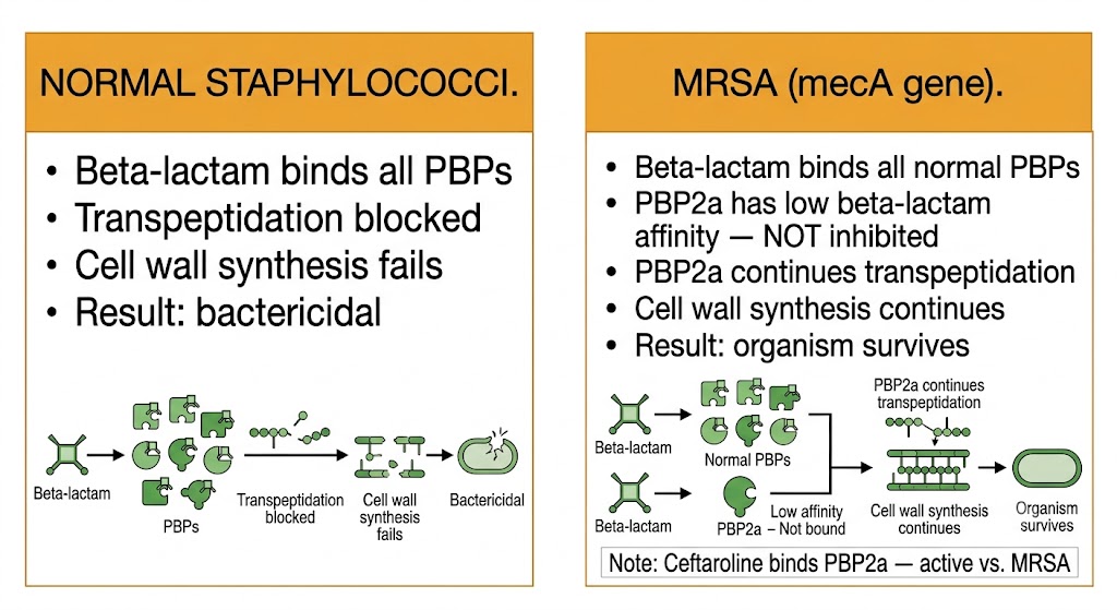
Beta-lactam antibiotics exert bactericidal activity by binding covalently to penicillin-binding proteins (PBPs), the transpeptidase enzymes responsible for cross-linking peptidoglycan. Methicillin-resistant Staphylococcus aureus resistance is mediated by the mecA gene, which encodes an alternative PBP designated PBP2a. PBP2a has greatly reduced affinity for all conventional beta-lactam antibiotics — while all other PBPs are occupied and inhibited by the drug, PBP2a continues to perform transpeptidation, enabling cell wall synthesis to proceed. The mecA gene is carried on a mobile genetic element integrated into the staphylococcal chromosome; different variants are associated with hospital-acquired and community-acquired methicillin-resistant Staphylococcus aureus strains.

Ceftaroline is the first beta-lactam with sufficient affinity for PBP2a to retain activity against methicillin-resistant Staphylococcus aureus, through greater affinity for PBP2a than conventional beta-lactams.

Two-panel diagram comparing normal staphylococci (beta-lactam binds all PBPs, transpeptidation blocked, bactericidal) versus MRSA with mecA gene (PBP2a has low affinity for beta-lactam and is not inhibited, cell wall synthesis continues, organism survives; ceftaroline binds PBP2a and is active against MRSA).
Figure 2. MRSA resistance mechanism: PBP2a encoded by mecA continues transpeptidation despite beta-lactam binding to all normal PBPs. Generated with Gemini AI.
Vancomycin-Resistant Enterococcus — D-Ala-D-Lac Substitution

Vancomycin resistance in enterococci is a target bypass mechanism. Vancomycin binds the D-alanyl-D-alanine terminus of the peptidoglycan precursor. Vancomycin-resistant enterococcus resistance, mediated primarily by the vanA and vanB gene clusters, reprograms cell wall biosynthesis to produce a precursor terminating in D-alanyl-D-lactate instead of D-alanyl-D-alanine. Vancomycin has approximately 1,000-fold lower affinity for the D-alanyl-D-lactate terminus, rendering it essentially ineffective. The vanA cluster confers high-level resistance to both vancomycin and teicoplanin and is transferable by conjugation. The vanB cluster confers variable resistance to vancomycin but not to teicoplanin. Transfer of vanA from vancomycin-resistant enterococcus to methicillin-resistant Staphylococcus aureus produces vancomycin-resistant Staphylococcus aureus — one of the most feared potential resistance developments in clinical infectious disease.

Ribosomal Methylation — Macrolide-Lincosamide-Streptogramin B Resistance

The erm gene family encodes methyltransferases that methylate the ribosomal binding site. This single methylation event simultaneously confers resistance to macrolides, lincosamides, and streptogramin B — the macrolide-lincosamide-streptogramin B phenotype. As noted in Module 10, erm expression can be constitutive (detectable by standard susceptibility testing) or inducible (appearing susceptible on routine testing but causing treatment failure with clindamycin, detectable by the D-zone test). Tetracycline resistance by ribosomal protection involves proteins that displace tetracycline from the ribosomal A-site, allowing translation to resume.

Fluoroquinolone Target Mutations

Fluoroquinolone resistance through target modification involves point mutations in the genes encoding DNA gyrase and topoisomerase IV, the two enzyme complexes targeted by fluoroquinolones. Single mutations in the primary target typically produce low- to moderate-level resistance; high-level resistance requires additional mutations in both primary and secondary targets. Because mutations in two independent genes must occur simultaneously for high-level resistance, fluoroquinolones can select for resistance through sequential single-step mutations during prolonged courses, with each mutation conferring incremental increases in minimum inhibitory concentration until the organism reaches clinical resistance.


Section 4

Efflux Pumps, Permeability Barriers, and Horizontal Gene Transfer

Active drug extrusion, outer membrane porin loss, multidrug resistance in Gram-negatives, and the epidemiology of global resistance gene dissemination

Efflux pumps and permeability barriers reduce intracellular drug concentrations by promoting drug exit or preventing entry, without chemically modifying the drug. These mechanisms are particularly important in Gram-negative organisms, where the outer membrane provides an intrinsic permeability barrier that synergizes with efflux to produce multidrug resistance affecting multiple structurally unrelated drug classes simultaneously.

Efflux Pumps

Bacterial efflux pumps are membrane transport proteins that actively extrude antibiotics from the bacterial cell interior. In Gram-negative bacteria, major efflux systems span both the inner and outer membranes, extruding drugs directly into the extracellular environment. These pumps simultaneously efflux fluoroquinolones, beta-lactams, tetracyclines, and chloramphenicol. Overexpression of these pumps through regulatory gene mutations is one of the most common mechanisms of fluoroquinolone resistance and simultaneously elevates resistance to multiple drug classes — producing multidrug resistance without any resistance gene acquisition.

Outer Membrane Permeability Reduction

Porins are outer membrane channel proteins through which hydrophilic antibiotics — including beta-lactams, fluoroquinolones, and carbapenems — diffuse into the periplasm of Gram-negative bacteria. Loss of specific porins through mutation significantly reduces antibiotic entry; when combined with efflux pump overexpression, porin loss produces synergistic resistance increases far exceeding what either mechanism alone confers. Loss of porins combined with extended-spectrum beta-lactamase or AmpC production is a mechanism of carbapenem resistance in organisms without a carbapenemase gene. In Pseudomonas aeruginosa, loss of a specific outer membrane porin combined with efflux overexpression produces selective imipenem resistance while meropenem may retain activity — explaining the clinical phenomenon of imipenem-resistant but meropenem-susceptible Pseudomonas aeruginosa.

Global Resistance Gene Dissemination

The epidemiology of resistance gene spread is best understood through successful plasmid-organism combinations that have spread globally. A globally disseminated lineage of Escherichia coli, which carries extended-spectrum beta-lactamase genes and is fluoroquinolone-resistant through chromosomal mutations, has become the dominant cause of community-onset ESBL-producing urinary tract infection in many countries without any link to healthcare exposure. Similarly, a specific Klebsiella pneumoniae lineage carrying KPC carbapenemases has established itself in healthcare institutions across multiple continents. These high-risk clones combine multiple resistance mechanisms with fitness attributes that enable prolonged silent colonization and transmission.

Agricultural antibiotic use creates selection pressure in animal gut flora that accelerates transfer and amplification of resistance genes subsequently transmissible to human pathogens through the food chain, water, and direct animal contact. The One Health framework, recognizing the interdependence of human, animal, and environmental health in resistance epidemiology, has become the dominant conceptual framework for global resistance control policy.

Antibiotic Stewardship — The Resistance Prevention Imperative

Every unnecessary antibiotic prescription amplifies selection pressure for resistance. The three foundational stewardship principles are: use antibiotics only when there is a genuine bacterial indication; choose the narrowest-spectrum agent effective for the likely pathogen; and use the shortest duration proven to be clinically effective. De-escalation from broad-spectrum empiric therapy to targeted narrow-spectrum therapy once culture and susceptibility results are available is both clinically appropriate and a direct resistance prevention intervention.


Suggested References

Suggested References

Author / Organization Title Source
Blair JMA, Webber MA, Baylay AJ, et al. Molecular mechanisms of antibiotic resistance Nat Rev Microbiol. 2015;13(1):42–51
Partridge SR, Kwong SM, Firth N, Jensen SO Mobile genetic elements associated with antimicrobial resistance Clin Microbiol Rev. 2018;31(4):e00088-17
Bush K, Bradford PA Beta-lactams and beta-lactamase inhibitors: an overview Cold Spring Harb Perspect Med. 2016;6(8):a025247
Pitout JDD, Laupland KB Extended-spectrum beta-lactamase-producing Enterobacteriaceae: an emerging public-health concern Lancet Infect Dis. 2008;8(3):159–166
van Duin D, Bonomo RA Ceftazidime/avibactam and ceftolozane/tazobactam: second-generation beta-lactam/beta-lactamase inhibitor combinations Clin Infect Dis. 2016;63(2):234–241
Ramirez MS, Tolmasky ME Aminoglycoside modifying enzymes Drug Resist Updat. 2010;13(6):151–171
Lowy FD Antimicrobial resistance: the example of Staphylococcus aureus J Clin Invest. 2003;111(9):1265–1273
Leclercq R Mechanisms of resistance to macrolides and lincosamides: nature of the resistance elements and their clinical implications Clin Infect Dis. 2002;34(4):482–492
Hooper DC, Jacoby GA Mechanisms of drug resistance: quinolone resistance Ann N Y Acad Sci. 2015;1354(1):12–31
Liu Y-Y, Wang Y, Walsh TR, et al. Emergence of plasmid-mediated colistin resistance mechanism MCR-1 in animals and human beings in China Lancet Infect Dis. 2016;16(2):161–168
Piddock LJV Clinically relevant chromosomally encoded multidrug resistance efflux pumps in bacteria Clin Microbiol Rev. 2006;19(2):382–402
Nikaido H Molecular basis of bacterial outer membrane permeability revisited Microbiol Mol Biol Rev. 2003;67(4):593–656
Tacconelli E, Carrara E, Savoldi A, et al. Discovery, research, and development of new antibiotics: the WHO priority list of antibiotic-resistant bacteria and tuberculosis Lancet Infect Dis. 2018;18(3):318–327
Antimicrobial Resistance Collaborators Global burden of bacterial antimicrobial resistance in 2019: a systematic analysis Lancet. 2022;399(10325):629–655
Cassini A, Hogberg LD, Plachouras D, et al. Attributable deaths and disability-adjusted life-years caused by infections with antibiotic-resistant bacteria in the EU and the European Economic Area in 2015 Lancet Infect Dis. 2019;19(1):56–66