CHAPTER 28  ·  ADRENOCORTICOSTEROID PHARMACOLOGY
Section 01
Metabolic Adverse Effects: Hyperglycemia, Dyslipidemia, and Steroid Myopathy
Glucocorticoid receptor-mediated gluconeogenesis, peripheral insulin resistance, dyslipidemia patterns, and pharmacological distinction of steroid myopathy from inflammatory myositis

The metabolic adverse effects of glucocorticoids arise from glucocorticoid receptor-mediated transactivation of metabolic gene programs that evolved to mobilize substrates during acute stress. Sustained pharmacological activation of these programs produces the constellation of iatrogenic Cushing syndrome: hyperglycemia, central adiposity, dyslipidemia, and skeletal muscle atrophy.

Hyperglycemia

Glucocorticoid-induced hyperglycemia results from simultaneous stimulation of hepatic glucose production and suppression of peripheral glucose utilization. In the liver, glucocorticoid receptor transactivation upregulates the gluconeogenic enzymes phosphoenolpyruvate carboxykinase and glucose-6-phosphatase, increasing glucose output from amino acid and glycerol substrates. In skeletal muscle and adipose tissue, glucocorticoids reduce glucose transporter type 4 (GLUT4) translocation to the plasma membrane in response to insulin, impairing insulin-stimulated glucose uptake independently of changes in circulating insulin.

The resulting pattern is predominantly postprandial hyperglycemia — fasting glucose is mildly elevated, but afternoon and evening excursions after meals are more pronounced. This differs from the predominantly fasting pattern of early type 2 diabetes, and means hemoglobin A1c underestimates postprandial dysglycemia if glucocorticoid therapy began within the preceding 8 to 12 weeks. Point-of-care glucose at 2 hours after the largest meal is the most sensitive monitoring strategy. In pre-existing diabetes, glucocorticoid therapy often requires acute escalation of antidiabetic medications.

Dyslipidemia and Steroid Myopathy

Glucocorticoid-induced dyslipidemia is characterized by elevated total cholesterol, low-density lipoprotein cholesterol, and triglycerides (via very low-density lipoprotein). The mechanism involves upregulation of hepatic lipogenic enzymes, increased free fatty acid flux from glucocorticoid-stimulated lipolysis in adipose tissue, and reduced low-density lipoprotein receptor expression. Lipid monitoring at baseline and every 3 months with statin initiation at standard cardiovascular risk thresholds is appropriate for patients on long-term therapy above a prednisone equivalent of 7.5 mg per day.

Steroid myopathy presents as symmetrical proximal limb weakness from glucocorticoid receptor-dependent upregulation of the ubiquitin ligases MuRF1 and MAFbx, which target myofibrillar proteins for proteasomal degradation, combined with suppression of muscle protein synthesis. Creatine kinase is normal or mildly elevated — in contrast to inflammatory myositis where creatine kinase is markedly elevated. This distinction matters: inflammatory myopathy is treated with increased glucocorticoids; steroid myopathy requires dose reduction. Fluorinated glucocorticoids (dexamethasone, triamcinolone) carry higher myopathy risk than non-fluorinated agents at equivalent anti-inflammatory doses.
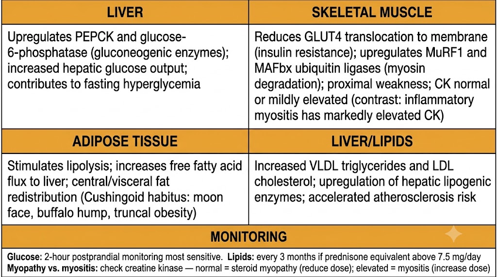
Summary diagram of glucocorticoid metabolic adverse effects organized by organ system
Figure 2. Glucocorticoid metabolic adverse effects by organ system. Hyperglycemia reflects simultaneous hepatic gluconeogenesis stimulation and peripheral insulin resistance. Steroid myopathy is distinguished from inflammatory myositis by normal or near-normal creatine kinase.

Section 02
Neuropsychiatric Effects and Ophthalmic Complications
Dose-dependent neuropsychiatric spectrum from euphoria to psychosis, posterior subcapsular cataract formation, and glucocorticoid-induced intraocular pressure elevation

Glucocorticoids affect the central nervous system and eyes through dose-dependent mechanisms that are underrecognized in clinical practice. Neuropsychiatric effects span from therapeutic mood elevation at low doses to frank psychosis at high doses. Ophthalmic complications are structural and require baseline evaluation before prolonged therapy.

Neuropsychiatric Effects

Neuropsychiatric adverse effects follow a dose-dependent spectrum reflecting glucocorticoid receptor density in limbic structures (hippocampus, amygdala, prefrontal cortex). At low to moderate doses (prednisone equivalent up to 20 mg per day), mild euphoria, improved energy, and insomnia are common — often experienced as beneficial short-term but contributing to difficulty tapering. At moderate to high doses (20 to 60 mg per day), irritability, emotional lability, anxiety, and significant insomnia emerge. At high doses (greater than 60 mg per day), serious psychiatric complications including major depression, mania, and frank psychosis (steroid psychosis) occur in approximately 5 to 10% of patients. Steroid psychosis typically resolves within days to weeks of dose reduction but may require temporary antipsychotic treatment.

Posterior Subcapsular Cataract

Posterior subcapsular cataract develops with both systemic and topical glucocorticoids (including inhaled and intranasal) through glucocorticoid receptor-dependent effects on lens epithelial cells that promote protein aggregate accumulation in the posterior subcapsular region. Development is slow (months to years), dose- and duration-dependent, and correlates with cumulative lifetime dose more than current dose. Early presentation is glare and difficulty with night driving; central vision is preserved until late stages. Baseline slit-lamp examination is recommended before initiating therapy anticipated to exceed 6 months, with annual examination thereafter.

Glucocorticoid-Induced Intraocular Pressure Elevation

Approximately 30 to 40% of patients on long-term systemic glucocorticoids develop elevated intraocular pressure, with 5 to 10% reaching levels sufficient to produce glaucomatous optic nerve damage. The mechanism is glucocorticoid receptor activation in trabecular meshwork cells, upregulating myocilin and extracellular matrix proteins that increase outflow resistance for aqueous humor. The intraocular pressure response is heritable and more common in individuals with primary open-angle glaucoma and their first-degree relatives. Intraocular pressure monitoring every 3 months during long-term therapy is standard; refer to ophthalmology for pressure greater than 21 mmHg.


Section 03
Glucocorticoid-Induced Osteoporosis: Mechanisms, Risk Assessment, and Prevention
RANKL/OPG imbalance, osteoblast suppression, fracture risk assessment with FRAX, bisphosphonate evidence base, and the roles of denosumab and teriparatide

Glucocorticoid-induced osteoporosis is the most common cause of secondary osteoporosis. Fracture risk exceeds what bone mineral density alone predicts because glucocorticoids impair bone quality through mechanisms beyond simple bone loss. Prevention is pharmacologically well-supported and should be initiated proactively.

Cellular Mechanisms

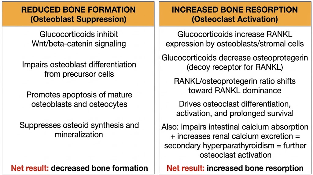
Glucocorticoids attack bone remodeling on both sides simultaneously. On the formation side, they suppress osteoblast differentiation by inhibiting Wnt/beta-catenin signaling and induce apoptosis of mature osteoblasts and osteocytes. On the resorption side, they increase RANKL expression by osteoblasts and stromal cells while suppressing osteoprotegerin (the decoy receptor that neutralizes RANKL), shifting the RANKL-to-osteoprotegerin ratio toward osteoclast activation. Additionally, glucocorticoids impair intestinal calcium absorption and increase renal calcium excretion, producing secondary hyperparathyroidism that further drives osteoclast activity.

Fracture Risk Assessment and Prevention

At any given bone mineral density T-score, glucocorticoid-treated patients have higher fracture probability than glucocorticoid-naive patients because bone quality deteriorates faster than density. The FRAX algorithm should be adjusted upward approximately 15% for major fracture probability and 20% for hip fracture probability in patients on prednisone greater than 7.5 mg per day for more than 3 months, to capture bone quality effects not measured by dual-energy X-ray absorptiometry.

For any patient initiating glucocorticoid therapy anticipated to last 3 or more months: start calcium 1000 to 1200 mg per day and vitamin D 600 to 800 international units per day universally. For medium or high fracture risk (FRAX-adjusted 10-year major fracture probability greater than 10 to 20%), add oral bisphosphonate therapy — alendronate 70 mg weekly or risedronate 35 mg weekly. Intravenous zoledronic acid annually is the alternative for patients with upper gastrointestinal intolerance or poor oral adherence.

Bone Protection Agents
Pharmacological Options in Glucocorticoid-Induced Osteoporosis
  • Bisphosphonates (alendronate, risedronate, zoledronic acid) — inhibit farnesyl pyrophosphate synthase in osteoclasts; 50–70% relative risk reduction for vertebral fractures; first-line for medium/high fracture risk; oral agents contraindicated if GFR below 30–35 mL/min/1.73 m²
  • Denosumab — monoclonal antibody targeting RANKL; directly neutralizes the key driver of osteoclast activation in glucocorticoid-induced osteoporosis; alternative when bisphosphonates are contraindicated; rebound bone loss occurs if doses are missed or therapy stops — transition to bisphosphonate before stopping
  • Teriparatide — recombinant parathyroid hormone (1–34); bone anabolic agent; stimulates osteoblast differentiation via PTH receptor 1; superior to alendronate for vertebral fracture prevention in glucocorticoid-induced osteoporosis; preferred at very high fracture risk (FRAX >20% or multiple vertebral fractures); 24-month treatment limit; transition to antiresorptive agent afterward
Diagram showing dual mechanism of glucocorticoid-induced osteoporosis affecting osteoblasts and osteoclasts
Figure 1. Dual mechanism of glucocorticoid-induced osteoporosis. Glucocorticoids simultaneously suppress bone formation by impairing osteoblast differentiation and increase bone resorption by shifting the RANKL-to-osteoprotegerin ratio toward osteoclast activation.

Section 04
Cardiovascular Effects and Infectious Complications
Pharmacological basis of glucocorticoid-induced hypertension, accelerated atherosclerosis, and the dose-dependent spectrum of infectious complications including Pneumocystis jirovecii pneumonia prophylaxis thresholds

Cardiovascular and infectious complications contribute substantially to the excess morbidity of long-term glucocorticoid therapy. Both are dose-dependent, mechanistically grounded, and partially preventable with targeted monitoring and prophylaxis.

Cardiovascular Effects

Glucocorticoid-induced hypertension arises through multiple parallel mechanisms: at pharmacological concentrations, glucocorticoids overwhelm the 11-beta-hydroxysteroid dehydrogenase type 2 enzyme that normally inactivates cortisol before it reaches the mineralocorticoid receptor in the distal nephron, causing sodium and water retention through epithelial sodium channel upregulation. Glucocorticoids also suppress endothelial nitric oxide synthase, reducing nitric oxide-mediated vasodilation, and increase vascular sensitivity to vasopressors including angiotensin II and catecholamines by upregulating their receptors on vascular smooth muscle. This mineralocorticoid overflow effect is most prominent with hydrocortisone and prednisolone, which have intrinsic mineralocorticoid activity.

Accelerated atherosclerosis is multifactorial: suppressed prostacyclin synthesis with preserved thromboxane A2 shifts the prostanoid balance toward platelet aggregation and vasoconstriction; central adiposity driven by glucocorticoid receptor-induced lipoprotein lipase upregulation in visceral fat generates a pro-inflammatory adipokine milieu; and compounding effects from hypertension, dyslipidemia, and hyperglycemia create a convergent cardiovascular risk burden. Atrial fibrillation risk is approximately doubled at high glucocorticoid doses, partly through hypokalemia from mineralocorticoid overflow and partly through direct glucocorticoid receptor effects on atrial cardiomyocyte electrical remodeling.

Infectious Complications

Common bacterial infections (pneumonia, urinary tract infection, skin and soft tissue infection) increase approximately 2-fold at prednisone equivalent doses above 10 mg per day. Opportunistic infections reflecting cell-mediated immunity impairment become clinically relevant above 20 mg per day for more than 4 weeks. Pneumocystis jirovecii pneumonia is the most consistently preventable opportunistic infection: prophylaxis with trimethoprim-sulfamethoxazole one double-strength tablet three times weekly is the standard when prednisone equivalent exceeds 20 mg per day for more than 4 weeks in glucocorticoid monotherapy, or 10 mg per day for more than 4 weeks when combined with additional immunosuppressants. For sulfonamide intolerance, alternatives are dapsone 100 mg daily or atovaquone 1500 mg daily.

Additional Prophylaxis Considerations

Herpes zoster reactivation is significantly increased in glucocorticoid-treated patients. Recombinant zoster vaccine (Shingrix) is recommended for patients 50 and older before initiating therapy or between cycles at the lowest immunosuppressive burden. The live attenuated varicella vaccine is contraindicated in significantly immunosuppressed patients. Tuberculosis reactivation requires interferon-gamma release assay or tuberculin skin test screening before long-term therapy in patients with risk factors; latent tuberculosis should be treated with isoniazid prophylaxis before beginning glucocorticoid therapy when possible. Pneumococcal vaccination is recommended for all adult patients on pharmacological glucocorticoid therapy regardless of age.


Section 05
Drug Interactions and Steroid-Sparing Strategies
CYP3A4 inducers and inhibitors, the glucocorticoid-warfarin and glucocorticoid-NSAID interactions, vaccination timing, and the pharmacological basis of steroid-sparing agents

Glucocorticoid drug interactions divide into pharmacokinetic interactions that alter plasma concentrations through cytochrome P450 3A4 modulation, and pharmacodynamic interactions that compound organ-specific toxicities. Steroid-sparing agents are pharmacologically necessary in any patient requiring prednisone equivalent above 7.5 mg per day for more than 3 months.

CYP3A4 Interactions

Cytochrome P450 3A4 inducers accelerate glucocorticoid metabolism and can reduce prednisolone exposure by 45 to 75%. The most clinically significant inducers are rifampin (which has caused acute transplant rejection by reducing glucocorticoid levels below the therapeutic threshold), phenytoin, carbamazepine, phenobarbital, and efavirenz. Patients on these combinations require higher glucocorticoid doses; when the inducer is discontinued, the previously compensatory dose may become toxic as normal enzyme activity returns over 2 to 4 weeks.

Cytochrome P450 3A4 inhibitors increase glucocorticoid concentrations and can produce iatrogenic Cushing syndrome at standard doses. Key inhibitors: azole antifungals (ketoconazole, itraconazole, voriconazole), clarithromycin, and ritonavir-boosted antiretroviral regimens. The ritonavir-fluticasone inhaled corticosteroid combination raises fluticasone levels approximately 350-fold and can cause Cushing syndrome even with standard inhaled doses; beclomethasone (not a cytochrome P450 3A4 substrate) is the preferred inhaled corticosteroid alternative in this population.

Warfarin and NSAID Interactions

The glucocorticoid-warfarin interaction produces variable international normalized ratio changes through effects on hepatic factor synthesis and protein binding competition; the international normalized ratio should be measured within 1 to 2 weeks of initiating or significantly changing a glucocorticoid dose in any patient on warfarin. The interaction with nonsteroidal anti-inflammatory drugs is pharmacodynamic: both independently suppress prostaglandin-mediated gastric mucosal defense, and their combination produces approximately 15-fold increased risk of peptic ulcer complications compared with neither drug alone (versus approximately 3-fold for each alone). Proton pump inhibitor prophylaxis is indicated when both agents are used simultaneously.

Steroid-Sparing Agents
Mechanism, Onset, and Principal Indications
  • Methotrexate — dihydrofolate reductase inhibition plus adenosine-mediated anti-inflammation; onset 6–12 weeks; permits 30–50% prednisone dose reduction; indications: rheumatoid arthritis, inflammatory myopathies, vasculitis, psoriatic arthritis; monitor liver function tests and complete blood count; folate supplementation required
  • Azathioprine — prodrug converted to 6-mercaptopurine; inhibits de novo purine synthesis in lymphocytes; onset 3–6 months; check thiopurine methyltransferase genotype before initiation (poor metabolizers at risk for myelosuppression); indications: inflammatory bowel disease, autoimmune hepatitis, myasthenia gravis, transplantation
  • Mycophenolate mofetil — selective inosine monophosphate dehydrogenase type II inhibition in activated lymphocytes; onset 4–8 weeks; indications: lupus nephritis (preferred first-line with hydroxychloroquine), transplantation, IgA nephropathy; teratogenic — pregnancy prevention required
  • Tocilizumab — interleukin-6 receptor antagonist; onset 4–8 weeks; proven steroid-sparing in giant cell arteritis (GiACTA trial); also used in rheumatoid arthritis; screen for tuberculosis and hepatitis B before initiating; monitor for serious infections and lipid elevation

Suggested References
Author / OrganizationTitleSource
Schacke H et al.Mechanisms involved in the side effects of glucocorticoidsPharmacol Ther. 2002;96(1):23–43
Liu D et al.A practical guide to the monitoring and management of the complications of systemic corticosteroid therapyAllergy Asthma Clin Immunol. 2013;9(1):30
Huscher D et al.Dose-related patterns of glucocorticoid-induced side effectsAnn Rheum Dis. 2009;68(7):1119–1124
Henzen C et al.Suppression and recovery of adrenal response after short-term, high-dose glucocorticoid treatmentLancet. 2000;355(9203):542–545
Warrington TP, Bostwick JMPsychiatric adverse effects of corticosteroidsMayo Clin Proc. 2006;81(10):1361–1367
Jones R, Rhee DJCorticosteroid-induced ocular hypertension and glaucomaCurr Opin Ophthalmol. 2006;17(2):163–167
Weinstein RSGlucocorticoid-induced bone diseaseN Engl J Med. 2011;365(1):62–70
Kanis JA et al.A meta-analysis of prior corticosteroid use and fracture riskJ Bone Miner Res. 2004;19(6):893–899
Buckley L et al.2017 ACR Guideline for the Prevention and Treatment of Glucocorticoid-Induced OsteoporosisArthritis Rheumatol. 2017;69(8):1521–1537
Saag KG et al.Teriparatide or alendronate in glucocorticoid-induced osteoporosisN Engl J Med. 2007;357(20):2028–2039
Souverein PC et al.Use of oral glucocorticoids and risk of cardiovascular and cerebrovascular diseaseHeart. 2004;90(8):859–865
Baddley JW et al.Non-viral opportunistic infections in new users of tumour necrosis factor inhibitor therapy (SABER study)Ann Rheum Dis. 2014;73(11):1942–1948
Czock D et al.Pharmacokinetics and pharmacodynamics of systemically administered glucocorticoidsClin Pharmacokinet. 2005;44(1):61–98
Stone JH et al.Trial of tocilizumab in giant-cell arteritisN Engl J Med. 2017;377(4):317–328